Medicină nucleară — Scintigrafie renală (MCB)
Sursă și prelucrare editorială
- Proveniență
- 1 referință declarată
- Fidelitatea preluării
- Nedocumentată
- Revizuire medicală
- Nedocumentată
Referințele declarate nu confirmă singure fidelitatea preluării sau aprobarea protocolului pentru utilizare locală.
Detalii despre surse și verificări
MCB Radiology — Renal Scan
- Ediție / versiune a sursei
- Neprecizată
- Pagină / secțiune
- Pagini: 1–3
- Relația cu protocolul
- Document original importat integral; atribuirea autorilor rămâne cea din PDF.
- Data consultării
- 2026-09-27
Prelucrarea acestei pagini
- Mod de prelucrare
- Titlu și navigare în română; documentul sursă în engleză.; PDF original integral, previzualizare și text extras automat; fără completarea parametrilor clinici lipsă.
- Adaptări față de sursă
- Nedocumentate; absența evidenței nu confirmă o preluare integrală.
- Responsabil pentru prelucrare
- Neprecizat
- Atribuire existentă în pagină
- MCB Radiology (portalul sursă). Această atribuire nu identifică automat autorul sursei sau revizorul medical.
- Versiunea paginii
- mcb-192e27b19b14
- Actualizare declarată a paginii
- 2026-09-27 — distinctă de revizuirea medicală.
Verificarea fidelității: Nedocumentată
Revizuirea medicală: Nedocumentată
Documentul original MCB — Scintigrafie renală
Protocol · 3 pagini · consultat la 2026-09-27.
Descarcă PDF-ul integral · Sursa MCB · Catalogul MCB pentru această modalitate
Instrucțiunile, valorile și ilustrațiile din documentul original sunt păstrate în engleză. Titlul și navigarea sunt în română.
Pagina 1
Pagina 2
Pagina 3
Textul documentului original
Pagina 1 — text în engleză
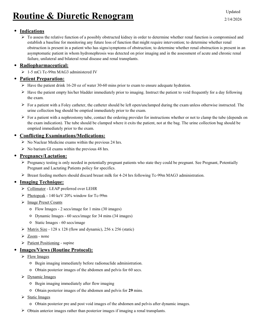
Updated Routine & Diuretic Renogram 2/14/2026 ●Indications To assess the relative function of a possibly obstructed kidney in order to determine whether renal function is compromised and establish a baseline for monitoring any future loss of function that might require intervention; to determine whether renal obstruction is present in a patient who has signs/symptoms of obstruction; to determine whether renal obstruction is present in an asymptomatic patient in whom hydronephrosis was detected on prior imaging and in the assessment of acute and chronic renal failure, unilateral and bilateral renal disease and renal transplants. ●Radiopharmaceutical: 1-5 mCi Tc-99m MAG3 administered IV ●Patient Preparation: Have the patient drink 16-20 oz of water 30-60 mins prior to exam to ensure adequate hydration. Have the patient empty his/her bladder immediately prior to imaging. Instruct the patient to void frequently for a day following the exam. For a patient with a Foley catheter, the catheter should be left open/unclamped during the exam unless otherwise instructed. The urine collection bag should be emptied immediately prior to the exam. For a patient with a nephrostomy tube, contact the ordering provider for instructions whether or not to clamp the tube (depends on the exam indication). The tube should be clamped where it exits the patient, not at the bag. The urine collection bag should be emptied immediately prior to the exam. ●Conflicting Examinations/Medications: No Nuclear Medicine exams within the previous 24 hrs. No barium GI exams within the previous 48 hrs. ●Pregnancy/Lactation: Pregnancy testing is only needed in potentially pregnant patients who state they could be pregnant. See Pregnant, Potentially Pregnant and Lactating Patients policy for specifics. Breast feeding mothers should discard breast milk for 4-24 hrs following Tc-99m MAG3 administration. ●Imaging Technique: Collimator - LEAP preferred over LEHR Photopeak - 140 keV 20% window for Tc-99m Image Preset Counts ◦Flow Images - 2 secs/image for 1 mins (30 images) ◦Dynamic Images - 60 secs/image for 34 mins (34 images) ◦Static Images - 60 secs/image Matrix Size - 128 x 128 (flow and dynamic), 256 x 256 (static) Zoom - none Patient Positioning - supine ●Images/Views (Routine Protocol): Flow Images ◦Begin imaging immediately before radionuclide administration. ◦Obtain posterior images of the abdomen and pelvis for 60 secs. Dynamic Images ◦Begin imaging immediately after flow imaging ◦Obtain posterior images of the abdomen and pelvis for 29 mins. Static Images ◦Obtain posterior pre and post void images of the abdomen and pelvis after dynamic images. Obtain anterior images rather than posterior images if imaging a renal transplants.
Pagina 2 — text în engleză
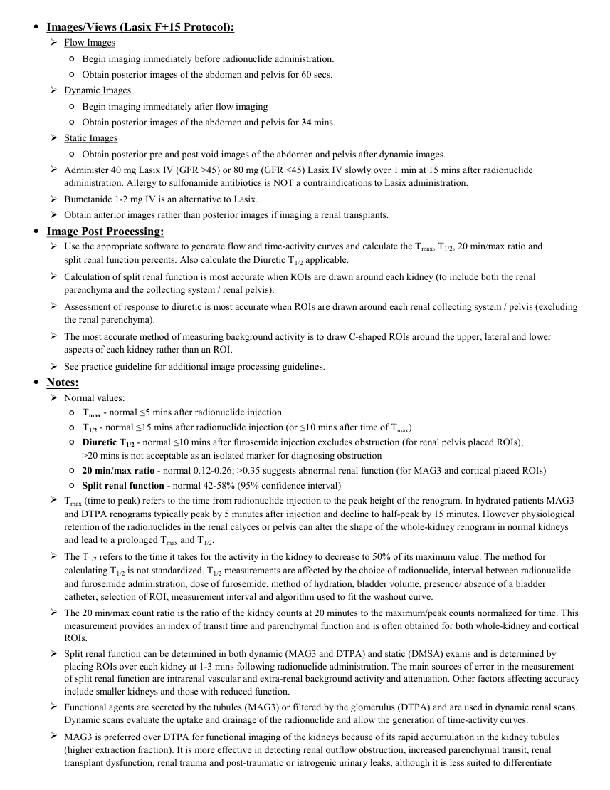
●Images/Views (Lasix F+15 Protocol): Flow Images ◦Begin imaging immediately before radionuclide administration. ◦Obtain posterior images of the abdomen and pelvis for 60 secs. Dynamic Images ◦Begin imaging immediately after flow imaging ◦Obtain posterior images of the abdomen and pelvis for 34 mins. Static Images ◦Obtain posterior pre and post void images of the abdomen and pelvis after dynamic images. Administer 40 mg Lasix IV (GFR >45) or 80 mg (GFR <45) Lasix IV slowly over 1 min at 15 mins after radionuclide administration. Allergy to sulfonamide antibiotics is NOT a contraindications to Lasix administration. Bumetanide 1-2 mg IV is an alternative to Lasix. Obtain anterior images rather than posterior images if imaging a renal transplants. ●Image Post Processing: Use the appropriate software to generate flow and time-activity curves and calculate the Tmax, T1/2, 20 min/max ratio and
split renal function percents. Also calculate the Diuretic T1/2 applicable. Calculation of split renal function is most accurate when ROIs are drawn around each kidney (to include both the renal parenchyma and the collecting system / renal pelvis). Assessment of response to diuretic is most accurate when ROIs are drawn around each renal collecting system / pelvis (excluding the renal parenchyma). The most accurate method of measuring background activity is to draw C-shaped ROIs around the upper, lateral and lower aspects of each kidney rather than an ROI. See practice guideline for additional image processing guidelines. ●Notes: Normal values: ◦Tmax - normal ≤5 mins after radionuclide injection ◦T1/2 - normal ≤15 mins after radionuclide injection (or ≤10 mins after time of Tmax) ◦ Diuretic T1/2 - normal ≤10 mins after furosemide injection excludes obstruction (for renal pelvis placed ROIs),
>20 mins is not acceptable as an isolated marker for diagnosing obstruction ◦20 min/max ratio - normal 0.12-0.26; >0.35 suggests abnormal renal function (for MAG3 and cortical placed ROIs) ◦Split renal function - normal 42-58% (95% confidence interval) Tmax (time to peak) refers to the time from radionuclide injection to the peak height of the renogram. In hydrated patients MAG3 and DTPA renograms typically peak by 5 minutes after injection and decline to half-peak by 15 minutes. However physiological retention of the radionuclides in the renal calyces or pelvis can alter the shape of the whole-kidney renogram in normal kidneys and lead to a prolonged Tmax and T1/2. The T1/2 refers to the time it takes for the activity in the kidney to decrease to 50% of its maximum value. The method for calculating T1/2 is not standardized. T1/2 measurements are affected by the choice of radionuclide, interval between radionuclide and furosemide administration, dose of furosemide, method of hydration, bladder volume, presence/ absence of a bladder catheter, selection of ROI, measurement interval and algorithm used to fit the washout curve. The 20 min/max count ratio is the ratio of the kidney counts at 20 minutes to the maximum/peak counts normalized for time. This measurement provides an index of transit time and parenchymal function and is often obtained for both whole-kidney and cortical ROIs. Split renal function can be determined in both dynamic (MAG3 and DTPA) and static (DMSA) exams and is determined by placing ROIs over each kidney at 1-3 mins following radionuclide administration. The main sources of error in the measurement of split renal function are intrarenal vascular and extra-renal background activity and attenuation. Other factors affecting accuracy include smaller kidneys and those with reduced function. Functional agents are secreted by the tubules (MAG3) or filtered by the glomerulus (DTPA) and are used in dynamic renal scans. Dynamic scans evaluate the uptake and drainage of the radionuclide and allow the generation of time-activity curves. MAG3 is preferred over DTPA for functional imaging of the kidneys because of its rapid accumulation in the kidney tubules (higher extraction fraction). It is more effective in detecting renal outflow obstruction, increased parenchymal transit, renal transplant dysfunction, renal trauma and post-traumatic or iatrogenic urinary leaks, although it is less suited to differentiate preserved perfusion in ATN (tubular retention is associated with a higher radiation dose).
split renal function percents. Also calculate the Diuretic T1/2 applicable. Calculation of split renal function is most accurate when ROIs are drawn around each kidney (to include both the renal parenchyma and the collecting system / renal pelvis). Assessment of response to diuretic is most accurate when ROIs are drawn around each renal collecting system / pelvis (excluding the renal parenchyma). The most accurate method of measuring background activity is to draw C-shaped ROIs around the upper, lateral and lower aspects of each kidney rather than an ROI. See practice guideline for additional image processing guidelines. ●Notes: Normal values: ◦Tmax - normal ≤5 mins after radionuclide injection ◦T1/2 - normal ≤15 mins after radionuclide injection (or ≤10 mins after time of Tmax) ◦ Diuretic T1/2 - normal ≤10 mins after furosemide injection excludes obstruction (for renal pelvis placed ROIs),
>20 mins is not acceptable as an isolated marker for diagnosing obstruction ◦20 min/max ratio - normal 0.12-0.26; >0.35 suggests abnormal renal function (for MAG3 and cortical placed ROIs) ◦Split renal function - normal 42-58% (95% confidence interval) Tmax (time to peak) refers to the time from radionuclide injection to the peak height of the renogram. In hydrated patients MAG3 and DTPA renograms typically peak by 5 minutes after injection and decline to half-peak by 15 minutes. However physiological retention of the radionuclides in the renal calyces or pelvis can alter the shape of the whole-kidney renogram in normal kidneys and lead to a prolonged Tmax and T1/2. The T1/2 refers to the time it takes for the activity in the kidney to decrease to 50% of its maximum value. The method for calculating T1/2 is not standardized. T1/2 measurements are affected by the choice of radionuclide, interval between radionuclide and furosemide administration, dose of furosemide, method of hydration, bladder volume, presence/ absence of a bladder catheter, selection of ROI, measurement interval and algorithm used to fit the washout curve. The 20 min/max count ratio is the ratio of the kidney counts at 20 minutes to the maximum/peak counts normalized for time. This measurement provides an index of transit time and parenchymal function and is often obtained for both whole-kidney and cortical ROIs. Split renal function can be determined in both dynamic (MAG3 and DTPA) and static (DMSA) exams and is determined by placing ROIs over each kidney at 1-3 mins following radionuclide administration. The main sources of error in the measurement of split renal function are intrarenal vascular and extra-renal background activity and attenuation. Other factors affecting accuracy include smaller kidneys and those with reduced function. Functional agents are secreted by the tubules (MAG3) or filtered by the glomerulus (DTPA) and are used in dynamic renal scans. Dynamic scans evaluate the uptake and drainage of the radionuclide and allow the generation of time-activity curves. MAG3 is preferred over DTPA for functional imaging of the kidneys because of its rapid accumulation in the kidney tubules (higher extraction fraction). It is more effective in detecting renal outflow obstruction, increased parenchymal transit, renal transplant dysfunction, renal trauma and post-traumatic or iatrogenic urinary leaks, although it is less suited to differentiate preserved perfusion in ATN (tubular retention is associated with a higher radiation dose).
Pagina 3 — text în engleză
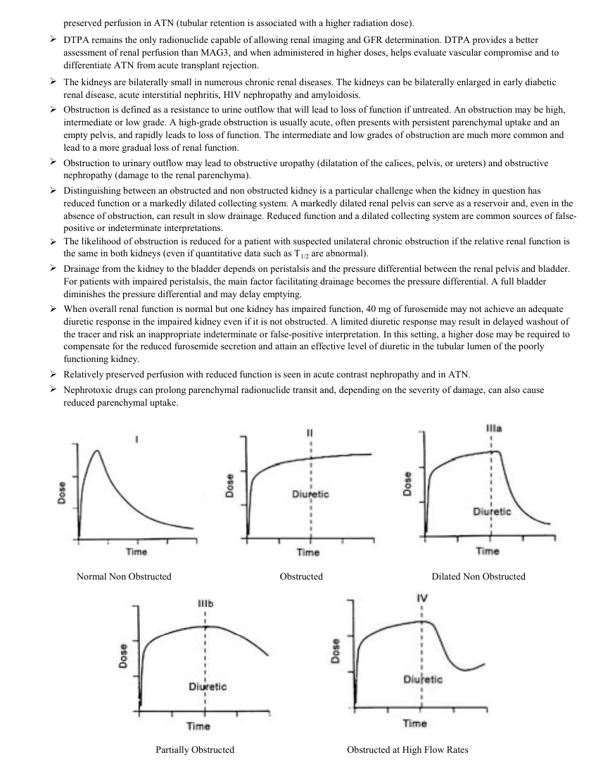
preserved perfusion in ATN (tubular retention is associated with a higher radiation dose). DTPA remains the only radionuclide capable of allowing renal imaging and GFR determination. DTPA provides a better assessment of renal perfusion than MAG3, and when administered in higher doses, helps evaluate vascular compromise and to differentiate ATN from acute transplant rejection. The kidneys are bilaterally small in numerous chronic renal diseases. The kidneys can be bilaterally enlarged in early diabetic renal disease, acute interstitial nephritis, HIV nephropathy and amyloidosis. Obstruction is defined as a resistance to urine outflow that will lead to loss of function if untreated. An obstruction may be high, intermediate or low grade. A high-grade obstruction is usually acute, often presents with persistent parenchymal uptake and an empty pelvis, and rapidly leads to loss of function. The intermediate and low grades of obstruction are much more common and lead to a more gradual loss of renal function. Obstruction to urinary outflow may lead to obstructive uropathy (dilatation of the calices, pelvis, or ureters) and obstructive nephropathy (damage to the renal parenchyma). Distinguishing between an obstructed and non obstructed kidney is a particular challenge when the kidney in question has reduced function or a markedly dilated collecting system. A markedly dilated renal pelvis can serve as a reservoir and, even in the absence of obstruction, can result in slow drainage. Reduced function and a dilated collecting system are common sources of false- positive or indeterminate interpretations. The likelihood of obstruction is reduced for a patient with suspected unilateral chronic obstruction if the relative renal function is the same in both kidneys (even if quantitative data such as T1/2 are abnormal). Drainage from the kidney to the bladder depends on peristalsis and the pressure differential between the renal pelvis and bladder. For patients with impaired peristalsis, the main factor facilitating drainage becomes the pressure differential. A full bladder diminishes the pressure differential and may delay emptying. When overall renal function is normal but one kidney has impaired function, 40 mg of furosemide may not achieve an adequate diuretic response in the impaired kidney even if it is not obstructed. A limited diuretic response may result in delayed washout of the tracer and risk an inappropriate indeterminate or false-positive interpretation. In this setting, a higher dose may be required to compensate for the reduced furosemide secretion and attain an effective level of diuretic in the tubular lumen of the poorly functioning kidney. Relatively preserved perfusion with reduced function is seen in acute contrast nephropathy and in ATN. Nephrotoxic drugs can prolong parenchymal radionuclide transit and, depending on the severity of damage, can also cause reduced parenchymal uptake. Normal Non Obstructed Obstructed Dilated Non Obstructed Partially Obstructed Obstructed at High Flow Rates