Instrucțiunile, valorile și ilustrațiile din documentul original sunt păstrate în engleză. Titlul și navigarea sunt în română.
Pagina 1
Pagina 2
Textul documentului original
Pagina 1 — text în engleză
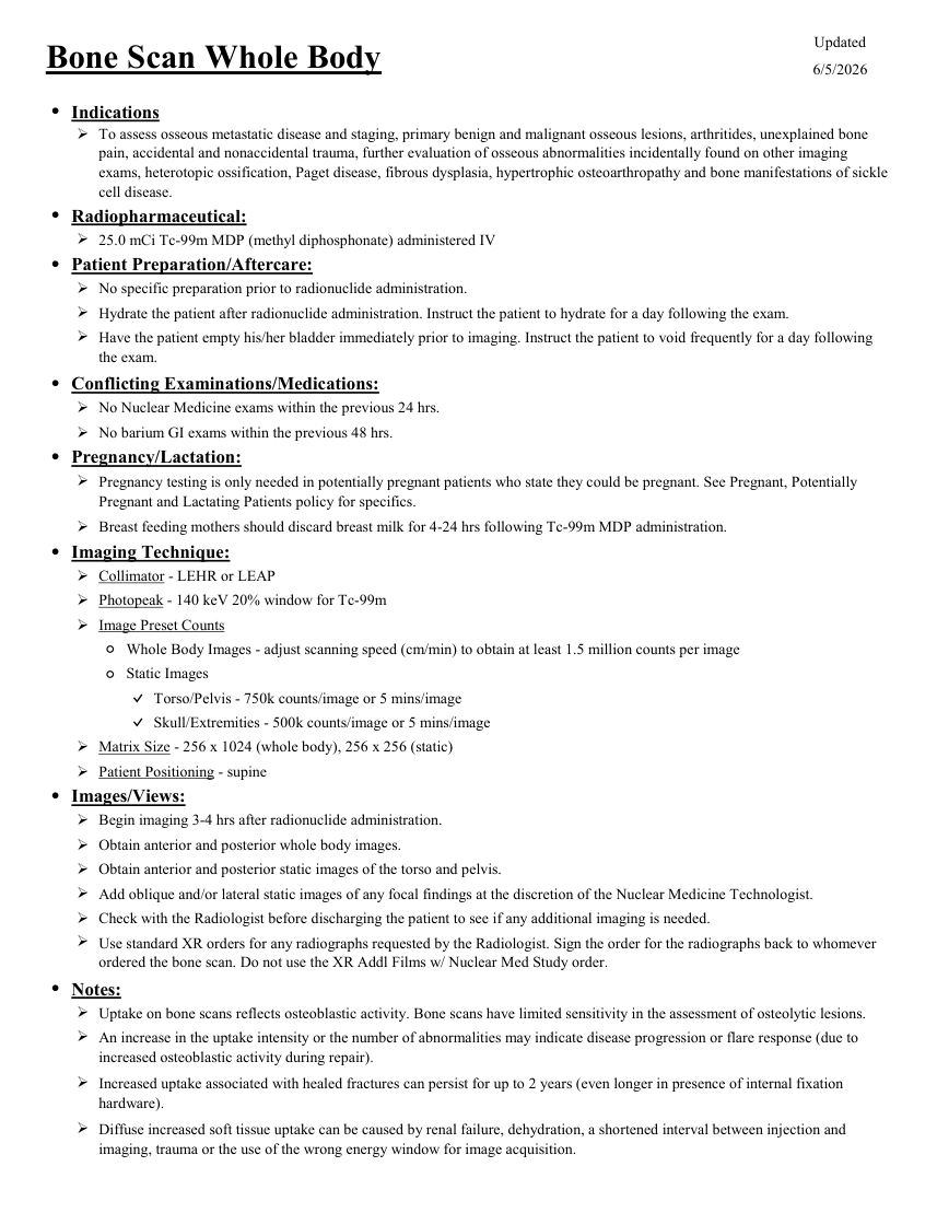
Updated Bone Scan Whole Body 6/5/2026 ●Indications To assess osseous metastatic disease and staging, primary benign and malignant osseous lesions, arthritides, unexplained bone pain, accidental and nonaccidental trauma, further evaluation of osseous abnormalities incidentally found on other imaging exams, heterotopic ossification, Paget disease, fibrous dysplasia, hypertrophic osteoarthropathy and bone manifestations of sickle cell disease. ●Radiopharmaceutical: 25.0 mCi Tc-99m MDP (methyl diphosphonate) administered IV ●Patient Preparation/Aftercare: No specific preparation prior to radionuclide administration. Hydrate the patient after radionuclide administration. Instruct the patient to hydrate for a day following the exam. Have the patient empty his/her bladder immediately prior to imaging. Instruct the patient to void frequently for a day following the exam. ●Conflicting Examinations/Medications: No Nuclear Medicine exams within the previous 24 hrs. No barium GI exams within the previous 48 hrs. ●Pregnancy/Lactation: Pregnancy testing is only needed in potentially pregnant patients who state they could be pregnant. See Pregnant, Potentially Pregnant and Lactating Patients policy for specifics. Breast feeding mothers should discard breast milk for 4-24 hrs following Tc-99m MDP administration. ●Imaging Technique: Collimator - LEHR or LEAP Photopeak - 140 keV 20% window for Tc-99m Image Preset Counts ◦Whole Body Images - adjust scanning speed (cm/min) to obtain at least 1.5 million counts per image ◦Static Images ✓Torso/Pelvis - 750k counts/image or 5 mins/image ✓Skull/Extremities - 500k counts/image or 5 mins/image Matrix Size - 256 x 1024 (whole body), 256 x 256 (static) Patient Positioning - supine ●Images/Views: Begin imaging 3-4 hrs after radionuclide administration. Obtain anterior and posterior whole body images. Obtain anterior and posterior static images of the torso and pelvis. Add oblique and/or lateral static images of any focal findings at the discretion of the Nuclear Medicine Technologist. Check with the Radiologist before discharging the patient to see if any additional imaging is needed. Use standard XR orders for any radiographs requested by the Radiologist. Sign the order for the radiographs back to whomever ordered the bone scan. Do not use the XR Addl Films w/ Nuclear Med Study order. ●Notes: Uptake on bone scans reflects osteoblastic activity. Bone scans have limited sensitivity in the assessment of osteolytic lesions. An increase in the uptake intensity or the number of abnormalities may indicate disease progression or flare response (due to increased osteoblastic activity during repair). Increased uptake associated with healed fractures can persist for up to 2 years (even longer in presence of internal fixation hardware). Diffuse increased soft tissue uptake can be caused by renal failure, dehydration, a shortened interval between injection and imaging, trauma or the use of the wrong energy window for image acquisition.
Pagina 2 — text în engleză
Focal increased soft tissue uptake can be caused by localized infection or inflammation, trauma, infarction and soft tissue metastasis (particularly from mucinous primary lesions). Diffuse decreased soft tissue uptake can reflect a superscan (diffusely increased uptake in bone) or result from a prolonged interval between injection and imaging (focal or diffuse uptake). Medications that interfere with uptake include aluminum-containing compounds, corticosteroids, iron, methotrexate, nifedipine (Procardia), hematopoietic growth factors (Epogen, Neupogen, Neulasta), androgen deprivation therapy, estrogens, bisphosphonates, medications that interfere with osteoblastic function, nephrotoxic chemotherapy and amino caproic acid.
Sinteza existentă în aplicație
Protocol oficial de Medicină Nucleară & Radiologie Nucleară integrat conform standardelor de calitate și radiofarmacie clinică ale MCB Radiology și ghidurilor internaționale SNMMI / EANM.
☢️ MEDICINĂ NUCLEARĂ • Clasa 2 (3 - 4 mSv)
Procedură diagnostică funcțională și moleculară. Evaluarea fiziologică in vivo a metabolismului tisular utilizând radiotrasori specifici de emisie gama sau pozitroni (PET/SPECT).
Cale de administrare: Intravenoasă, inhalatorie sau orală conform protocolului specific.
Măsuri de radioprotecție: Hidratare abundentă post-procedură pentru favorizarea eliminării urinare a radiofarmaceuticului nefixat. Evitarea contactului prelungit cu femei gravide și copii mici timp de 24 ore.
3. Pregătirea Pacientului
Hidratare abundentă (1-1.5 litri apă) în intervalul de 2-4 ore dintre injectare și scanare. Golirea completă a vezicii urinare imediat înainte de examinare.
4. Protocol Tehnic de Achiziție Imagistică
Scanare corp întreg antero-posterioară la 2.5 - 4 ore post-injectare. Viteză scanare 12-15 cm/minut. Incidențe statice suplimentare pentru craniu, bazin, torace sau extremități la nevoie.
5. Criterii de Interpretare Diagnostică
Fixare simetrică normală a scheletului axial și apendicular. Metastaze osoase: focare multiple asimetrice hipercaptante distribuite predominant în scheletul axial (coloană, coaste, bazin).