Instrucțiunile, valorile și ilustrațiile din documentul original sunt păstrate în engleză. Titlul și navigarea sunt în română.
Pagina 1
Pagina 2
Textul documentului original
Pagina 1 — text în engleză
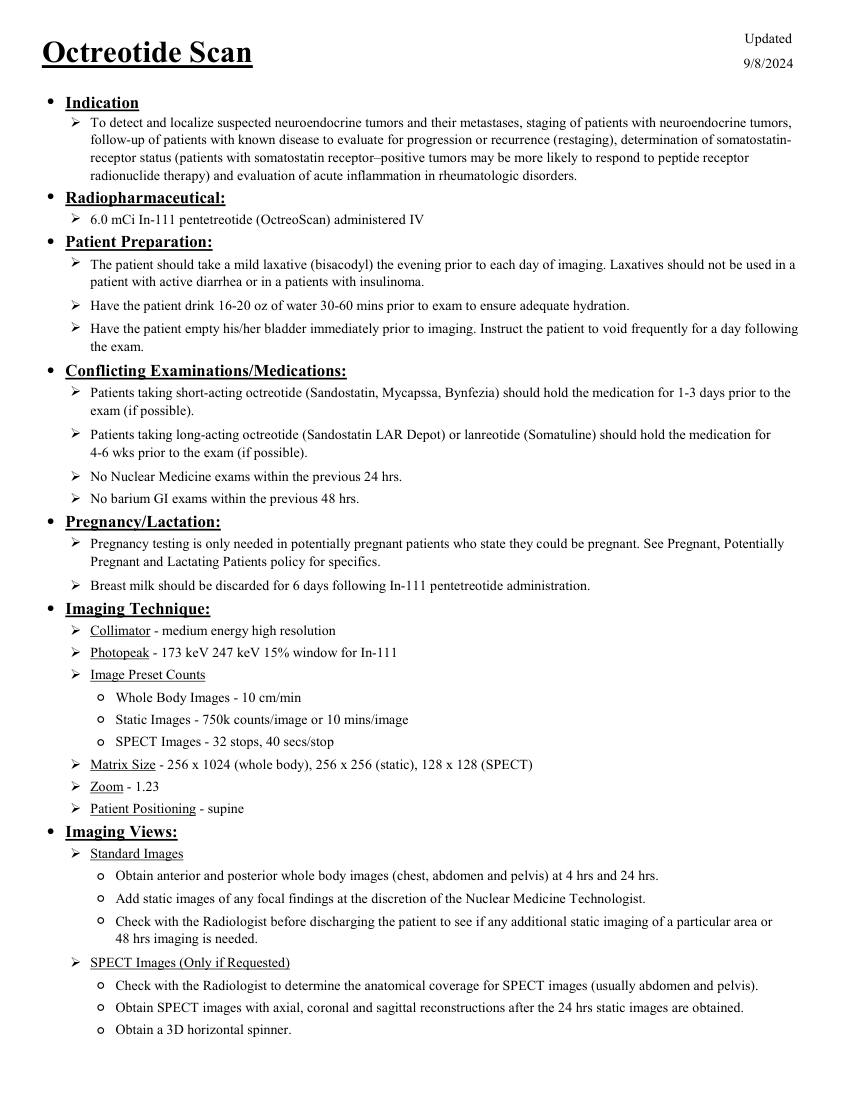
Updated Octreotide Scan 9/8/2024 ●Indication To detect and localize suspected neuroendocrine tumors and their metastases, staging of patients with neuroendocrine tumors, follow-up of patients with known disease to evaluate for progression or recurrence (restaging), determination of somatostatin- receptor status (patients with somatostatin receptor–positive tumors may be more likely to respond to peptide receptor radionuclide therapy) and evaluation of acute inflammation in rheumatologic disorders. ●Radiopharmaceutical: 6.0 mCi In-111 pentetreotide (OctreoScan) administered IV ●Patient Preparation: The patient should take a mild laxative (bisacodyl) the evening prior to each day of imaging. Laxatives should not be used in a patient with active diarrhea or in a patients with insulinoma. Have the patient drink 16-20 oz of water 30-60 mins prior to exam to ensure adequate hydration. Have the patient empty his/her bladder immediately prior to imaging. Instruct the patient to void frequently for a day following the exam. ●Conflicting Examinations/Medications: Patients taking short-acting octreotide (Sandostatin, Mycapssa, Bynfezia) should hold the medication for 1-3 days prior to the exam (if possible). Patients taking long-acting octreotide (Sandostatin LAR Depot) or lanreotide (Somatuline) should hold the medication for 4-6 wks prior to the exam (if possible). No Nuclear Medicine exams within the previous 24 hrs. No barium GI exams within the previous 48 hrs. ●Pregnancy/Lactation: Pregnancy testing is only needed in potentially pregnant patients who state they could be pregnant. See Pregnant, Potentially Pregnant and Lactating Patients policy for specifics. Breast milk should be discarded for 6 days following In-111 pentetreotide administration. ●Imaging Technique: Collimator - medium energy high resolution Photopeak - 173 keV 247 keV 15% window for In-111 Image Preset Counts ◦Whole Body Images - 10 cm/min ◦Static Images - 750k counts/image or 10 mins/image ◦SPECT Images - 32 stops, 40 secs/stop Matrix Size - 256 x 1024 (whole body), 256 x 256 (static), 128 x 128 (SPECT) Zoom - 1.23 Patient Positioning - supine ●Imaging Views: Standard Images ◦Obtain anterior and posterior whole body images (chest, abdomen and pelvis) at 4 hrs and 24 hrs. ◦Add static images of any focal findings at the discretion of the Nuclear Medicine Technologist. ◦ Check with the Radiologist before discharging the patient to see if any additional static imaging of a particular area or 48 hrs imaging is needed. SPECT Images (Only if Requested) ◦Check with the Radiologist to determine the anatomical coverage for SPECT images (usually abdomen and pelvis). ◦Obtain SPECT images with axial, coronal and sagittal reconstructions after the 24 hrs static images are obtained. ◦Obtain a 3D horizontal spinner.
Pagina 2 — text în engleză
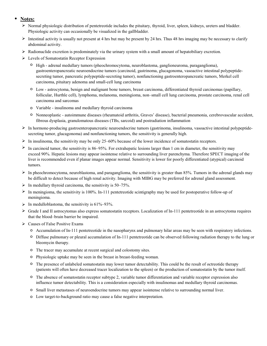
●Notes: Normal physiologic distribution of pentetreotide includes the pituitary, thyroid, liver, spleen, kidneys, ureters and bladder. Physiologic activity can occasionally be visualized in the gallbladder. Intestinal activity is usually not present at 4 hrs but may be present by 24 hrs. Thus 48 hrs imaging may be necessary to clarify abdominal activity. Radionuclide excretion is predominately via the urinary system with a small amount of hepatobiliary excretion. Levels of Somatostatin Receptor Expression ◦ High - adrenal medullary tumors (pheochromocytoma, neuroblastoma, ganglioneuroma, paraganglioma), gastroenteropancreatic neuroendocrine tumors (carcinoid, gastrinoma, glucagonoma, vasoactive intestinal polypeptide- secreting tumor, pancreatic polypeptide-secreting tumor), nonfunctioning gastroenteropancreatic tumors, Merkel cell carcinoma, pituitary adenoma and small-cell lung carcinoma ◦ Low - astrocytoma, benign and malignant bone tumors, breast carcinoma, differentiated thyroid carcinomas (papillary, follicular, Hurthle cell), lymphoma, melanoma, meningioma, non–small cell lung carcinoma, prostate carcinoma, renal cell carcinoma and sarcomas ◦Variable - insulinoma and medullary thyroid carcinoma ◦ Nonneoplastic - autoimmune diseases (rheumatoid arthritis, Graves’ disease), bacterial pneumonia, cerebrovascular accident, fibrous dysplasia, granulomatous diseases (TBs, sarcoid) and postradiation inflammation In hormone-producing gastroenteropancreatic neuroendocrine tumors (gastrinoma, insulinoma, vasoactive intestinal polypeptide- secreting tumor, glucagonoma) and nonfunctioning tumors, the sensitivity is generally high. In insulinoma, the sensitivity may be only 25–60% because of the lower incidence of somatostatin receptors. In carcinoid tumor, the sensitivity is 86–95%. For extrahepatic lesions larger than 1 cm in diameter, the sensitivity may exceed 90%. Hepatic lesions may appear isointense relative to surrounding liver parenchyma. Therefore SPECT imaging of the liver is recommended even if planar images appear normal. Sensitivity is lower for poorly differentiated (atypical) carcinoid tumors. In pheochromocytoma, neuroblastoma, and paraganglioma, the sensitivity is greater than 85%. Tumors in the adrenal glands may be difficult to detect because of high renal activity. Imaging with MIBG may be preferred for adrenal gland assessment. In medullary thyroid carcinoma, the sensitivity is 50–75%. In meningioma, the sensitivity is 100%. In-111 pentetreotide scintigraphy may be used for postoperative follow-up of meningioma. In medulloblastoma, the sensitivity is 61%–93%. Grade I and II astrocytomas also express somatostatin receptors. Localization of In-111 pentetreotide in an astrocytoma requires that the blood–brain barrier be impaired. Causes of False Positive Exams ◦Accumulation of In-111 pentetreotide in the nasopharynx and pulmonary hilar areas may be seen with respiratory infections. ◦ Diffuse pulmonary or pleural accumulation of In-111 pentetreotide can be observed following radiation therapy to the lung or bleomycin therapy. ◦The tracer may accumulate at recent surgical and colostomy sites. ◦Physiologic uptake may be seen in the breast in breast-feeding woman. ◦ The presence of unlabeled somatostatin may lower tumor detectability. This could be the result of octreotide therapy (patients will often have decreased tracer localization to the spleen) or the production of somatostatin by the tumor itself. ◦ The absence of somatostatin receptor subtype 2, variable tumor differentiation and variable receptor expression also influence tumor detectability. This is a consideration especially with insulinomas and medullary thyroid carcinomas. ◦Small liver metastases of neuroendocrine tumors may appear isointense relative to surrounding normal liver. ◦Low target-to-background ratio may cause a false negative interpretation.
Sinteza existentă în aplicație
Protocol oficial de Medicină Nucleară & Radiologie Nucleară integrat conform standardelor de calitate și radiofarmacie clinică ale MCB Radiology și ghidurilor internaționale SNMMI / EANM.
☢️ MEDICINĂ NUCLEARĂ • Clasa 3 (8 - 10 mSv)
Procedură diagnostică funcțională și moleculară. Evaluarea fiziologică in vivo a metabolismului tisular utilizând radiotrasori specifici de emisie gama sau pozitroni (PET/SPECT).
Cale de administrare: Intravenoasă, inhalatorie sau orală conform protocolului specific.
Măsuri de radioprotecție: Hidratare abundentă post-procedură pentru favorizarea eliminării urinare a radiofarmaceuticului nefixat. Evitarea contactului prelungit cu femei gravide și copii mici timp de 24 ore.
3. Pregătirea Pacientului
Hidratare bună. Oprirea analogilor de somatostatină cu acțiune scurtă (Octreotid subcutanat) cu 24-48 ore înainte, iar a formelor retard (LAR) cu 3-4 săptămâni înainte (dacă este tolerat clinic). Laxativ ușor în seara anterioară scanării de 24 ore pentru clearance-ul intestinal.
4. Protocol Tehnic de Achiziție Imagistică
Imagini planare la 4 ore și 24 ore post-injectare (opțional 48 ore). Colimator MEGP. SPECT sau SPECT/CT obligatoriu pentru abdomen și pelvis la 24 ore.
5. Criterii de Interpretare Diagnostică
Fixare intensă în tumorile neuroendocrine bogate în receptori de somatostatină. Evaluare Krenning score (grad 1-4) pentru stabilirea indicației de radioterapie PRRT.