Instrucțiunile, valorile și ilustrațiile din documentul original sunt păstrate în engleză. Titlul și navigarea sunt în română.
Pagina 1
Pagina 2
Pagina 3
Textul documentului original
Pagina 1 — text în engleză
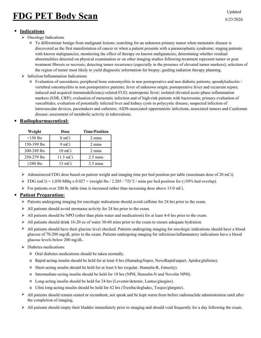
Updated FDG PET Body Scan 6/23/2026 ●Indications Oncology Indications ◦ To differentiate benign from malignant lesions; searching for an unknown primary tumor when metastatic disease is discovered as the first manifestation of cancer or when a patient presents with a paraneoplastic syndrome; staging patients with known malignancies; monitoring the effect of therapy on known malignancies; determining whether residual abnormalities detected on physical examination or on other imaging studies following treatment represent tumor or post treatment fibrosis or necrosis; detecting tumor recurrence (especially in the presence of elevated tumor markers); selection of the region of tumor most likely to yield diagnostic information for biopsy; guiding radiation therapy planning. Infection/Inflammation Indications ◦ Evaluation of sarcoidosis; peripheral bone osteomyelitis in non postoperative and non diabetic patients; spondylodiscitis / vertebral osteomyelitis in non postoperative patients; fever of unknown origin; postoperative fever and recurrent sepsis; induced and acquired immunodeficiency-related FUO; neutropenic fever; isolated elevated acute-phase inflammation markers (ESR, CRP); evaluation of metastatic infection and of high-risk patients with bacteremia; primary evaluation of vasculitides; evaluation of potentially infected liver and kidney cysts in polycystic disease; suspected infection of intravascular devices, pacemakers and catheters; AIDS-associated opportunistic infections, associated tumors and Castleman disease; assessment of metabolic activity in tuberculosis. ●Radiopharmaceutical: Weight Dose Time/Position 2 mins <150 lbs 8 mCi 2 mins 150-199 lbs 9 mCi 200-249 lbs 10 mCi 2 mins 250-279 lbs 11.5 mCi 2.5 mins 13 mCi ≥280 lbs 2.5 mins Administered FDG dose based on patient weight and imaging time per bed position per table (maximum dose of 20 mCi). FDG (mCi) = 1,050 MBq x 0.027 × (weight lbs / 2.205 / 75)^2 / mins per bed position for (≤30% bed overlap). For patients over 200 lb, table time is increased rather than increasing dose above 15.0 mCi. ●Patient Preparation: Patients undergoing imaging for oncologic indications should avoid caffeine for 24 hrs prior to the exam. All patients should avoid strenuous activity for 24 hrs prior to the exam. All patients should be NPO (other than plain water and medications) for at least 4-6 hrs prior to the exam. All patients should drink 16-20 oz of water 30-60 mins prior to the exam to ensure adequate hydration. All patients should have their glucose level checked. Patients undergoing imaging for oncologic indications should have a blood glucose of 70-200 mg/dL prior to the exam. Patients undergoing imaging for infectious/inflammatory indications have a blood glucose levels below 200 mg/dL. Diabetes medications: Ultra long-acting insulin should be held for 42 hrs (Tresiba/degludec, Toujeo/glargine). ◦Oral diabetes medications should be taken normally. ◦Rapid-acting insulin should be held for at least 4 hrs (Humalog/lispro, NovoRapid/aspart, Apidra/glulisine). ◦Short-acting insulin should be held for at least 6 hrs (regular, Humulin-R, Entuzity). ◦Intermediate-acting insulin should be held for 18 hrs (NPH, Humulin-N and Novolin NPH). ◦Long-acting insulin should be held for 24 hrs (Levemir/detemir, Lantus/glargine). ◦ All patients should remain seated or recumbent, not speak and be kept warm from before radionuclide administration until after the completion of imaging, All patients should empty their bladder immediately prior to imaging and should void frequently for a day following the exam.
Pagina 2 — text în engleză
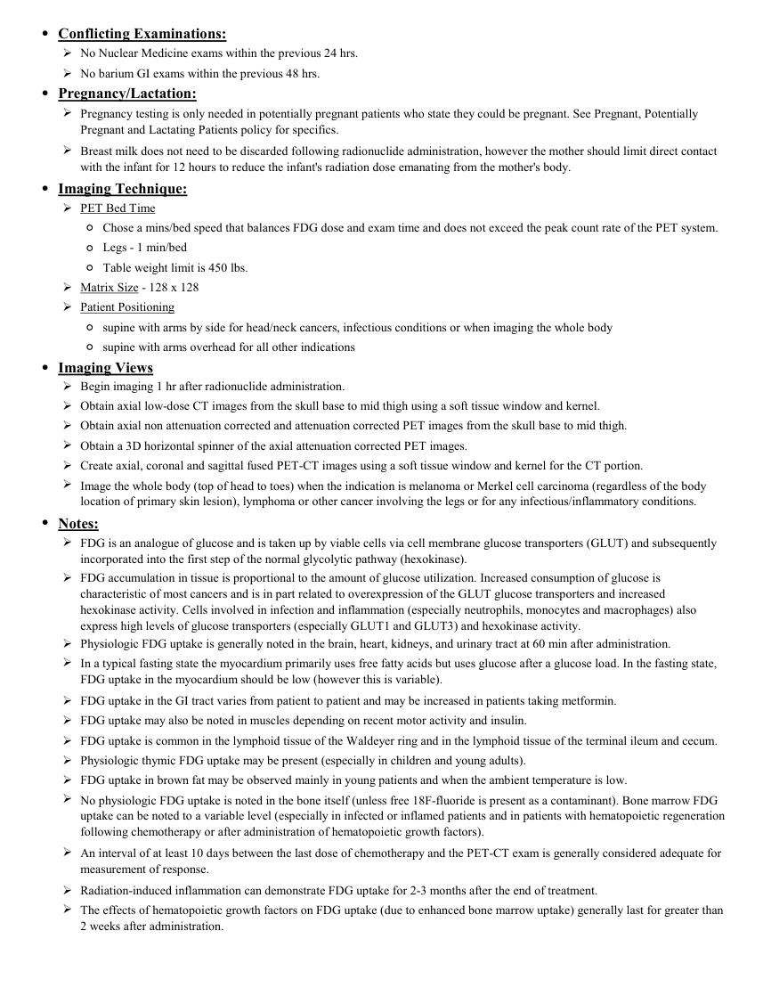
●Conflicting Examinations: No Nuclear Medicine exams within the previous 24 hrs. No barium GI exams within the previous 48 hrs. ●Pregnancy/Lactation: Pregnancy testing is only needed in potentially pregnant patients who state they could be pregnant. See Pregnant, Potentially Pregnant and Lactating Patients policy for specifics. Breast milk does not need to be discarded following radionuclide administration, however the mother should limit direct contact with the infant for 12 hours to reduce the infant's radiation dose emanating from the mother's body. ●Imaging Technique: PET Bed Time ◦Chose a mins/bed speed that balances FDG dose and exam time and does not exceed the peak count rate of the PET system. ◦Legs - 1 min/bed ◦Table weight limit is 450 lbs. Matrix Size - 128 x 128 Patient Positioning ◦supine with arms by side for head/neck cancers, infectious conditions or when imaging the whole body ◦supine with arms overhead for all other indications ●Imaging Views Begin imaging 1 hr after radionuclide administration. Obtain axial low-dose CT images from the skull base to mid thigh using a soft tissue window and kernel. Obtain axial non attenuation corrected and attenuation corrected PET images from the skull base to mid thigh. Obtain a 3D horizontal spinner of the axial attenuation corrected PET images. Create axial, coronal and sagittal fused PET-CT images using a soft tissue window and kernel for the CT portion. Image the whole body (top of head to toes) when the indication is melanoma or Merkel cell carcinoma (regardless of the body location of primary skin lesion), lymphoma or other cancer involving the legs or for any infectious/inflammatory conditions. ●Notes: FDG is an analogue of glucose and is taken up by viable cells via cell membrane glucose transporters (GLUT) and subsequently incorporated into the first step of the normal glycolytic pathway (hexokinase). FDG accumulation in tissue is proportional to the amount of glucose utilization. Increased consumption of glucose is characteristic of most cancers and is in part related to overexpression of the GLUT glucose transporters and increased hexokinase activity. Cells involved in infection and inflammation (especially neutrophils, monocytes and macrophages) also express high levels of glucose transporters (especially GLUT1 and GLUT3) and hexokinase activity. Physiologic FDG uptake is generally noted in the brain, heart, kidneys, and urinary tract at 60 min after administration. In a typical fasting state the myocardium primarily uses free fatty acids but uses glucose after a glucose load. In the fasting state, FDG uptake in the myocardium should be low (however this is variable). FDG uptake in the GI tract varies from patient to patient and may be increased in patients taking metformin. FDG uptake may also be noted in muscles depending on recent motor activity and insulin. FDG uptake is common in the lymphoid tissue of the Waldeyer ring and in the lymphoid tissue of the terminal ileum and cecum. Physiologic thymic FDG uptake may be present (especially in children and young adults). FDG uptake in brown fat may be observed mainly in young patients and when the ambient temperature is low. No physiologic FDG uptake is noted in the bone itself (unless free 18F-fluoride is present as a contaminant). Bone marrow FDG uptake can be noted to a variable level (especially in infected or inflamed patients and in patients with hematopoietic regeneration following chemotherapy or after administration of hematopoietic growth factors). An interval of at least 10 days between the last dose of chemotherapy and the PET-CT exam is generally considered adequate for measurement of response. Radiation-induced inflammation can demonstrate FDG uptake for 2-3 months after the end of treatment. The effects of hematopoietic growth factors on FDG uptake (due to enhanced bone marrow uptake) generally last for greater than 2 weeks after administration.
Pagina 3 — text în engleză
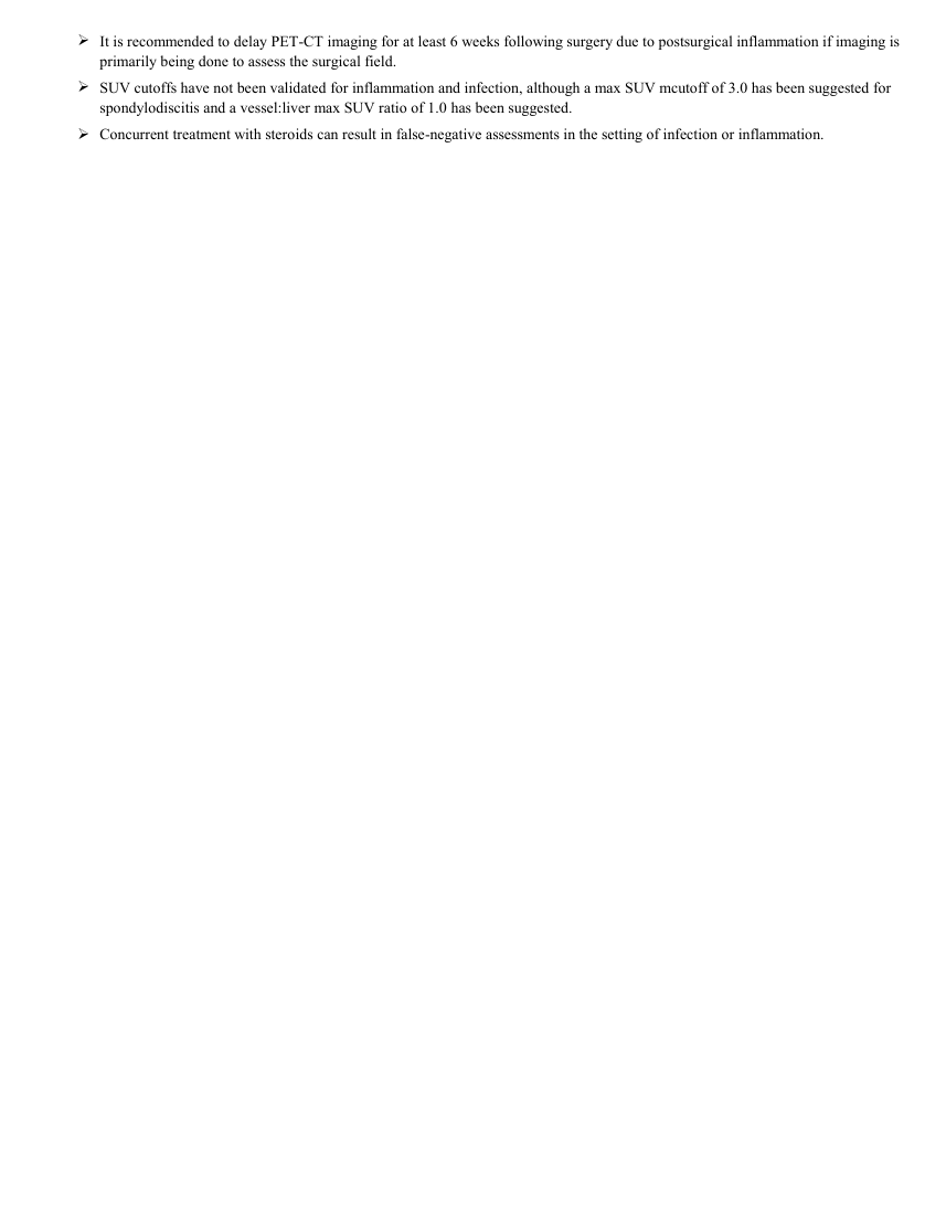
It is recommended to delay PET-CT imaging for at least 6 weeks following surgery due to postsurgical inflammation if imaging is primarily being done to assess the surgical field. SUV cutoffs have not been validated for inflammation and infection, although a max SUV mcutoff of 3.0 has been suggested for spondylodiscitis and a vessel:liver max SUV ratio of 1.0 has been suggested. Concurrent treatment with steroids can result in false-negative assessments in the setting of infection or inflammation.
Sinteza existentă în aplicație
Protocol oficial de Medicină Nucleară & Radiologie Nucleară integrat conform standardelor de calitate și radiofarmacie clinică ale MCB Radiology și ghidurilor internaționale SNMMI / EANM.
☢️ MEDICINĂ NUCLEARĂ • Clasa 3 (7 - 12 mSv)
Procedură diagnostică funcțională și moleculară. Evaluarea fiziologică in vivo a metabolismului tisular utilizând radiotrasori specifici de emisie gama sau pozitroni (PET/SPECT).
Stadializarea inițială, restadializarea și evaluarea eficienței terapeutice (chimioterapie, radioterapie, imunoterapie) în limfoame, cancer bronhopulmonar, cancer colo-rectal, melanom, cancere ORL, esofagiene etc.
Detectarea recidivelor tumorale la pacienți cu markeri tumorali serici în creștere și imagistică convențională negativă
Cale de administrare: Intravenoasă, inhalatorie sau orală conform protocolului specific.
Măsuri de radioprotecție: Hidratare abundentă post-procedură pentru favorizarea eliminării urinare a radiofarmaceuticului nefixat. Evitarea contactului prelungit cu femei gravide și copii mici timp de 24 ore.
3. Pregătirea Pacientului
Post alimentar strict de minim 6 ore. Hidratare cu apă plată (fără zahăr). Glicemie < 150-180 mg/dL înainte de injectare. Repaus fizic absolut și mediu cald timp de 60 min post-injectare pentru prevenirea captării în mușchi și grăsimea brună.
4. Protocol Tehnic de Achiziție Imagistică
Timp de biodistribuție: 60 ± 10 minute în cameră de liniște. Scanare hibridă PET-CT de la vertex până la jumătatea coapselor (sau din creștet până în tălpi pentru melanom), combinând CT cu doză joasă pentru corecție de atenuare și localizare anatomică cu achiziție 3D PET (1.5 - 3 min/pat).
5. Criterii de Interpretare Diagnostică
Calculul valorii standardizate a captării (SUVmax). Zone de hipermetabolism glucidic suspecte oncologic corelate cu leziunile anatomice CT. Criterii RECIST / PERCIST pentru răspuns terapeutic.