Medicină nucleară — Limfoscintigrafie ginecologică (MCB)
Sursă și prelucrare editorială
- Proveniență
- 1 referință declarată
- Fidelitatea preluării
- Nedocumentată
- Revizuire medicală
- Nedocumentată
Referințele declarate nu confirmă singure fidelitatea preluării sau aprobarea protocolului pentru utilizare locală.
Detalii despre surse și verificări
MCB Radiology — Lympho Gynecologic
- Ediție / versiune a sursei
- Neprecizată
- Pagină / secțiune
- Pagini: 1–2
- Relația cu protocolul
- Document original importat integral; atribuirea autorilor rămâne cea din PDF.
- Data consultării
- 2026-09-27
Prelucrarea acestei pagini
- Mod de prelucrare
- Titlu și navigare în română; documentul sursă în engleză.; PDF original integral, previzualizare și text extras automat; fără completarea parametrilor clinici lipsă.
- Adaptări față de sursă
- Nedocumentate; absența evidenței nu confirmă o preluare integrală.
- Responsabil pentru prelucrare
- Neprecizat
- Atribuire existentă în pagină
- MCB Radiology (portalul sursă). Această atribuire nu identifică automat autorul sursei sau revizorul medical.
- Versiunea paginii
- mcb-e39e2a14baf8
- Actualizare declarată a paginii
- 2026-09-27 — distinctă de revizuirea medicală.
Verificarea fidelității: Nedocumentată
Revizuirea medicală: Nedocumentată
Documentul original MCB — Limfoscintigrafie ginecologică
Protocol · 2 pagini · consultat la 2026-09-27.
Descarcă PDF-ul integral · Sursa MCB · Catalogul MCB pentru această modalitate
Instrucțiunile, valorile și ilustrațiile din documentul original sunt păstrate în engleză. Titlul și navigarea sunt în română.
Pagina 1
Pagina 2

Textul documentului original
Pagina 1 — text în engleză
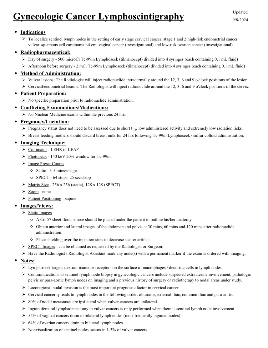
Updated Gynecologic Cancer Lymphoscintigraphy 9/8/2024 ●Indications To localize sentinel lymph nodes in the setting of early stage cervical cancer, stage 1 and 2 high-risk endometrial cancer,
vulvar squamous cell carcinoma <4 cm, vaginal cancer (investigational) and low-risk ovarian cancer (investigational). ●Radiopharmaceutical: Day of surgery - 500 microCi Tc-99m Lymphoseek (tilmanocept) divided into 4 syringes (each containing 0.1 mL fluid) Afternoon before surgery - 2 mCi Tc-99m Lymphoseek (tilmanocept) divided into 4 syringes (each containing 0.1 mL fluid) ●Method of Administration: Cervical/endometrial lesions: The Radiologist will inject radionuclide around the 12, 3, 6 and 9 o'clock positions of the cervix. Vulvar lesions: The Radiologist will inject radionuclide intradermally around the 12, 3, 6 and 9 o'clock positions of the lesion.
●Patient Preparation: No specific preparation prior to radionuclide administration. ●Conflicting Examinations/Medications: No Nuclear Medicine exams within the previous 24 hrs. ●Pregnancy/Lactation: Pregnancy status does not need to be assessed due to short t1/2, low administered activity and extremely low radiation risks. Breast feeding mothers should discard breast milk for 24 hrs following Tc-99m Lymphoseek / sulfur colloid administration. ●Imaging Technique: Collimator - LEHR or LEAP Photopeak - 140 keV 20% window for Tc-99m Image Preset Counts ◦Static - 3-5 mins/image ◦SPECT - 64 stops, 25 secs/stop Matrix Size - 256 x 256 (static), 128 x 128 (SPECT) Zoom - none Patient Positioning - supine ●Images/Views: Static Images ◦A Co-57 sheet flood source should be placed under the patient to outline his/her anatomy. ◦ Obtain anterior and lateral images of the abdomen and pelvis at 30 mins, 60 mins and 120 mins after radionuclide administration. ◦Place shielding over the injection sites to decrease scatter artifact. SPECT Images - can be obtained as requested by the Radiologist or Surgeon. Have the Radiologist / Radiologist Assistant mark any node(s) with a permanent marker if the exam is ordered with imaging. ●Notes: Lymphoseek targets dextran-mannose receptors on the surface of macrophages / dendritic cells in lymph nodes. Contraindications to sentinel lymph node biopsy in gynecologic cancers include suspected extrauterine involvement, pathologic pelvic or para-aortic lymph nodes on imaging and a previous history of surgery or radiotherapy to nodal areas under study. Locoregional nodal invasion is the most important prognostic factor in cervical cancer. Cervical cancer spreads to lymph nodes in the following order: obturator, external iliac, common iliac and para-aortic. 80% of nodal metastases are ipsilateral when vulvar cancers are unilateral. Inguinofemoral lymphadenectomy in vulvar cancers is only performed when there is sentinel lymph node involvement. 55% of vaginal cancers drain to bilateral lymph nodes (most frequently inguinal nodes). 64% of ovarian cancers drain to bilateral lymph nodes. Nonvisualization of sentinel nodes occurs in 1-3% of vulvar cancers.
vulvar squamous cell carcinoma <4 cm, vaginal cancer (investigational) and low-risk ovarian cancer (investigational). ●Radiopharmaceutical: Day of surgery - 500 microCi Tc-99m Lymphoseek (tilmanocept) divided into 4 syringes (each containing 0.1 mL fluid) Afternoon before surgery - 2 mCi Tc-99m Lymphoseek (tilmanocept) divided into 4 syringes (each containing 0.1 mL fluid) ●Method of Administration: Cervical/endometrial lesions: The Radiologist will inject radionuclide around the 12, 3, 6 and 9 o'clock positions of the cervix. Vulvar lesions: The Radiologist will inject radionuclide intradermally around the 12, 3, 6 and 9 o'clock positions of the lesion.
●Patient Preparation: No specific preparation prior to radionuclide administration. ●Conflicting Examinations/Medications: No Nuclear Medicine exams within the previous 24 hrs. ●Pregnancy/Lactation: Pregnancy status does not need to be assessed due to short t1/2, low administered activity and extremely low radiation risks. Breast feeding mothers should discard breast milk for 24 hrs following Tc-99m Lymphoseek / sulfur colloid administration. ●Imaging Technique: Collimator - LEHR or LEAP Photopeak - 140 keV 20% window for Tc-99m Image Preset Counts ◦Static - 3-5 mins/image ◦SPECT - 64 stops, 25 secs/stop Matrix Size - 256 x 256 (static), 128 x 128 (SPECT) Zoom - none Patient Positioning - supine ●Images/Views: Static Images ◦A Co-57 sheet flood source should be placed under the patient to outline his/her anatomy. ◦ Obtain anterior and lateral images of the abdomen and pelvis at 30 mins, 60 mins and 120 mins after radionuclide administration. ◦Place shielding over the injection sites to decrease scatter artifact. SPECT Images - can be obtained as requested by the Radiologist or Surgeon. Have the Radiologist / Radiologist Assistant mark any node(s) with a permanent marker if the exam is ordered with imaging. ●Notes: Lymphoseek targets dextran-mannose receptors on the surface of macrophages / dendritic cells in lymph nodes. Contraindications to sentinel lymph node biopsy in gynecologic cancers include suspected extrauterine involvement, pathologic pelvic or para-aortic lymph nodes on imaging and a previous history of surgery or radiotherapy to nodal areas under study. Locoregional nodal invasion is the most important prognostic factor in cervical cancer. Cervical cancer spreads to lymph nodes in the following order: obturator, external iliac, common iliac and para-aortic. 80% of nodal metastases are ipsilateral when vulvar cancers are unilateral. Inguinofemoral lymphadenectomy in vulvar cancers is only performed when there is sentinel lymph node involvement. 55% of vaginal cancers drain to bilateral lymph nodes (most frequently inguinal nodes). 64% of ovarian cancers drain to bilateral lymph nodes. Nonvisualization of sentinel nodes occurs in 1-3% of vulvar cancers.
Pagina 2 — text în engleză
Nonvisualization of sentinel nodes occurs in 10-15% of cervical and endometrial cancers.