IRM – Sân: integritatea implanturilor (MCB)
Sursă și prelucrare editorială
- Proveniență
- 1 referință declarată
- Fidelitatea preluării
- Nedocumentată
- Revizuire medicală
- Nedocumentată
Referințele declarate nu confirmă singure fidelitatea preluării sau aprobarea protocolului pentru utilizare locală.
Detalii despre surse și verificări
2 Implant Integrity
- Ediție / versiune a sursei
- Neprecizată
- Pagină / secțiune
- Pagini: 1
- Relația cu protocolul
- Document instituțional original importat integral
- Data consultării
- 2026-09-27
Prelucrarea acestei pagini
- Mod de prelucrare
- Titlu și etichete de parametri în română; textul clinic original păstrat în engleză.; Import PDF integral, imagini ale tuturor paginilor și extragere automată a parametrilor secvențelor.
- Adaptări față de sursă
- Nedocumentate; absența evidenței nu confirmă o preluare integrală.
- Responsabil pentru prelucrare
- Neprecizat
- Atribuire existentă în pagină
- MCB Radiology (documentul sursă). Această atribuire nu identifică automat autorul sursei sau revizorul medical.
- Versiunea paginii
- mcb-mri-df6aa44decb4
- Actualizare declarată a paginii
- 2026-09-27 — distinctă de revizuirea medicală.
Verificarea fidelității: Nedocumentată
Revizuirea medicală: Nedocumentată
Catalog MCB · Descarcă PDF-ul complet · Sursa MCB
Documentul MCB este disponibil integral mai jos, inclusiv notele, condițiile de utilizare a contrastului și imaginile de planificare. Textul clinic al sursei este în engleză; titlul și etichetele parametrilor sunt în română.
Protocolul original
Pagina 1
Textul paginii (engleză)
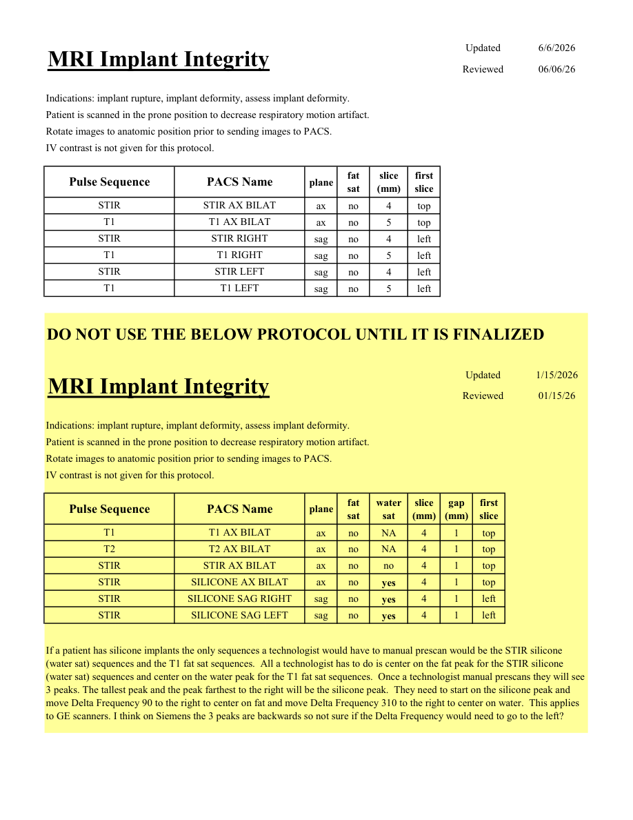
Updated 6/6/2026 MRI Implant Integrity Reviewed 06/06/26 Indications: implant rupture, implant deformity, assess implant deformity. Patient is scanned in the prone position to decrease respiratory motion artifact. Rotate images to anatomic position prior to sending images to PACS. IV contrast is not given for this protocol. slice
Pulse Sequence PACS Name plane fat
sat (mm) first
slice STIR STIR AX BILAT ax no 4 top T1 T1 AX BILAT ax no 5 top STIR STIR RIGHT sag no 4 left T1 T1 RIGHT sag no 5 left STIR STIR LEFT sag no 4 left T1 T1 LEFT sag no 5 left DO NOT USE THE BELOW PROTOCOL UNTIL IT IS FINALIZED Updated 1/15/2026 MRI Implant Integrity Reviewed 01/15/26 Indications: implant rupture, implant deformity, assess implant deformity. Patient is scanned in the prone position to decrease respiratory motion artifact. Rotate images to anatomic position prior to sending images to PACS. IV contrast is not given for this protocol. water
slice
gap
Pulse Sequence PACS Name plane fat
sat sat (mm) (mm) first
slice T1 T1 AX BILAT ax no NA 4 1 top T2 T2 AX BILAT ax no NA 4 1 top STIR STIR AX BILAT ax no no 4 1 top STIR SILICONE AX BILAT ax no yes 4 1 top STIR SILICONE SAG RIGHT sag no yes 4 1 left STIR SILICONE SAG LEFT sag no yes 4 1 left If a patient has silicone implants the only sequences a technologist would have to manual prescan would be the STIR silicone (water sat) sequences and the T1 fat sat sequences. All a technologist has to do is center on the fat peak for the STIR silicone (water sat) sequences and center on the water peak for the T1 fat sat sequences. Once a technologist manual prescans they will see 3 peaks. The tallest peak and the peak farthest to the right will be the silicone peak. They need to start on the silicone peak and move Delta Frequency 90 to the right to center on fat and move Delta Frequency 310 to the right to center on water. This applies to GE scanners. I think on Siemens the 3 peaks are backwards so not sure if the Delta Frequency would need to go to the left?
Pulse Sequence PACS Name plane fat
sat (mm) first
slice STIR STIR AX BILAT ax no 4 top T1 T1 AX BILAT ax no 5 top STIR STIR RIGHT sag no 4 left T1 T1 RIGHT sag no 5 left STIR STIR LEFT sag no 4 left T1 T1 LEFT sag no 5 left DO NOT USE THE BELOW PROTOCOL UNTIL IT IS FINALIZED Updated 1/15/2026 MRI Implant Integrity Reviewed 01/15/26 Indications: implant rupture, implant deformity, assess implant deformity. Patient is scanned in the prone position to decrease respiratory motion artifact. Rotate images to anatomic position prior to sending images to PACS. IV contrast is not given for this protocol. water
slice
gap
Pulse Sequence PACS Name plane fat
sat sat (mm) (mm) first
slice T1 T1 AX BILAT ax no NA 4 1 top T2 T2 AX BILAT ax no NA 4 1 top STIR STIR AX BILAT ax no no 4 1 top STIR SILICONE AX BILAT ax no yes 4 1 top STIR SILICONE SAG RIGHT sag no yes 4 1 left STIR SILICONE SAG LEFT sag no yes 4 1 left If a patient has silicone implants the only sequences a technologist would have to manual prescan would be the STIR silicone (water sat) sequences and the T1 fat sat sequences. All a technologist has to do is center on the fat peak for the STIR silicone (water sat) sequences and center on the water peak for the T1 fat sat sequences. Once a technologist manual prescans they will see 3 peaks. The tallest peak and the peak farthest to the right will be the silicone peak. They need to start on the silicone peak and move Delta Frequency 90 to the right to center on fat and move Delta Frequency 310 to the right to center on water. This applies to GE scanners. I think on Siemens the 3 peaks are backwards so not sure if the Delta Frequency would need to go to the left?
Parametrii secvențelor
Tabele extrase din PDF. Variantele, secvențele condiționate și momentele administrării contrastului se citesc împreună cu notele de pe pagina originală indicată.
Tabelul 1 — pagina 1
| Secvență | Denumire PACS | Plan | Supresie grăsime | Grosime (mm) | Prima secțiune |
|---|---|---|---|---|---|
| STIR | STIR AX BILAT | Axial | Nu | 4 | Superior |
| T1 | T1 AX BILAT | Axial | Nu | 5 | Superior |
| STIR | STIR RIGHT | Sagital | Nu | 4 | Stânga |
| T1 | T1 RIGHT | Sagital | Nu | 5 | Stânga |
| STIR | STIR LEFT | Sagital | Nu | 4 | Stânga |
| T1 | T1 LEFT | Sagital | Nu | 5 | Stânga |
Tabelul 2 — pagina 1
| Secvență | Denumire PACS | Plan | Supresie grăsime | Supresie apă | Grosime (mm) | Interval (mm) | Prima secțiune |
|---|---|---|---|---|---|---|---|
| T1 | T1 AX BILAT | Axial | Nu | NA | 4 | 1 | Superior |
| T2 | T2 AX BILAT | Axial | Nu | NA | 4 | 1 | Superior |
| STIR | STIR AX BILAT | Axial | Nu | Nu | 4 | 1 | Superior |
| STIR | SILICONE AX BILAT | Axial | Nu | Da | 4 | 1 | Superior |
| STIR | SILICONE SAG RIGHT | Sagital | Nu | Da | 4 | 1 | Stânga |
| STIR | SILICONE SAG LEFT | Sagital | Nu | Da | 4 | 1 | Stânga |