Ecografie — Tromboză venoasă profundă: membru superior (MCB)
Sursă și prelucrare editorială
- Proveniență
- 1 referință declarată
- Fidelitatea preluării
- Nedocumentată
- Revizuire medicală
- Nedocumentată
Referințele declarate nu confirmă singure fidelitatea preluării sau aprobarea protocolului pentru utilizare locală.
Detalii despre surse și verificări
MCB Radiology — Arm DVT
- Ediție / versiune a sursei
- Neprecizată
- Pagină / secțiune
- Pagini: 1–2
- Relația cu protocolul
- Document original importat integral; atribuirea autorilor rămâne cea din PDF.
- Data consultării
- 2026-09-27
Prelucrarea acestei pagini
- Mod de prelucrare
- Titlu și navigare în română; documentul sursă în engleză.; PDF original integral, previzualizare și text extras automat; fără completarea parametrilor clinici lipsă.
- Adaptări față de sursă
- Nedocumentate; absența evidenței nu confirmă o preluare integrală.
- Responsabil pentru prelucrare
- Neprecizat
- Atribuire existentă în pagină
- MCB Radiology (portalul sursă). Această atribuire nu identifică automat autorul sursei sau revizorul medical.
- Versiunea paginii
- mcb-26ec8aec0ddc
- Actualizare declarată a paginii
- 2026-09-27 — distinctă de revizuirea medicală.
Verificarea fidelității: Nedocumentată
Revizuirea medicală: Nedocumentată
Documentul original MCB — Tromboză venoasă profundă: membru superior
Protocol · 2 pagini · consultat la 2026-09-27.
Descarcă PDF-ul integral · Sursa MCB · Catalogul MCB pentru această modalitate
Instrucțiunile, valorile și ilustrațiile din documentul original sunt păstrate în engleză. Titlul și navigarea sunt în română.
Pagina 1
Pagina 2
Textul documentului original
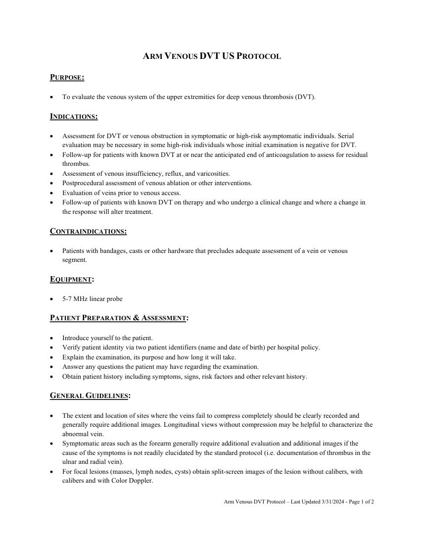
Pagina 1 — text în engleză
PURPOSE:
To evaluate the venous system of the upper extremities for deep venous thrombosis (DVT).
INDICATIONS:
Assessment for DVT or venous obstruction in symptomatic or high-risk asymptomatic individuals. Serial evaluation may be necessary in some high-risk individuals whose initial examination is negative for DVT. Follow-up for patients with known DVT at or near the anticipated end of anticoagulation to assess for residual thrombus. Assessment of venous insufficiency, reflux, and varicosities. Postprocedural assessment of venous ablation or other interventions. Evaluation of veins prior to venous access. Follow-up of patients with known DVT on therapy and who undergo a clinical change and where a change in the response will alter treatment.
CONTRAINDICATIONS:
Patients with bandages, casts or other hardware that precludes adequate assessment of a vein or venous segment.
EQUIPMENT:
5-7 MHz linear probe
PATIENT PREPARATION & ASSESSMENT:
Introduce yourself to the patient.
Verify patient identity via two patient identifiers (name and date of birth) per hospital policy. Explain the examination, its purpose and how long it will take. Answer any questions the patient may have regarding the examination. Obtain patient history including symptoms, signs, risk factors and other relevant history.
GENERAL GUIDELINES:
The extent and location of sites where the veins fail to compress completely should be clearly recorded and generally require additional images. Longitudinal views without compression may be helpful to characterize the abnormal vein. Symptomatic areas such as the forearm generally require additional evaluation and additional images if the cause of the symptoms is not readily elucidated by the standard protocol (i.e. documentation of thrombus in the ulnar and radial vein). For focal lesions (masses, lymph nodes, cysts) obtain split-screen images of the lesion without calibers, with calibers and with Color Doppler.
Arm Venous DVT Protocol – Last Updated 3/31/2024 - Page 1 of 2
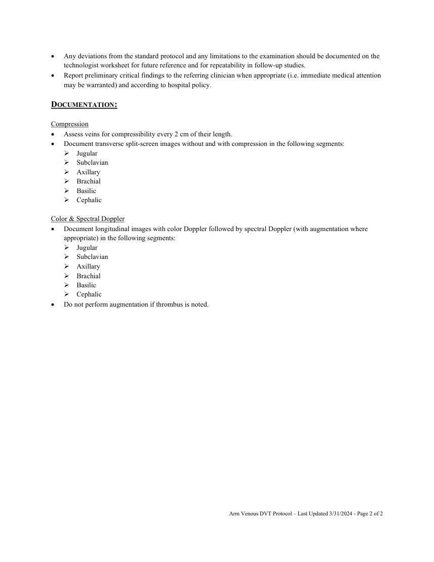
Pagina 2 — text în engleză
DOCUMENTATION:
Compression Assess veins for compressibility every 2 cm of their length.
Document transverse split-screen images without and with compression in the following segments: Jugular Subclavian Axillary Brachial Basilic Cephalic
Color & Spectral Doppler Document longitudinal images with color Doppler followed by spectral Doppler (with augmentation where appropriate) in the following segments: Jugular Subclavian Axillary Brachial Basilic Cephalic Do not perform augmentation if thrombus is noted.
Arm Venous DVT Protocol – Last Updated 3/31/2024 - Page 2 of 2