Ecografie — Presiuni segmentare: membru superior (MCB)
Sursă și prelucrare editorială
- Proveniență
- 1 referință declarată
- Fidelitatea preluării
- Nedocumentată
- Revizuire medicală
- Nedocumentată
Referințele declarate nu confirmă singure fidelitatea preluării sau aprobarea protocolului pentru utilizare locală.
Detalii despre surse și verificări
MCB Radiology — Arm Segmentals
- Ediție / versiune a sursei
- Neprecizată
- Pagină / secțiune
- Pagini: 1–6
- Relația cu protocolul
- Document original importat integral; atribuirea autorilor rămâne cea din PDF.
- Data consultării
- 2026-09-27
Prelucrarea acestei pagini
- Mod de prelucrare
- Titlu și navigare în română; documentul sursă în engleză.; PDF original integral, previzualizare și text extras automat; fără completarea parametrilor clinici lipsă.
- Adaptări față de sursă
- Nedocumentate; absența evidenței nu confirmă o preluare integrală.
- Responsabil pentru prelucrare
- Neprecizat
- Atribuire existentă în pagină
- MCB Radiology (portalul sursă). Această atribuire nu identifică automat autorul sursei sau revizorul medical.
- Versiunea paginii
- mcb-fc666fb0954d
- Actualizare declarată a paginii
- 2026-09-27 — distinctă de revizuirea medicală.
Verificarea fidelității: Nedocumentată
Revizuirea medicală: Nedocumentată
Documentul original MCB — Presiuni segmentare: membru superior
Protocol · 6 pagini · consultat la 2026-09-27.
Descarcă PDF-ul integral · Sursa MCB · Catalogul MCB pentru această modalitate
Instrucțiunile, valorile și ilustrațiile din documentul original sunt păstrate în engleză. Titlul și navigarea sunt în română.
Pagina 1

Pagina 2
Pagina 3
Pagina 4
Pagina 5
Pagina 6
Textul documentului original
Pagina 1 — text în engleză
PURPOSE:
To detect the presence of peripheral arterial disease and categorize the overall severity and general location of stenoses using indirect testing modalities.
INDICATION:
Peripheral vascular disease. Claudication (pain with exertion). Rest pain. Hand or finger ulceration or gangrene. Absent palpable pulses. Suspected acute limb ischemia. Follow-up of decreased wrist-brachial or finger-brachial index from a prior examination. Post-operative evaluation or follow-up.
CONTRAINDICATIONS:
Patients with suspected or known acute deep venous thrombosis (DVT) may need exams limited to waveform analysis and use of a finger-brachial index. Any site of recent surgery, trauma, ulceration, casts or dressings that cannot or should not be compressed by pressure cuffs. Patients with incompressible arteries due to medial calcification (finger-brachial index may be used). Patients who have significant tremors or involuntary movement may render waveform collection suboptimal or unreliable.
EQUIPMENT/SUPPLIES:
Unetixs System 5-8 MHz linear probe Automatic inflation device Blood pressure cuffs of varying widths and lengths
PATIENT PREPARATION & ASSESSMENT:
Introduce yourself to patient. Verify patient identity via two patient identifiers (name and date of birth) per hospital policy. Explain the examination, its purpose and how long it will take. Answer any questions the patient may have regarding the examination. Obtain patient history including symptoms, signs, risk factors and other relevant history. Document signs and symptoms of peripheral vascular disease, bruits, hypertension, claudication, rest pain, ulceration, gangrene, ischemia, hair loss, coolness, pallor, dependent rubor and any prior revascularizations. Verify that the requested examination is appropriate based on the patient’s clinical presentation and history.
Arm Artery Segmental Protocol – Last Updated 9/1/2024 - Page 1 of 6
Pagina 2 — text în engleză
Efforts should be made to keep the patient’s limbs covered and warm during the study to prevent vasoconstriction.
GENERAL GUIDELINES:
Limited exams for recurring indications may be performed as indicated. Any deviations from the standard protocol and any limitations to the examination should be documented on the technologist worksheet for future reference and for repeatability in follow-up studies. Report preliminary critical findings to the referring clinician when appropriate (i.e. immediate medical attention may be warranted) and according to hospital policy.
PROPER BLOOD PRESSURE CUFF PLACEMENT: Blood pressure cuff sizes should be based on limb circumference: Arm = 12 x 44.5 cm or 10 x 44.5 cm Forearm = 12 x 44.5 cm or 10 x 44.5 cm Thigh = 12 x 44.5 cm Thigh (long) = 12 x 85 cm Calf = 12 x 44.5 cm or 10 x 44.5 cm Ankle = 12 x 44.5 cm or 10 x 44.5 cm Metatarsal = 7 x 40 cm Index Finger or Great Toe = 2.5 x 9 cm The most important technical consideration for accurate results is to make sure that the cuff is appropriately sized, tightly wrapped and in close contact with the extremity. The recommended cuff size is 20% wider than the diameter of the limb where the cuff is placed. The length of the cuff bladder should be twice its width. If the cuff is too small, it will result in erroneously high pressures. If it is too wide, the measurement will be erroneously too low. For instance, in small women and in children, it may be more appropriate to use the 10 cm cuff or even an 8 mm cuff to measure arm pressure. Accurate pressure measurements can be obtained only when the head of pressure generated by the cuff bladder can reach the artery being examined. When the bladder fails to encircle the limb, the bladder must be placed over the artery. The bladder should be placed on the posterior–medial aspect of the limb, bringing the cuff one full wrap. Then pull upward and across to tighten. In addition, the bladder must compress soft tissue not bony structures.
Cuffs should be placed straight rather than angled, even though the latter may better conform to the limb.
Patients should not lift their arm to assist with placing the cuff. As soon as they relax their muscles, the cuff becomes loose, and a loose cuff can affect the accuracy of the pressure. The arms should be supported by placing a pillow under the heel to prevent cuffs from being compressed by the bed, being careful not to elevate the limb. Failure to adhere to the guidelines will produce falsely elevated pressure readings.
Arm Artery Segmental Protocol – Last Updated 9/1/2024 - Page 2 of 6
Pagina 3 — text în engleză
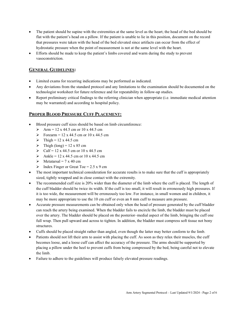
Continuous Wave Doppler Analysis
Select Continuous wave Doppler from the Unetix arterial study menu. Care must be taken to assure the recording of the optimal signal. Sufficient acoustic gel must be used to eliminate any air between the probe and the skin.
The Doppler beam must be aligned with the long axis of the vessel. An angle of 60 degrees is usually the best angle of insonation.
The Doppler beam is positioned to exclude interference from the adjacent vein. Signals are optimized by sliding the transducer slowly from medial to lateral across the artery until the strongest and best signal is obtained. The transducer is then adjusted so that an approximately 45-60 degree angle with the skin is achieved. This is varied slightly so that an accurate waveform with a maximum deflection is achieved. Modest probe pressure should be used to ensure good contact while not distorting the signal. For most sites, an 8 MHz transducer permits adequate depth penetration. However, the 5 MHz probe may be used if the signal is attenuated due to depth or calcification. Gain settings should be maximized to define waveform morphology at each level.
At least three representative Doppler waveforms (i.e. cardiac cycles) are obtained (at a paper speed of 25 mm/sec) from the following arteries bilaterally:
Subclavian
Axillary
Brachial
Radial
Ulnar
Pulse Volume Recording Waveforms (PVR or VPR) Air plethysmographic devices record changes in segmental limb volume.
Place pressure cuffs at the following locations with the bladder of the cuff directly over the artery:
Brachial Forearm Index finger (if appropriate) Connect the cuffs to the corresponding air hoses (right are clear, left are blue). Select Pulse Volume Recording (PVR) from the Unetix arterial study menu. Press and release the inflate button on the remote. Calibration of the cuffs is performed by the machine. Inflate each cuff to 55-65 mm Hg. Standardized inflation pressures must be used in all cuffs.
After the cuffs have stabilized, waveforms will appear on the screen. Choose satisfactory waveforms then press the capture button on your remote to freeze the screen.
Record at least three waveforms from each location.
If necessary, scroll to group diagnostic waveforms and press capture. This will save the waveforms and advance to the next test site. Repeat these steps for each of the remaining test sites. Press the capture button to advance to the Segmental Pressures examination. A photoplethysmographic (PPG) technique may be necessary for the index finger pulse if the continuous wave Doppler pulse cannot be found.
Arm Artery Segmental Protocol – Last Updated 9/1/2024 - Page 3 of 6
Pagina 4 — text în engleză
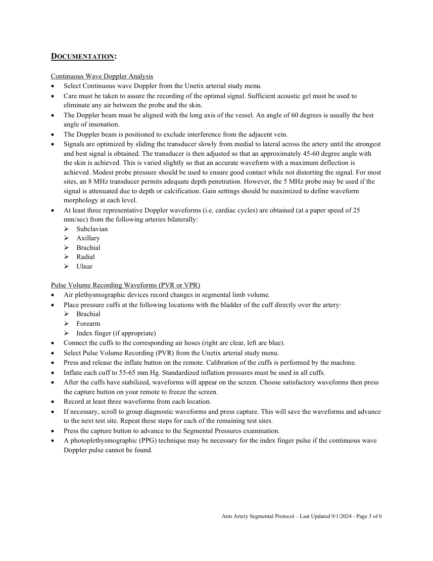
Systolic pressures obtained correspond to the vessels at the site of the pressure cuff and not to the vessel at the level of the transducer that is recording the pressure signals. Variability in systolic pressure measurements should always be considered.
Place pressure cuffs at the following locations with the bladder of the cuff directly over the artery:
Brachial Forearm Index finger (if appropriate) The Doppler transducer is positioned with the transducer directed proximally so that arterial flow will move directly into the ultrasound beam at an angle of 45-60 degrees. Care must be taken not to compress the underlying artery with the Doppler transducer. Systolic blood pressures are recorded using the Doppler transducer. Press and hold the inflate button on the remote control to inflate the intended pressure cuff. The cuff is inflated until the audible signal is no longer heard and there is no pulsatility to the waveform. In order to assure complete cessation of blood flow, the cuffs should be inflated 20-30 mmHg beyond the last audible Doppler arterial signal.
Press the deflate button once. This will slowly release the pressure from the cuff at a rate of 2-4 mmHg per second.
When the Doppler signal returns, press the capture button to record the systolic pressure.
Using the scroll return to the first deflection and press capture to advance to next segment. If pressure measurements need to be repeated, the cuff should be fully deflated for approximately one minute prior to repeat measurement. Record the systolic blood pressures for the following arteries: Right brachial Right ulnar Right radial Left brachial Left ulnar Left radial Wrist-brachial index is calculated by the Unetixs Multilab automatically using the highest systolic wrist pressure on each side (ulnar or radial) divided by the brachial pressure on that same arm.
If ABIs are less than 0.75 proceed to the Arterial Duplex Imaging Protocol.
If ABIs are unable to be obtained due to incompressible vessels proceed to Arterial Duplex Imaging Protocol.
Systolic pressures are invalid in the presence of calcified, incompressible vessels and interpretation must rely on waveform analysis and finger-brachial waveforms. If neither brachial pressure is normal, the brachial pressure would not be an accurate indicator of central arterial pressure, and calculation of indices would underestimate the disease. In this case neither the wrist- brachial or finger-brachial indices would be accurate, so interpretation would have to be by waveform analysis and duplex examination. Finger-brachial indices are calculated using the finger pressure divided by the highest brachial pressure.
The finger pressure can be obtained using photoplethysmography (see technique above for application) or using PVR.
Exercise Testing Exercise testing is not typically performed in the upper extremities.
Arm Artery Segmental Protocol – Last Updated 9/1/2024 - Page 4 of 6
Pagina 5 — text în engleză
The Doppler transducer is cleaned with germicidal disinfectant wipes. All cuffs are cleaned after each examination with germicidal disinfectant wipes as per the hospital cleaning protocol. DISEASE GRADING:
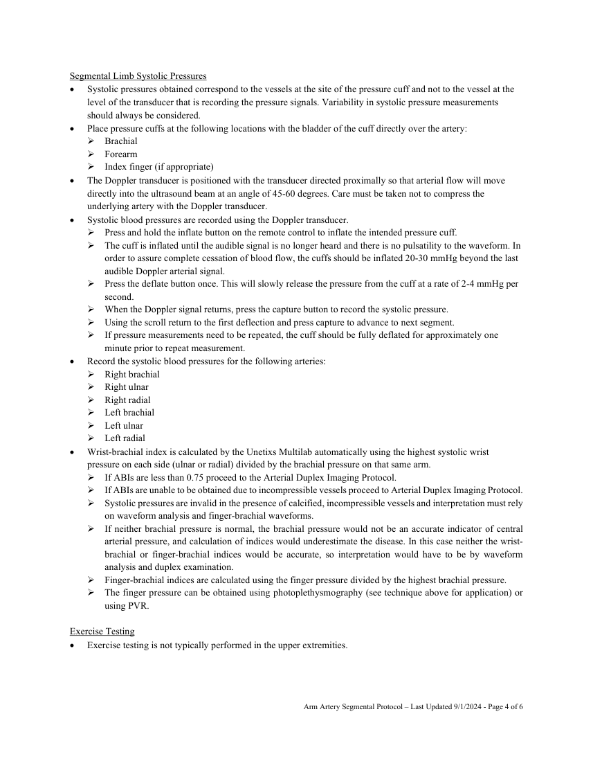
Upper and lower extremity velocities and ratios:
Stenosis PSV PSV Ratio None <150 cm/sec <1.5 Mild (30-49%) 150-200 cm/sec 1.5 to 2.0 Moderate (50-75%) 200-400 cm/sec 2.0 to 4.0 Severe (>75%) >400 cm/sec >4.0 Occlusion no flow NA
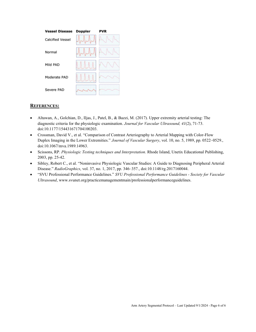
Upper extremity segmental pressures, Doppler waveforms and PVRs Greater than 10 mmHg segment to segment pressure change indicates intervening stenosis. Pressures at same level between right and left should be within 20 mmHg. Pressures between the radial and ulnar arteries should be within 5-10 mmHg. A pressure difference of ≥20 mmHg indicates stenosis in the vessel with the lower pressure. An absolute finger pressure of <70 mmHg or a brachial-finger pressure gradient of >35 mmHg indicates a stenosis between the brachial artery and finger. A wrist-to-finger pressure gradient of ≥30 mmHg suggests distal digit ischemia.
Degree Wrist-Brachial Digital-Brachial Normal ≥0.90 ≥0.86 Mild 0.75-0.89 0.70-0.85 Moderate 0.60-0.74 0.50-0.69 Severe 0.40-0.59 0.30-0.49 Critical ≤0.39 ≤0.29
Arm Artery Segmental Protocol – Last Updated 9/1/2024 - Page 5 of 6
Pagina 6 — text în engleză
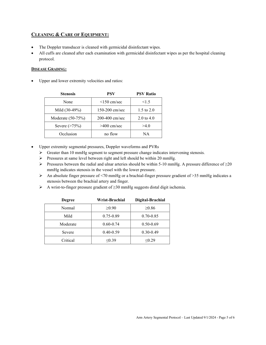
REFERENCES: Altawan, A., Golchian, D., Iljas, J., Patel, B., & Bazzi, M. (2017). Upper extremity arterial testing: The diagnostic criteria for the physiologic examination. Journal for Vascular Ultrasound, 41(2), 71-73. doi:10.1177/154431671704100203. Crossman, David V., et al. “Comparison of Contrast Arteriography to Arterial Mapping with Color-Flow Duplex Imaging in the Lower Extremities.” Journal of Vascular Surgery, vol. 10, no. 5, 1989, pp. 0522–0529., doi:10.1067/mva.1989.14963.
Scissons, RP. Physiologic Testing techniques and Interpretation. Rhode Island, Unetix Educational Publishing, 2003, pp. 25-42. Sibley, Robert C., et al. “Noninvasive Physiologic Vascular Studies: A Guide to Diagnosing Peripheral Arterial Disease.” RadioGraphics, vol. 37, no. 1, 2017, pp. 346–357., doi:10.1148/rg.2017160044.
“SVU Professional Performance Guidelines.” SVU Professional Performance Guidelines - Society for Vascular Ultrasound, www.svunet.org/practicemanagementmain/professionalperformanceguidelines.
Arm Artery Segmental Protocol – Last Updated 9/1/2024 - Page 6 of 6