Ecografie — Cartografiere venoasă: membru inferior (MCB)
Sursă și prelucrare editorială
- Proveniență
- 1 referință declarată
- Fidelitatea preluării
- Nedocumentată
- Revizuire medicală
- Nedocumentată
Referințele declarate nu confirmă singure fidelitatea preluării sau aprobarea protocolului pentru utilizare locală.
Detalii despre surse și verificări
MCB Radiology — Leg Vein Mapping
- Ediție / versiune a sursei
- Neprecizată
- Pagină / secțiune
- Pagini: 1–2
- Relația cu protocolul
- Document original importat integral; atribuirea autorilor rămâne cea din PDF.
- Data consultării
- 2026-09-27
Prelucrarea acestei pagini
- Mod de prelucrare
- Titlu și navigare în română; documentul sursă în engleză.; PDF original integral, previzualizare și text extras automat; fără completarea parametrilor clinici lipsă.
- Adaptări față de sursă
- Nedocumentate; absența evidenței nu confirmă o preluare integrală.
- Responsabil pentru prelucrare
- Neprecizat
- Atribuire existentă în pagină
- MCB Radiology (portalul sursă). Această atribuire nu identifică automat autorul sursei sau revizorul medical.
- Versiunea paginii
- mcb-834af1cf665f
- Actualizare declarată a paginii
- 2026-09-27 — distinctă de revizuirea medicală.
Verificarea fidelității: Nedocumentată
Revizuirea medicală: Nedocumentată
Documentul original MCB — Cartografiere venoasă: membru inferior
Protocol · 2 pagini · consultat la 2026-09-27.
Descarcă PDF-ul integral · Sursa MCB · Catalogul MCB pentru această modalitate
Instrucțiunile, valorile și ilustrațiile din documentul original sunt păstrate în engleză. Titlul și navigarea sunt în română.
Pagina 1

Pagina 2
Textul documentului original
Pagina 1 — text în engleză
PURPOSE:
To evaluate the patency, size, condition (such as calcification or thickening), and the course of superficial veins for surgical planning prior to vein harvesting or AV fistula creation
EQUIPMENT:
5-7 MHz linear probe
PATIENT PREPARATION & ASSESSMENT:
Introduce yourself to the patient.
Verify patient identity via two patient identifiers (name and date of birth) per hospital policy. Explain the examination, its purpose and how long it will take. Answer any questions the patient may have regarding the examination. Obtain patient history including symptoms, signs, risk factors and other relevant history.
GENERAL GUIDELINES:
If thrombus is noted in the great saphenous vein, measure how close it comes to the saphenofemoral junction. Thrombus extending to within 2 cm of the junction is treated as a DVT. The extent and location of sites where the veins fail to compress completely should be clearly recorded and generally require additional images. Longitudinal views without compression may be helpful to characterize the abnormal vein. For focal lesions (masses, lymph nodes, popliteal cysts) obtain split-screen images of the lesion without calibers, with calibers and with Color Doppler.
Arm preference for AVFs is nondominate arm over dominate arm and forearm over upper arm. Order preference for AVF site is radial artery to cephalic vein, brachial artery to cephalic vein, brachial artery to basilic vein and brachial artery to brachial vein. Then move to PTFE grafts in order brachial artery to antecubital vein, brachial artery to basilic vein and axillary artery to axillary vein. Then move to thigh PTFE grafts femoral artery to great saphenous vein or common femoral vein. Any deviations from the standard protocol and any limitations to the examination should be documented on the technologist worksheet for future reference and for repeatability in follow-up studies. Report preliminary critical findings to the referring clinician when appropriate (i.e. immediate medical attention may be warranted) and according to hospital policy.
DOCUMENTATION:
DVT Evaluation Assess veins for compressibility every 2 cm of their length.
Document transverse split-screen images without and with compression and transverse color flow images of the following:
Leg Vein Mapping Protocol – Last Updated 3/31/2024 - Page 1 of 2
Pagina 2 — text în engleză
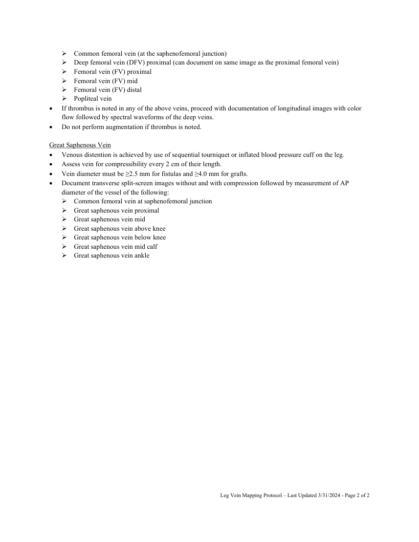
Great Saphenous Vein Venous distention is achieved by use of sequential tourniquet or inflated blood pressure cuff on the leg. Assess vein for compressibility every 2 cm of their length.
Vein diameter must be ≥2.5 mm for fistulas and ≥4.0 mm for grafts. Document transverse split-screen images without and with compression followed by measurement of AP diameter of the vessel of the following: Common femoral vein at saphenofemoral junction Great saphenous vein proximal Great saphenous vein mid Great saphenous vein above knee Great saphenous vein below knee Great saphenous vein mid calf Great saphenous vein ankle
Leg Vein Mapping Protocol – Last Updated 3/31/2024 - Page 2 of 2