Ecografie — Acces vascular pentru hemodializă (MCB)
Sursă și prelucrare editorială
- Proveniență
- 1 referință declarată
- Fidelitatea preluării
- Nedocumentată
- Revizuire medicală
- Nedocumentată
Referințele declarate nu confirmă singure fidelitatea preluării sau aprobarea protocolului pentru utilizare locală.
Detalii despre surse și verificări
MCB Radiology — Hemodialysis Access
- Ediție / versiune a sursei
- Neprecizată
- Pagină / secțiune
- Pagini: 1–4
- Relația cu protocolul
- Document original importat integral; atribuirea autorilor rămâne cea din PDF.
- Data consultării
- 2026-09-27
Prelucrarea acestei pagini
- Mod de prelucrare
- Titlu și navigare în română; documentul sursă în engleză.; PDF original integral, previzualizare și text extras automat; fără completarea parametrilor clinici lipsă.
- Adaptări față de sursă
- Nedocumentate; absența evidenței nu confirmă o preluare integrală.
- Responsabil pentru prelucrare
- Neprecizat
- Atribuire existentă în pagină
- MCB Radiology (portalul sursă). Această atribuire nu identifică automat autorul sursei sau revizorul medical.
- Versiunea paginii
- mcb-4f8ffd19c550
- Actualizare declarată a paginii
- 2026-09-27 — distinctă de revizuirea medicală.
Verificarea fidelității: Nedocumentată
Revizuirea medicală: Nedocumentată
Documentul original MCB — Acces vascular pentru hemodializă
Protocol · 4 pagini · consultat la 2026-09-27.
Descarcă PDF-ul integral · Sursa MCB · Catalogul MCB pentru această modalitate
Instrucțiunile, valorile și ilustrațiile din documentul original sunt păstrate în engleză. Titlul și navigarea sunt în română.
Pagina 1
Pagina 2
Pagina 3
Pagina 4
Textul documentului original
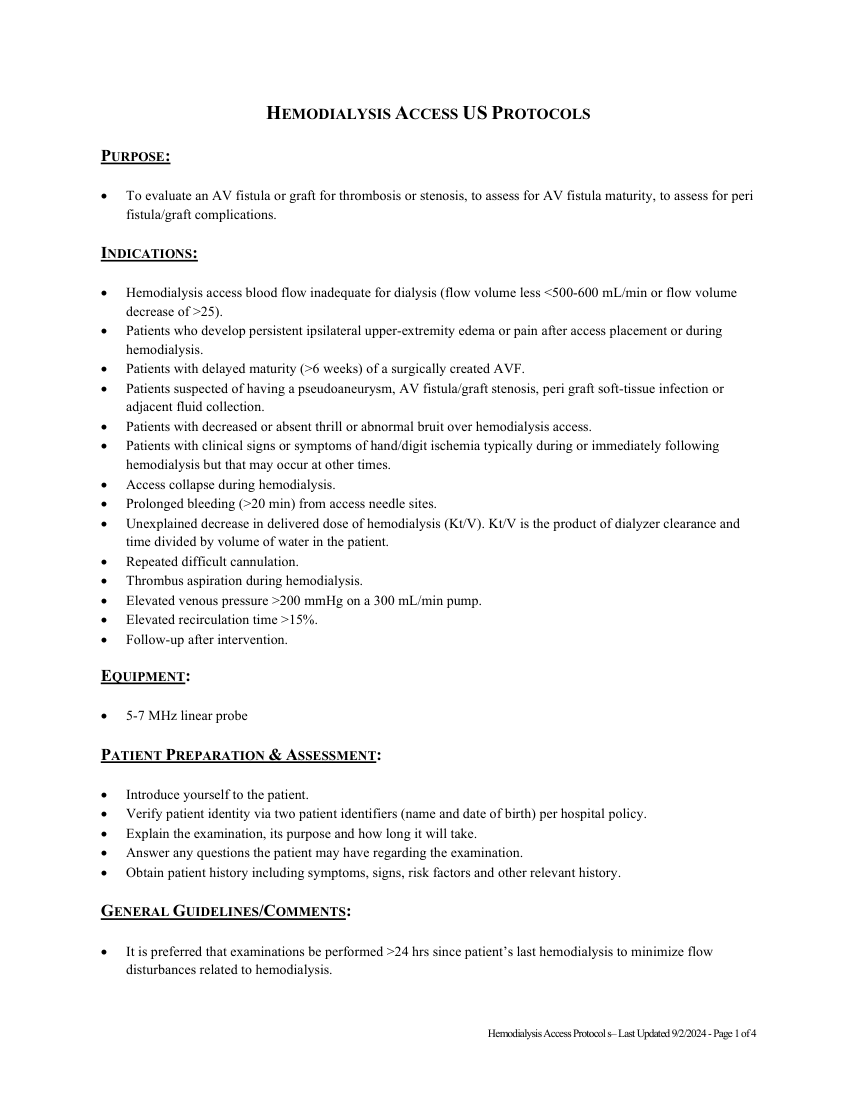
Pagina 1 — text în engleză
PURPOSE:
To evaluate an AV fistula or graft for thrombosis or stenosis, to assess for AV fistula maturity, to assess for peri fistula/graft complications.
INDICATIONS:
Hemodialysis access blood flow inadequate for dialysis (flow volume less <500-600 mL/min or flow volume decrease of >25).
Patients who develop persistent ipsilateral upper-extremity edema or pain after access placement or during hemodialysis.
Patients with delayed maturity (>6 weeks) of a surgically created AVF.
Patients suspected of having a pseudoaneurysm, AV fistula/graft stenosis, peri graft soft-tissue infection or adjacent fluid collection. Patients with decreased or absent thrill or abnormal bruit over hemodialysis access.
Patients with clinical signs or symptoms of hand/digit ischemia typically during or immediately following hemodialysis but that may occur at other times.
Access collapse during hemodialysis.
Prolonged bleeding (>20 min) from access needle sites.
Unexplained decrease in delivered dose of hemodialysis (Kt/V). Kt/V is the product of dialyzer clearance and time divided by volume of water in the patient.
Repeated difficult cannulation.
Thrombus aspiration during hemodialysis.
Elevated venous pressure >200 mmHg on a 300 mL/min pump.
Elevated recirculation time >15%.
Follow-up after intervention.
EQUIPMENT:
5-7 MHz linear probe
PATIENT PREPARATION & ASSESSMENT:
Introduce yourself to the patient.
Verify patient identity via two patient identifiers (name and date of birth) per hospital policy. Explain the examination, its purpose and how long it will take. Answer any questions the patient may have regarding the examination. Obtain patient history including symptoms, signs, risk factors and other relevant history.
GENERAL GUIDELINES/COMMENTS:
It is preferred that examinations be performed >24 hrs since patient’s last hemodialysis to minimize flow disturbances related to hemodialysis.
Hemodialysis Access Protocol s– Last Updated 9/2/2024 - Page 1 of 4
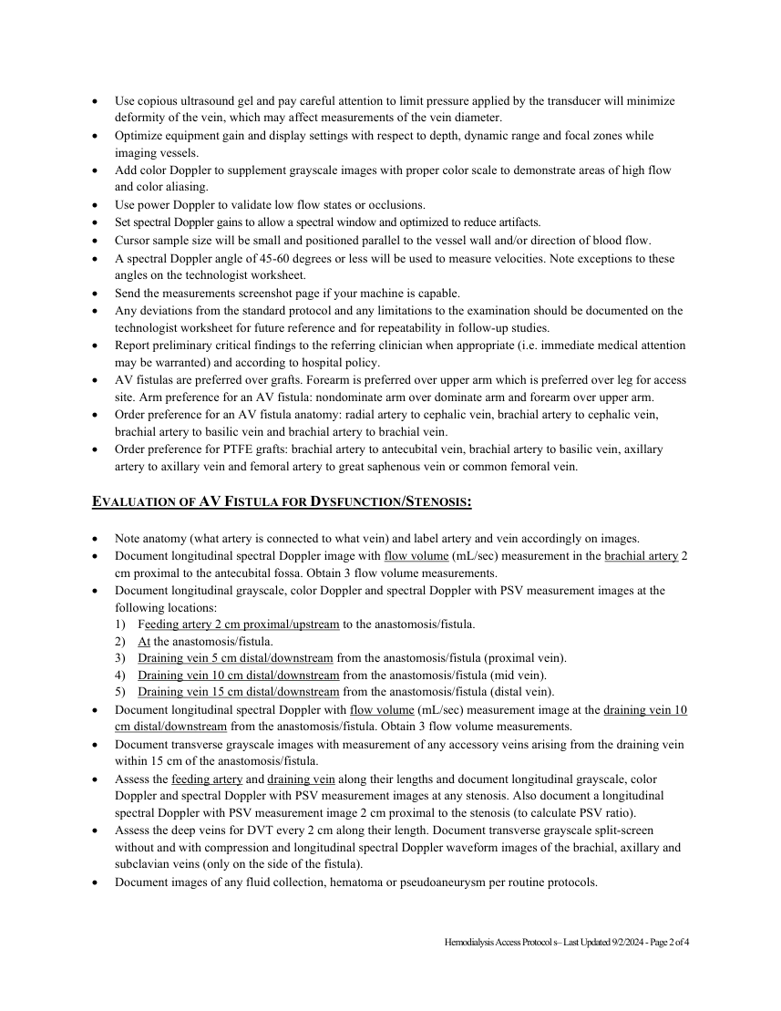
Pagina 2 — text în engleză
EVALUATION OF AV FISTULA FOR DYSFUNCTION/STENOSIS:
Note anatomy (what artery is connected to what vein) and label artery and vein accordingly on images. Document longitudinal spectral Doppler image with flow volume (mL/sec) measurement in the brachial artery 2 cm proximal to the antecubital fossa. Obtain 3 flow volume measurements. Document longitudinal grayscale, color Doppler and spectral Doppler with PSV measurement images at the following locations: 1) Feeding artery 2 cm proximal/upstream to the anastomosis/fistula. 2) At the anastomosis/fistula. 3) Draining vein 5 cm distal/downstream from the anastomosis/fistula (proximal vein). 4) Draining vein 10 cm distal/downstream from the anastomosis/fistula (mid vein). 5) Draining vein 15 cm distal/downstream from the anastomosis/fistula (distal vein). Document longitudinal spectral Doppler with flow volume (mL/sec) measurement image at the draining vein 10 cm distal/downstream from the anastomosis/fistula. Obtain 3 flow volume measurements. Document transverse grayscale images with measurement of any accessory veins arising from the draining vein within 15 cm of the anastomosis/fistula. Assess the feeding artery and draining vein along their lengths and document longitudinal grayscale, color Doppler and spectral Doppler with PSV measurement images at any stenosis. Also document a longitudinal spectral Doppler with PSV measurement image 2 cm proximal to the stenosis (to calculate PSV ratio). Assess the deep veins for DVT every 2 cm along their length. Document transverse grayscale split-screen without and with compression and longitudinal spectral Doppler waveform images of the brachial, axillary and subclavian veins (only on the side of the fistula). Document images of any fluid collection, hematoma or pseudoaneurysm per routine protocols.
Hemodialysis Access Protocol s– Last Updated 9/2/2024 - Page 2 of 4
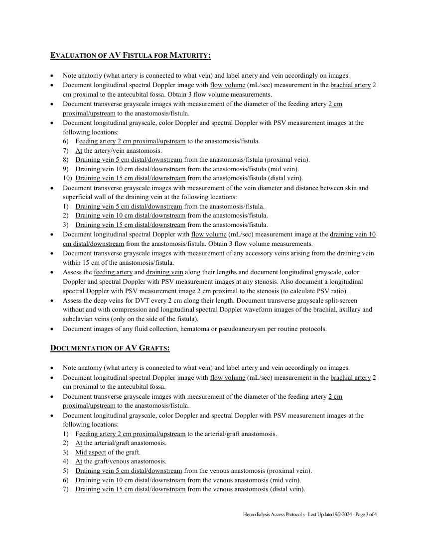
Pagina 3 — text în engleză
Note anatomy (what artery is connected to what vein) and label artery and vein accordingly on images. Document longitudinal spectral Doppler image with flow volume (mL/sec) measurement in the brachial artery 2 cm proximal to the antecubital fossa. Obtain 3 flow volume measurements. Document transverse grayscale images with measurement of the diameter of the feeding artery 2 cm proximal/upstream to the anastomosis/fistula. Document longitudinal grayscale, color Doppler and spectral Doppler with PSV measurement images at the following locations: 6) Feeding artery 2 cm proximal/upstream to the anastomosis/fistula. 7) At the artery/vein anastomosis. 8) Draining vein 5 cm distal/downstream from the anastomosis/fistula (proximal vein). 9) Draining vein 10 cm distal/downstream from the anastomosis/fistula (mid vein). 10) Draining vein 15 cm distal/downstream from the anastomosis/fistula (distal vein). Document transverse grayscale images with measurement of the vein diameter and distance between skin and superficial wall of the draining vein at the following locations: 1) Draining vein 5 cm distal/downstream from the anastomosis/fistula. 2) Draining vein 10 cm distal/downstream from the anastomosis/fistula. 3) Draining vein 15 cm distal/downstream from the anastomosis/fistula. Document longitudinal spectral Doppler with flow volume (mL/sec) measurement image at the draining vein 10 cm distal/downstream from the anastomosis/fistula. Obtain 3 flow volume measurements. Document transverse grayscale images with measurement of any accessory veins arising from the draining vein within 15 cm of the anastomosis/fistula. Assess the feeding artery and draining vein along their lengths and document longitudinal grayscale, color Doppler and spectral Doppler with PSV measurement images at any stenosis. Also document a longitudinal spectral Doppler with PSV measurement image 2 cm proximal to the stenosis (to calculate PSV ratio). Assess the deep veins for DVT every 2 cm along their length. Document transverse grayscale split-screen without and with compression and longitudinal spectral Doppler waveform images of the brachial, axillary and subclavian veins (only on the side of the fistula). Document images of any fluid collection, hematoma or pseudoaneurysm per routine protocols.
DOCUMENTATION OF AV GRAFTS:
Note anatomy (what artery is connected to what vein) and label artery and vein accordingly on images. Document longitudinal spectral Doppler image with flow volume (mL/sec) measurement in the brachial artery 2 cm proximal to the antecubital fossa.
Document transverse grayscale images with measurement of the diameter of the feeding artery 2 cm proximal/upstream to the anastomosis/fistula. Document longitudinal grayscale, color Doppler and spectral Doppler with PSV measurement images at the following locations: 1) Feeding artery 2 cm proximal/upstream to the arterial/graft anastomosis. 2) At the arterial/graft anastomosis. 3) Mid aspect of the graft. 4) At the graft/venous anastomosis. 5) Draining vein 5 cm distal/downstream from the venous anastomosis (proximal vein). 6) Draining vein 10 cm distal/downstream from the venous anastomosis (mid vein). 7) Draining vein 15 cm distal/downstream from the venous anastomosis (distal vein).
Hemodialysis Access Protocol s– Last Updated 9/2/2024 - Page 3 of 4
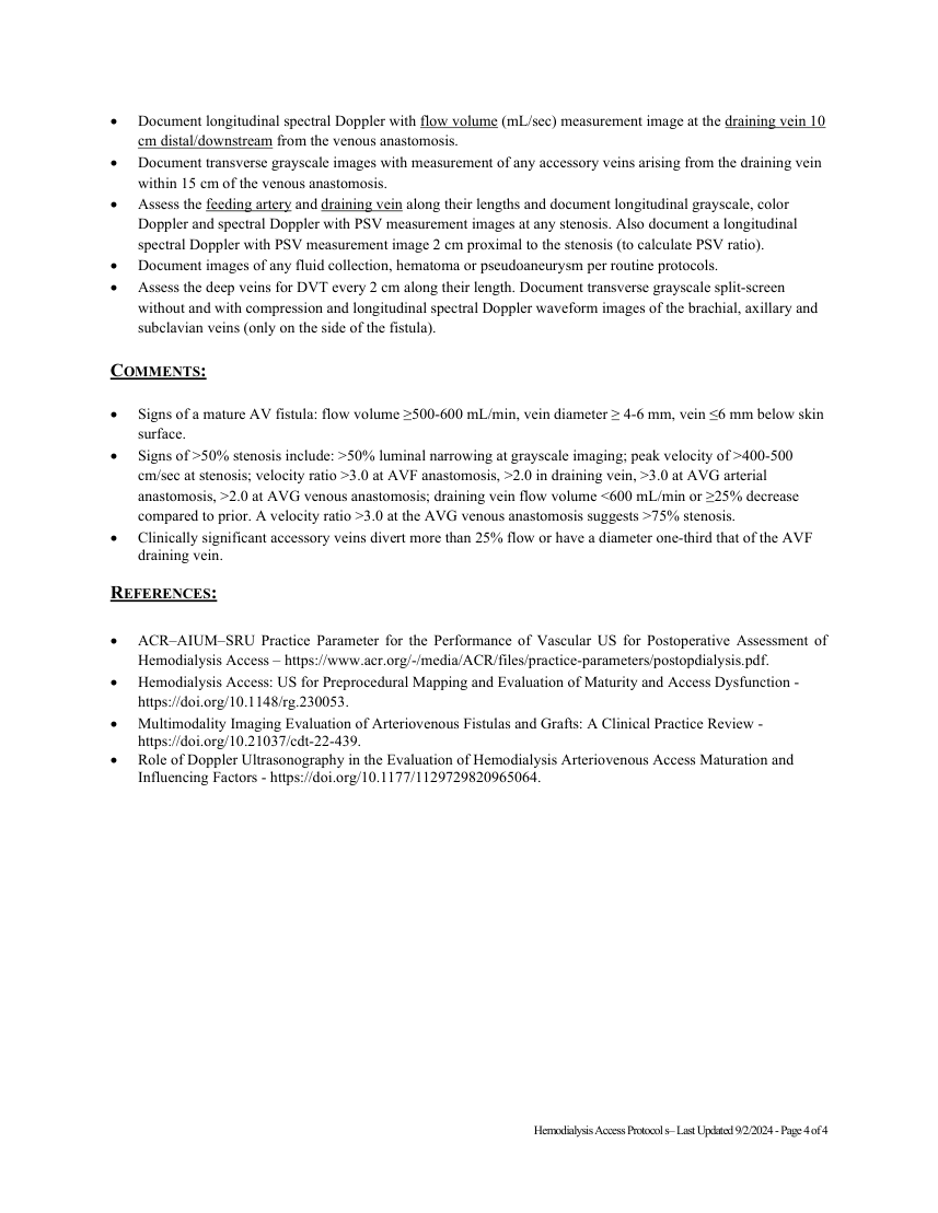
Pagina 4 — text în engleză
Document transverse grayscale images with measurement of any accessory veins arising from the draining vein within 15 cm of the venous anastomosis. Assess the feeding artery and draining vein along their lengths and document longitudinal grayscale, color Doppler and spectral Doppler with PSV measurement images at any stenosis. Also document a longitudinal spectral Doppler with PSV measurement image 2 cm proximal to the stenosis (to calculate PSV ratio). Document images of any fluid collection, hematoma or pseudoaneurysm per routine protocols. Assess the deep veins for DVT every 2 cm along their length. Document transverse grayscale split-screen without and with compression and longitudinal spectral Doppler waveform images of the brachial, axillary and subclavian veins (only on the side of the fistula).
COMMENTS:
Signs of a mature AV fistula: flow volume ≥500-600 mL/min, vein diameter ≥ 4-6 mm, vein ≤6 mm below skin surface. Signs of >50% stenosis include: >50% luminal narrowing at grayscale imaging; peak velocity of >400-500 cm/sec at stenosis; velocity ratio >3.0 at AVF anastomosis, >2.0 in draining vein, >3.0 at AVG arterial anastomosis, >2.0 at AVG venous anastomosis; draining vein flow volume <600 mL/min or ≥25% decrease compared to prior. A velocity ratio >3.0 at the AVG venous anastomosis suggests >75% stenosis. Clinically significant accessory veins divert more than 25% flow or have a diameter one-third that of the AVF draining vein.
REFERENCES:
ACR–AIUM–SRU Practice Parameter for the Performance of Vascular US for Postoperative Assessment of Hemodialysis Access – https://www.acr.org/-/media/ACR/files/practice-parameters/postopdialysis.pdf. Hemodialysis Access: US for Preprocedural Mapping and Evaluation of Maturity and Access Dysfunction - https://doi.org/10.1148/rg.230053. Multimodality Imaging Evaluation of Arteriovenous Fistulas and Grafts: A Clinical Practice Review - https://doi.org/10.21037/cdt-22-439. Role of Doppler Ultrasonography in the Evaluation of Hemodialysis Arteriovenous Access Maturation and Influencing Factors - https://doi.org/10.1177/1129729820965064.
Hemodialysis Access Protocol s– Last Updated 9/2/2024 - Page 4 of 4