Ecografie — Sân și axilă (MCB)
Sursă și prelucrare editorială
- Proveniență
- 1 referință declarată
- Fidelitatea preluării
- Nedocumentată
- Revizuire medicală
- Nedocumentată
Referințele declarate nu confirmă singure fidelitatea preluării sau aprobarea protocolului pentru utilizare locală.
Detalii despre surse și verificări
MCB Radiology — Breast & Axilla
- Ediție / versiune a sursei
- Neprecizată
- Pagină / secțiune
- Pagini: 1–2
- Relația cu protocolul
- Document original importat integral; atribuirea autorilor rămâne cea din PDF.
- Data consultării
- 2026-09-27
Prelucrarea acestei pagini
- Mod de prelucrare
- Titlu și navigare în română; documentul sursă în engleză.; PDF original integral, previzualizare și text extras automat; fără completarea parametrilor clinici lipsă.
- Adaptări față de sursă
- Nedocumentate; absența evidenței nu confirmă o preluare integrală.
- Responsabil pentru prelucrare
- Neprecizat
- Atribuire existentă în pagină
- MCB Radiology (portalul sursă). Această atribuire nu identifică automat autorul sursei sau revizorul medical.
- Versiunea paginii
- mcb-b6c1321145f9
- Actualizare declarată a paginii
- 2026-09-27 — distinctă de revizuirea medicală.
Verificarea fidelității: Nedocumentată
Revizuirea medicală: Nedocumentată
Documentul original MCB — Sân și axilă
Protocol · 2 pagini · consultat la 2026-09-27.
Descarcă PDF-ul integral · Sursa MCB · Catalogul MCB pentru această modalitate
Instrucțiunile, valorile și ilustrațiile din documentul original sunt păstrate în engleză. Titlul și navigarea sunt în română.
Pagina 1

Pagina 2
Textul documentului original
Pagina 1 — text în engleză
BREAST & AXILLA PROTOCOL
PURPOSE:
To evaluate patients with indeterminate findings on other imaging modalities and symptomatic patients.
INDICATIONS:
Indeterminate breast findings on other imaging modalities and patients with breast symptoms.
EQUIPMENT:
10-18 MHz linear probe
PATIENT PREPARATION & ASSESSMENT:
Introduce yourself to the patient.
Verify patient identity via two patient identifiers (name and date of birth) per hospital policy. Explain the examination, its purpose and how long it will take. Answer any questions the patient may have regarding the examination. Obtain patient history including symptoms, signs, risk factors and other relevant history.
GENERAL GUIDELINES:
Optimize equipment gain and display settings with respect to depth, dynamic range and focal zones while imaging vessels. Add color Doppler to supplement grayscale images with proper color scale to demonstrate areas of high flow and color aliasing. Set spectral Doppler gains to allow a spectral window and optimized to reduce artifacts. Cursor sample size will be small and positioned parallel to the vessel wall and/or direction of blood flow. A spectral Doppler angle of 45-60 degrees or less will be used to measure velocities. Note exceptions to these angles on the technologist worksheet. Send the measurements screenshot page if your machine is capable. Any deviations from the standard protocol and any limitations to the examination should be documented on the technologist worksheet for future reference and for repeatability in follow-up studies. Report preliminary critical findings to the referring clinician when appropriate (i.e. immediate medical attention may be warranted) and according to hospital policy. Whole breast screening US (handheld or automated) is not performed at any St Vincents or Optimal Imaging facility.
DOCUMENTATION:
Focal Breast or Axillary Finding The breast clock and centimeter from the nipple location or axilla location must be annotated on all images. Document radial and antiradial grayscale images of the finding without and with measurement calibers.
Breast & Axilla Protocol – Last Updated 8/19/2026 - Page 1 of 2
Pagina 2 — text în engleză
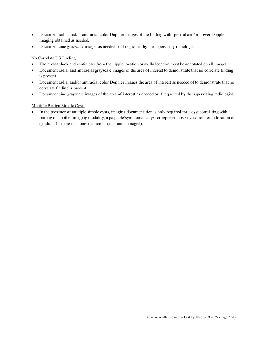
Document radial and/or antiradial color Doppler images of the finding with spectral and/or power Doppler imaging obtained as needed. Document cine grayscale images as needed or if requested by the supervising radiologist.
No Correlate US Finding The breast clock and centimeter from the nipple location or axilla location must be annotated on all images. Document radial and antiradial grayscale images of the area of interest to demonstrate that no correlate finding is present. Document radial and/or antiradial color Doppler images the area of interest as needed of to demonstrate that no correlate finding is present. Document cine grayscale images of the area of interest as needed or if requested by the supervising radiologist.
Multiple Benign Simple Cysts In the presence of multiple simple cysts, imaging documentation is only required for a cyst correlating with a finding on another imaging modality, a palpable/symptomatic cyst or representative cysts from each location or quadrant (if more than one location or quadrant is imaged).
Breast & Axilla Protocol – Last Updated 8/19/2026 - Page 2 of 2