Ecografie — Tiroidă (MCB)
Sursă și prelucrare editorială
- Proveniență
- 1 referință declarată
- Fidelitatea preluării
- Nedocumentată
- Revizuire medicală
- Nedocumentată
Referințele declarate nu confirmă singure fidelitatea preluării sau aprobarea protocolului pentru utilizare locală.
Detalii despre surse și verificări
MCB Radiology — Thyroid
- Ediție / versiune a sursei
- Neprecizată
- Pagină / secțiune
- Pagini: 1–9
- Relația cu protocolul
- Document original importat integral; atribuirea autorilor rămâne cea din PDF.
- Data consultării
- 2026-09-27
Prelucrarea acestei pagini
- Mod de prelucrare
- Titlu și navigare în română; documentul sursă în engleză.; PDF original integral, previzualizare și text extras automat; fără completarea parametrilor clinici lipsă.
- Adaptări față de sursă
- Nedocumentate; absența evidenței nu confirmă o preluare integrală.
- Responsabil pentru prelucrare
- Neprecizat
- Atribuire existentă în pagină
- MCB Radiology (portalul sursă). Această atribuire nu identifică automat autorul sursei sau revizorul medical.
- Versiunea paginii
- mcb-33dc40e56c45
- Actualizare declarată a paginii
- 2026-09-27 — distinctă de revizuirea medicală.
Verificarea fidelității: Nedocumentată
Revizuirea medicală: Nedocumentată
Documentul original MCB — Tiroidă
Protocol · 9 pagini · consultat la 2026-09-27.
Descarcă PDF-ul integral · Sursa MCB · Catalogul MCB pentru această modalitate
Instrucțiunile, valorile și ilustrațiile din documentul original sunt păstrate în engleză. Titlul și navigarea sunt în română.
Pagina 1
Pagina 2
Pagina 3
Pagina 4
Pagina 5
Pagina 6
Pagina 7
Pagina 8
Pagina 9
Textul documentului original
Pagina 1 — text în engleză
PURPOSE:
To evaluate the size of the thyroid, to assess for suspicious nodules and to assess parenchymal heterogeneity and vascularity. INDICATIONS:
Evaluation of the location and characteristics of palpable neck masses, including an enlarged thyroid. Evaluation of abnormalities detected by other imaging examinations. Evaluation of abnormal thyroid function tests. Evaluation of the presence, size and location of the thyroid gland. Evaluation of patients at high risk for occult thyroid malignancy. Follow-up imaging of previously detected thyroid nodules, when indicated. Evaluation for regional nodal metastases in patients with proven or suspected thyroid carcinoma prior to thyroidectomy. Evaluation of the thyroid gland for suspicious nodules prior to neck surgery for nonthyroid disease. Evaluation of the thyroid gland for suspicious nodules prior to radioiodine ablation. Identification and localization of parathyroid abnormalities in patients with known or suspected hyperparathyroidism. Assessment of the number and size of enlarged parathyroid glands in patients who have undergone previous parathyroid surgery or ablative therapy with recurrent symptoms of hyperparathyroidism. Localization of thyroid/parathyroid abnormalities or adjacent cervical lymph nodes for biopsy, ablation or other interventional procedures. Localization of autologous parathyroid implants. EQUIPMENT: 5-15 MHz linear probe PATIENT PREPARATION & ASSESSMENT: Introduce yourself to the patient.
Verify patient identity via two patient identifiers (name and date of birth) per hospital policy. Explain the examination, its purpose and how long it will take. Answer any questions the patient may have regarding the examination. Obtain patient history including symptoms, signs, risk factors and other relevant history. GENERAL GUIDELINES: Send the measurements screenshot page if your machine is capable. For focal lesions (masses, cysts, nodules, lymph nodes, fibroids) obtain split-screen images of the lesion without calibers, with calibers and with Color Doppler.
Specifically for thyroid nodules, obtain anterior/posterior (AP) and transverse (TR) size measurements on the transverse image and craniocaudal (CC) measurement on the longitudinal image. Any deviations from the standard protocol and any limitations to the examination should be documented on the technologist worksheet for future reference and for repeatability in follow-up studies. Report preliminary critical findings to the referring clinician when appropriate (i.e. immediate medical attention may be warranted) and according to hospital policy.
Thyroid Protocol – Last Updated 12/25/2024 - Page 1 of 9
Pagina 2 — text în engleză
Document transverse images of the lobe: Sweeping grayscale images from superior to inferior. Measure the AP and TR dimensions of the lobe at its thickest (normal 1.3-1.8 cm AP). Document color Doppler flow image of the lobe at its thickest site.
Isthmus Document transverse grayscale images without and with AP thickness (normal <6 mm). Dual Comparison Document transverse grayscale and color Doppler images showing both lobes.
Thyroid Nodules Measure up to 3 nodules per lobe and 1 nodule in the isthmus. Specifically for thyroid nodules, obtain anterior/posterior (AP) and transverse (TR) size measurements on the transverse image and craniocaudal (CC) measurement on the longitudinal image. Features that increase the likelihood of a nodule being malignant: hypoechoic, hypoechoic (relative to strap muscles), taller than wide shape, irregular margins, extension outside thyroid capsule or bulging of the thyroid capsule, punctate microcalcifications or rim calcification with nodule tissue extending beyond the rim.
Entirely cystic or almost entirely thyroid nodules are essentially never malignant and do not need to be specifically measured unless that nodule is large and causing mass effect. These nodules are occasionally aspirated for symptomatic relief.
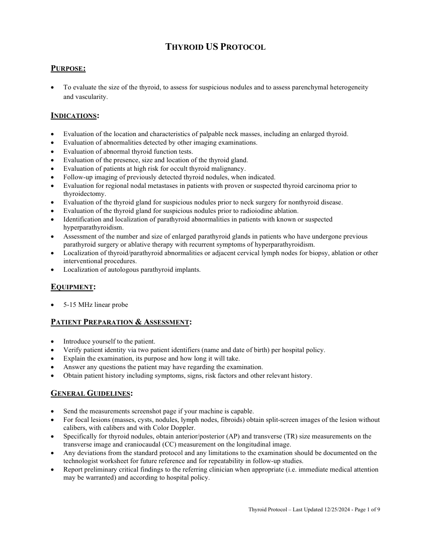
TI-RADS DESCRIPTORS FOR THYROID NODULES: Composition Solid – Composed entirely or nearly entirely of soft tissue with only a few cystic spaces.
Thyroid Protocol – Last Updated 12/25/2024 - Page 2 of 9
Pagina 3 — text în engleză
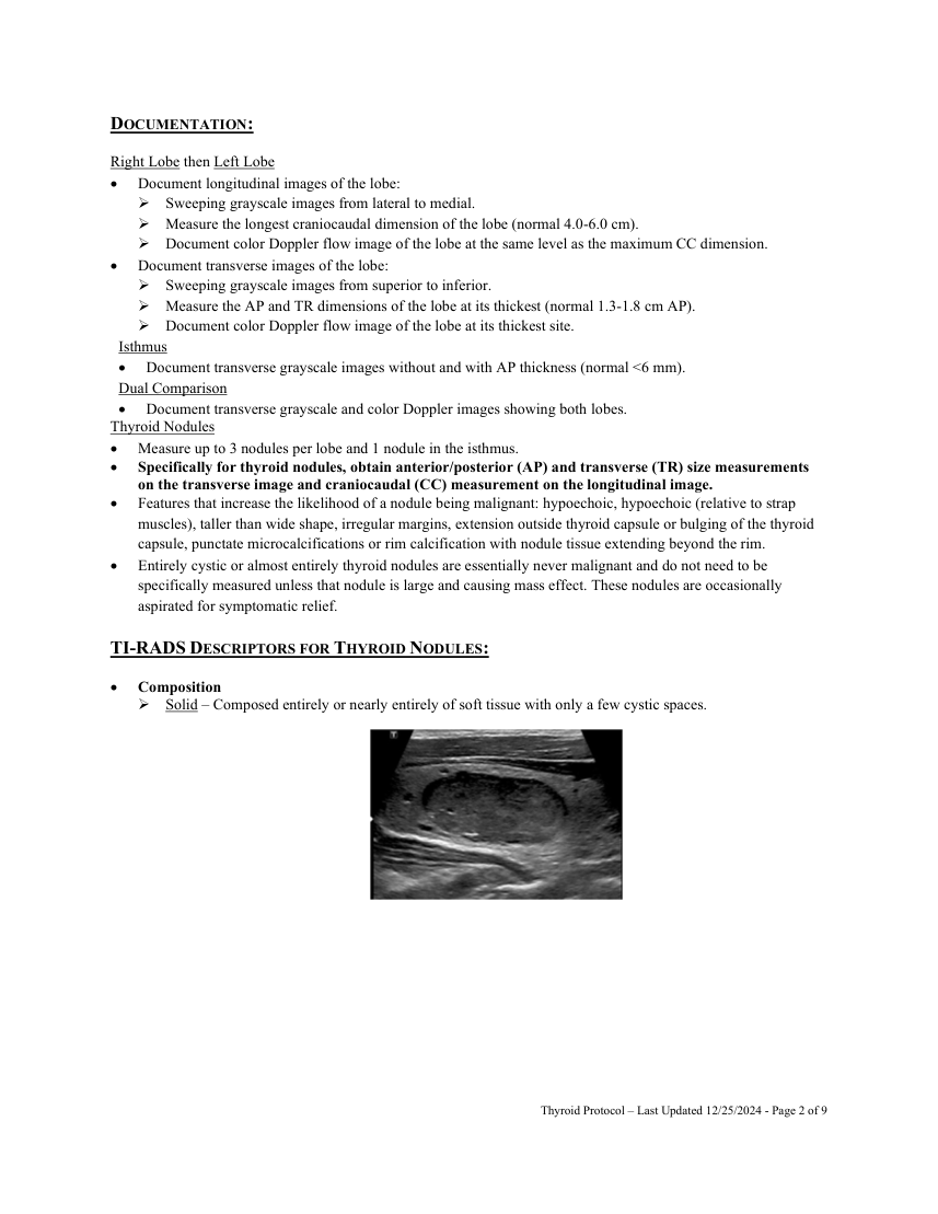
Predominately Cystic – Composed of soft tissue components occupying less than 50% of the volume of the nodule.
Spongiform – Composed predominately of tiny cystic space.
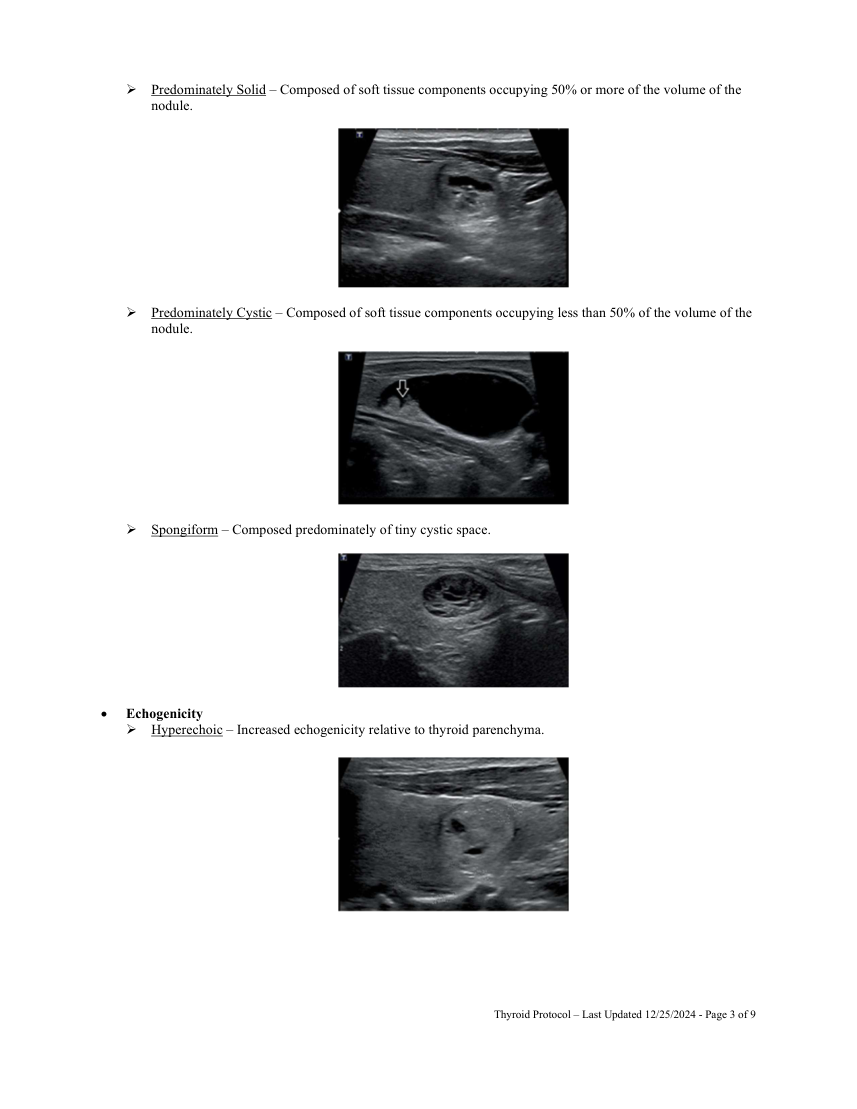
Echogenicity Hyperechoic – Increased echogenicity relative to thyroid parenchyma.
Thyroid Protocol – Last Updated 12/25/2024 - Page 3 of 9
Pagina 4 — text în engleză
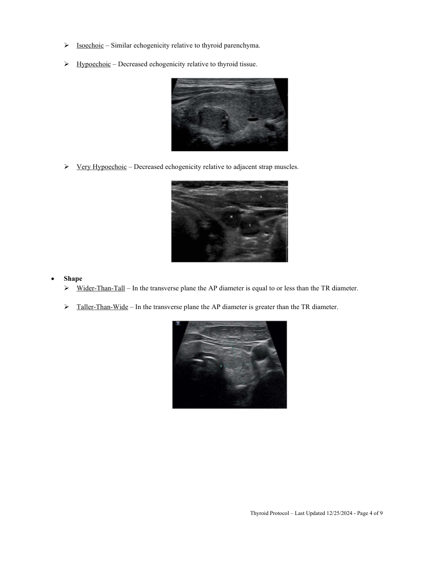
Hypoechoic – Decreased echogenicity relative to thyroid tissue.
Very Hypoechoic – Decreased echogenicity relative to adjacent strap muscles.
Shape Wider-Than-Tall – In the transverse plane the AP diameter is equal to or less than the TR diameter.
Taller-Than-Wide – In the transverse plane the AP diameter is greater than the TR diameter.
Thyroid Protocol – Last Updated 12/25/2024 - Page 4 of 9
Pagina 5 — text în engleză
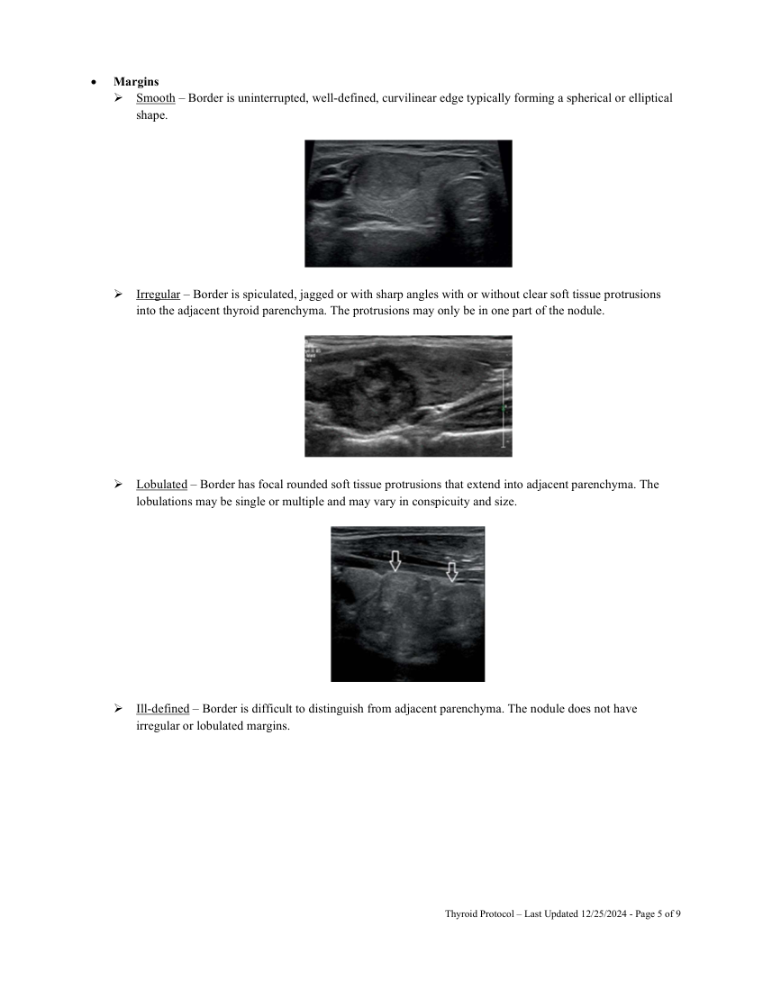
Irregular – Border is spiculated, jagged or with sharp angles with or without clear soft tissue protrusions into the adjacent thyroid parenchyma. The protrusions may only be in one part of the nodule.
Lobulated – Border has focal rounded soft tissue protrusions that extend into adjacent parenchyma. The lobulations may be single or multiple and may vary in conspicuity and size.
Ill-defined – Border is difficult to distinguish from adjacent parenchyma. The nodule does not have irregular or lobulated margins.
Thyroid Protocol – Last Updated 12/25/2024 - Page 5 of 9
Pagina 6 — text în engleză
Echogenic Foci Macrocalcifications – Calcifications large enough to cause posterior shadowing. Macrocalcifications may be irregular in shape.
Peripheral calcifications – Calcifications occupy the peripheral of the nodule. The calcification may or may not be continuous.
Thyroid Protocol – Last Updated 12/25/2024 - Page 6 of 9
Pagina 7 — text în engleză
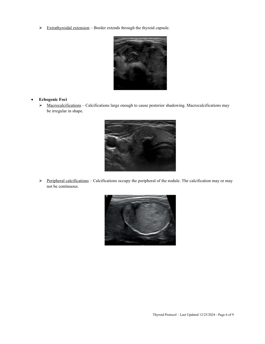
Punctate Echogenic Foci – Dot-like foci (<1 mm) having no posterior reverberation or shadowing.
CERVICAL LYMPH NODES: If there is any thyroid nodule with at least one of the suspicious characteristics noted in the above section, evaluation of the bilateral central and lateral cervical nodal compartments must be performed. Do NOT measure every normal lymph node. Measure the 1-2 largest nodes on each side of the neck, then focus on assessing for lymph nodes with suspicious features. The technologist does not need to document every nodal level on each side.
Features that favor a benign lymph node include: less than 10 mm in short-axis and elongated or kidney bean shaped (although level II nodes can measure up to 15 mm in short-axis and be more rounded) and have a thin homogeneous hypoechoic cortex surrounding an echogenic hilum. In small nodes the echogenic hilum can be difficult to visualized, however these nodes will maintain an elongated shape. Benign nodes usually demonstrate central/hilum vascularity if detectable. Features that favor a malignant lymph node include: larger in size, more rounded in shape, eccentric cortical thickening, irregular or nodular borders, loss of the echogenic hilum, increased echogenicity relative to adjacent muscle and peripheral blood flow. Lymph nodes containing calcifications or areas of cystic change are almost always malignant.
Thyroid Protocol – Last Updated 12/25/2024 - Page 7 of 9
Pagina 8 — text în engleză
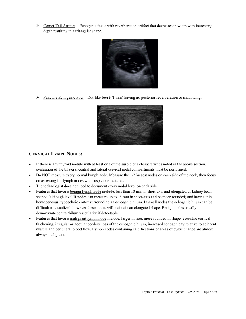
Metastatic nodal involvement in differentiated thyroid cancer (i.e. papillary and follicular subtypes) is on the same side of the thyroid cancer in 67% of cases and involves more than one node in 50% of cases. Metastatic nodule involvement most frequently involves levels III and IV followed by levels VI and V. Level II nodes are less frequently involved.
EVALUATION FOR PARATHYROID ADENOMAS: The examination should be performed with the neck hyperextended and should include longitudinal and transverse images from the carotid arteries to the midline bilaterally and extending from the carotid artery bifurcation superiorly to the thoracic inlet inferiorly.
As parathyroid glands may be hidden below the clavicles in the lower neck and upper mediastinum, it may also be helpful to have the patient swallow during the examination with constant real-time observation.
The upper mediastinum may be imaged with an appropriate probe by angling under the sternum from the sternal notch. Rarely, parathyroid adenomas may also be intrathyroidal.
Thyroid Protocol – Last Updated 12/25/2024 - Page 8 of 9
Pagina 9 — text în engleză
Thyroid Protocol – Last Updated 12/25/2024 - Page 9 of 9