Referințele declarate nu confirmă singure fidelitatea preluării sau aprobarea protocolului pentru utilizare locală.
Detalii despre surse și verificări
MCB Radiology — Histotripsy Pre Scan
Ediție / versiune a sursei
Neprecizată
Pagină / secțiune
Pagini: 1–6
Relația cu protocolul
Document original importat integral; atribuirea autorilor rămâne cea din PDF.
Data consultării
2026-09-27
Prelucrarea acestei pagini
Mod de prelucrare
Titlu și navigare în română; documentul sursă în engleză.; PDF original integral, previzualizare și text extras automat; fără completarea parametrilor clinici lipsă.
Adaptări față de sursă
Nedocumentate; absența evidenței nu confirmă o preluare integrală.
Responsabil pentru prelucrare
Neprecizat
Atribuire existentă în pagină
MCB Radiology (portalul sursă). Această atribuire nu identifică automat autorul sursei sau revizorul medical.
Instrucțiunile, valorile și ilustrațiile din documentul original sunt păstrate în engleză. Titlul și navigarea sunt în română.
Pagina 1
Pagina 2
Pagina 3
Pagina 4
Pagina 5
Pagina 6
Textul documentului original
Pagina 1 — text în engleză
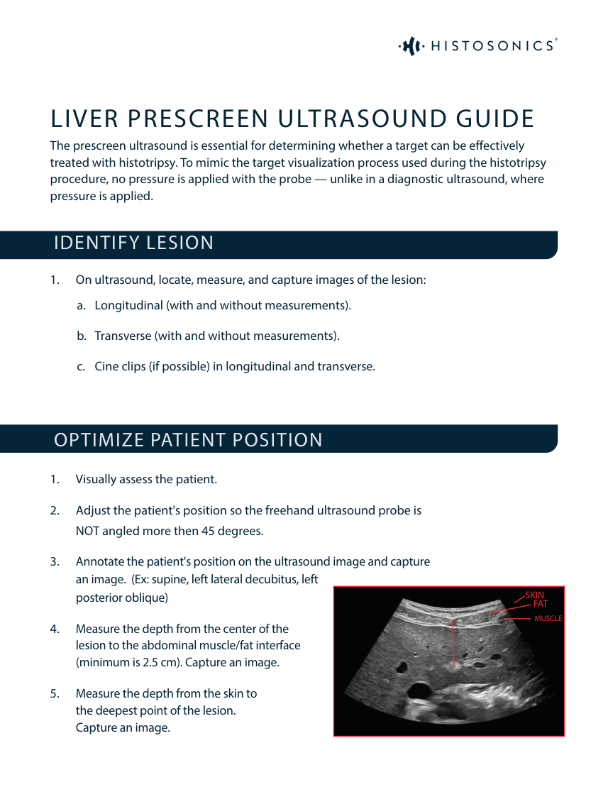
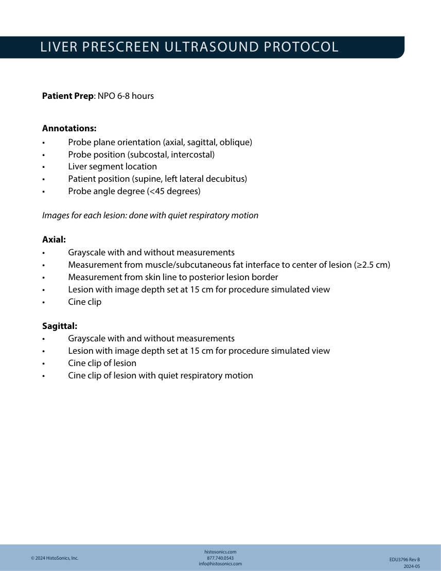
LIVER PRESCREEN ULTRASOUND GUIDE The prescreen ultrasound is essential for determining whether a target can be effectively treated with histotripsy. To mimic the target visualization process used during the histotripsy procedure, no pressure is applied with the probe — unlike in a diagnostic ultrasound, where pressure is applied. IDENTIFY LESION 1. On ultrasound, locate, measure, and capture images of the lesion: a. Longitudinal (with and without measurements). b. Transverse (with and without measurements). c. Cine clips (if possible) in longitudinal and transverse. OPTIMIZE PATIENT POSITION 1. Visually assess the patient. 2. Adjust the patient's position so the freehand ultrasound probe is NOT angled more then 45 degrees. SKIN 3. Annotate the patient's position on the ultrasound image and capture an image. (Ex: supine, left lateral decubitus, left posterior oblique) MUSCLE FAT 4. Measure the depth from the center of the lesion to the abdominal muscle/fat interface (minimum is 2.5 cm). Capture an image. 5. Measure the depth from the skin to the deepest point of the lesion. Capture an image.
Pagina 2 — text în engleză
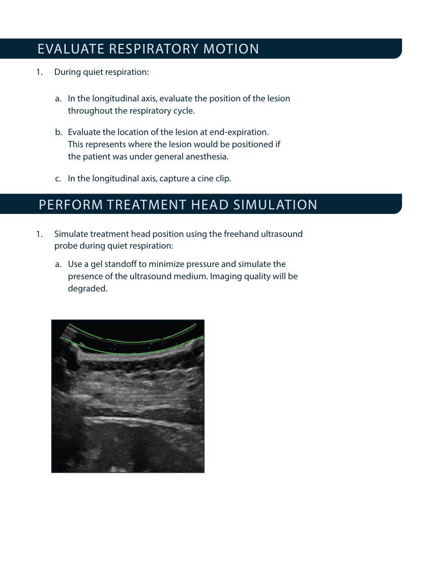
EVALUATE RESPIRATORY MOTION 1. During quiet respiration: a. In the longitudinal axis, evaluate the position of the lesion throughout the respiratory cycle. b. Evaluate the location of the lesion at end-expiration. This represents where the lesion would be positioned if the patient was under general anesthesia. c. In the longitudinal axis, capture a cine clip. PERFORM TREATMENT HEAD SIMULATION 1. Simulate treatment head position using the freehand ultrasound probe during quiet respiration: a. Use a gel standoff to minimize pressure and simulate the presence of the ultrasound medium. Imaging quality will be degraded.
Pagina 3 — text în engleză
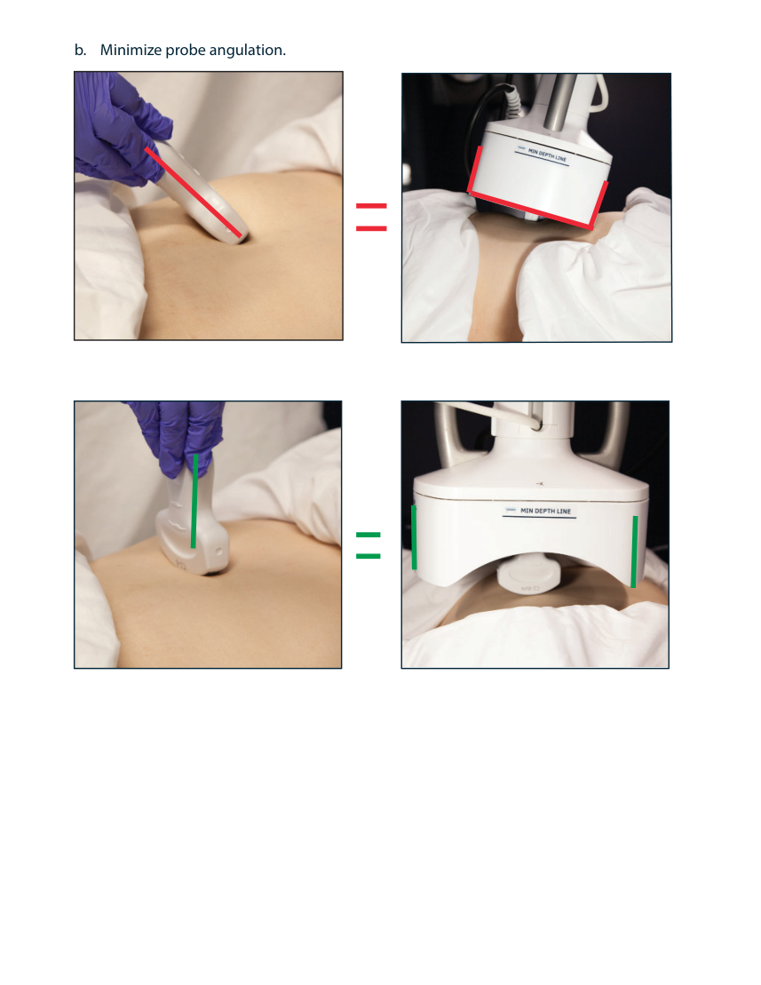
b. Minimize probe angulation.
Pagina 4 — text în engleză
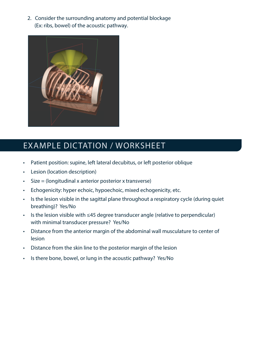
2. Consider the surrounding anatomy and potential blockage (Ex: ribs, bowel) of the acoustic pathway. EXAMPLE DICTATION / WORKSHEET • Patient position: supine, left lateral decubitus, or left posterior oblique • Lesion (location description) • Size = (longitudinal x anterior posterior x transverse) • Echogenicity: hyper echoic, hypoechoic, mixed echogenicity, etc. • Is the lesion visible in the sagittal plane throughout a respiratory cycle (during quiet breathing)? Yes/No • Is the lesion visible with ≤45 degree transducer angle (relative to perpendicular) with minimal transducer pressure? Yes/No • Distance from the anterior margin of the abdominal wall musculature to center of lesion • Distance from the skin line to the posterior margin of the lesion • Is there bone, bowel, or lung in the acoustic pathway? Yes/No
Pagina 5 — text în engleză
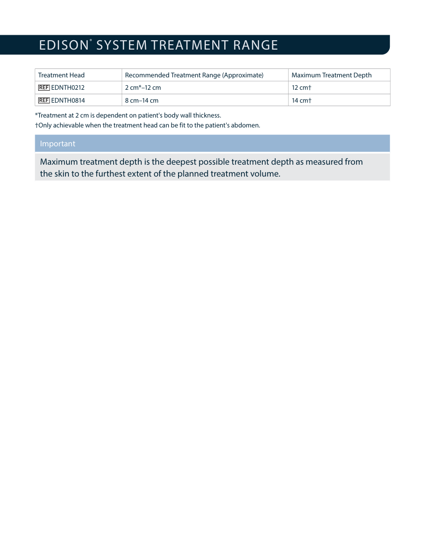
EDISON ® SYSTEM TREATMENT RANGE Treatment Head
Recommended Treatment Range (Approximate) Maximum Treatment Depth EDNTH0212 2 cm–12 cm 12 cm† EDNTH0814 8 cm–14 cm 14 cm† Treatment at 2 cm is dependent on patient's body wall thickness. †Only achievable when the treatment head can be fit to the patient's abdomen. Important Maximum treatment depth is the deepest possible treatment depth as measured from the skin to the furthest extent of the planned treatment volume.