CT — Genunchi Zimmer (MCB)
Sursă și prelucrare editorială
- Proveniență
- 1 referință declarată
- Fidelitatea preluării
- Nedocumentată
- Revizuire medicală
- Nedocumentată
Referințele declarate nu confirmă singure fidelitatea preluării sau aprobarea protocolului pentru utilizare locală.
Detalii despre surse și verificări
MCB Radiology — 18 Zimmer Knee
- Ediție / versiune a sursei
- Neprecizată
- Pagină / secțiune
- Pagini: 1–8
- Relația cu protocolul
- Document original importat integral; atribuirea autorilor rămâne cea din PDF.
- Data consultării
- 2026-09-27
Prelucrarea acestei pagini
- Mod de prelucrare
- Titlu și navigare în română; documentul sursă în engleză.; PDF original integral, previzualizare și text extras automat; fără completarea parametrilor clinici lipsă.
- Adaptări față de sursă
- Nedocumentate; absența evidenței nu confirmă o preluare integrală.
- Responsabil pentru prelucrare
- Neprecizat
- Atribuire existentă în pagină
- MCB Radiology (portalul sursă). Această atribuire nu identifică automat autorul sursei sau revizorul medical.
- Versiunea paginii
- mcb-eeb256101a56
- Actualizare declarată a paginii
- 2026-09-27 — distinctă de revizuirea medicală.
Verificarea fidelității: Nedocumentată
Revizuirea medicală: Nedocumentată
Documentul original MCB — Genunchi Zimmer
Protocol · 8 pagini · consultat la 2026-09-27.
Descarcă PDF-ul integral · Sursa MCB · Catalogul MCB pentru această modalitate
Instrucțiunile, valorile și ilustrațiile din documentul original sunt păstrate în engleză. Titlul și navigarea sunt în română.
Pagina 1
Pagina 2
Pagina 3
Pagina 4
Pagina 5
Pagina 6
Pagina 7
Pagina 8

Textul documentului original
Pagina 1 — text în engleză
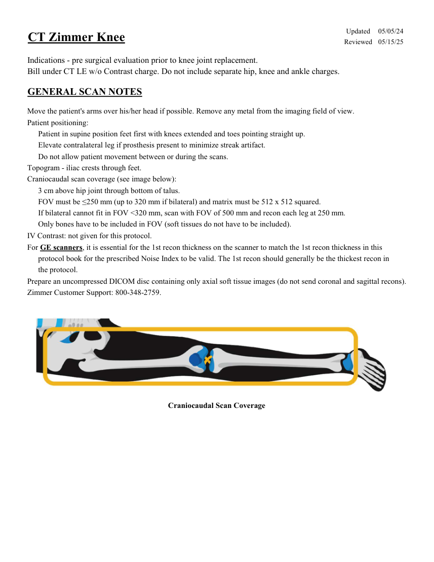
Updated 05/05/24 CT Zimmer Knee Reviewed 05/15/25 Indications - pre surgical evaluation prior to knee joint replacement. Bill under CT LE w/o Contrast charge. Do not include separate hip, knee and ankle charges. GENERAL SCAN NOTES Move the patient's arms over his/her head if possible. Remove any metal from the imaging field of view. Patient positioning: Patient in supine position feet first with knees extended and toes pointing straight up. Elevate contralateral leg if prosthesis present to minimize streak artifact. Do not allow patient movement between or during the scans. Topogram - iliac crests through feet. Craniocaudal scan coverage (see image below): 3 cm above hip joint through bottom of talus. FOV must be ≤250 mm (up to 320 mm if bilateral) and matrix must be 512 x 512 squared. If bilateral cannot fit in FOV <320 mm, scan with FOV of 500 mm and recon each leg at 250 mm. Only bones have to be included in FOV (soft tissues do not have to be included). IV Contrast: not given for this protocol. For GE scanners, it is essential for the 1st recon thickness on the scanner to match the 1st recon thickness in this protocol book for the prescribed Noise Index to be valid. The 1st recon should generally be the thickest recon in the protocol. Prepare an uncompressed DICOM disc containing only axial soft tissue images (do not send coronal and sagittal recons). Zimmer Customer Support: 800-348-2759. Craniocaudal Scan Coverage
Pagina 2 — text în engleză
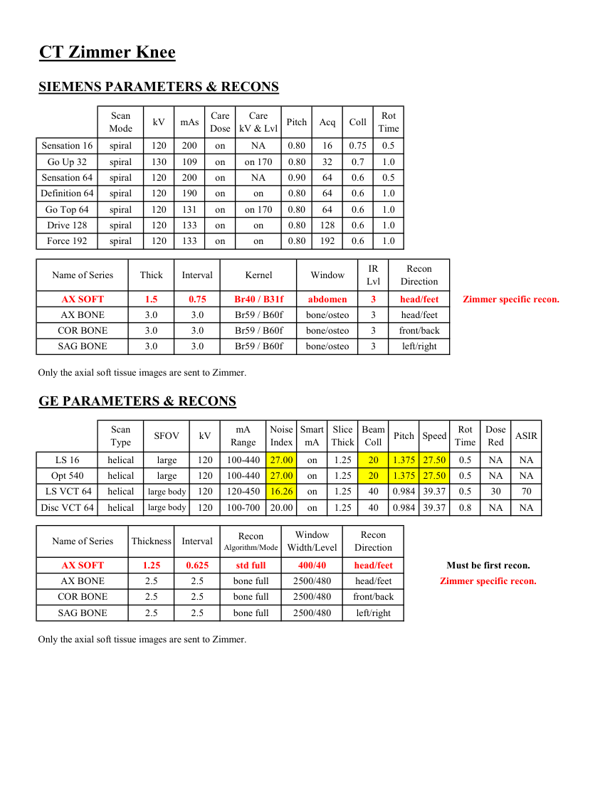
CT Zimmer Knee SIEMENS PARAMETERS & RECONS Scan
Care
Mode kV mAs Care
Dose kV & Lvl Pitch Acq Coll Rot Time Sensation 16 spiral 120 200 on NA 0.80 16 0.75 0.5 Go Up 32 spiral 130 109 on on 170 0.80 32 0.7 1.0 Sensation 64 spiral 120 200 on NA 0.90 64 0.6 0.5 Definition 64 spiral 120 190 on 0.6 1.0 on 0.80 64 Go Top 64 spiral 120 131 on on 170 0.80 64 0.6 1.0 Drive 128 spiral 120 133 on 0.80 128 0.6 1.0 on Force 192 spiral 120 133 on on 0.80 192 0.6 1.0 IR
Recon
Name of Series Thick Interval Kernel Window Lvl Direction AX SOFT 1.5 0.75 Br40 / B31f abdomen 3 head/feet Zimmer specific recon. AX BONE head/feet 3.0 3.0 Br59 / B60f bone/osteo 3 COR BONE front/back 3.0 3.0 Br59 / B60f bone/osteo 3 SAG BONE left/right 3.0 3.0 Br59 / B60f bone/osteo 3 Only the axial soft tissue images are sent to Zimmer. GE PARAMETERS & RECONS Smart
Slice
Beam
Dose
Pitch Speed Rot
Type SFOV kV mA
Red ASIR Scan
Range Noise
Index mA Thick Coll Time on 1.25 20 1.375 27.50 0.5 LS 16 helical large 120 100-440 27.00 NA NA Opt 540 helical large 120 100-440 27.00 on 1.25 20 1.375 27.50 0.5 NA NA LS VCT 64 helical large body 120 120-450 16.26 on 1.25 40 0.984 39.37 0.5 30 70 0.984 39.37 0.8 NA NA Disc VCT 64 helical large body 120 100-700 20.00 on 1.25 40 Window
Recon Name of Series Thickness Interval Recon
Algorithm/Mode Width/Level Direction AX SOFT 1.25 0.625 std full 400/40 head/feet Must be first recon. AX BONE 2.5 2.5 bone full 2500/480 head/feet Zimmer specific recon. COR BONE 2.5 2.5 bone full 2500/480 front/back SAG BONE 2.5 2.5 bone full 2500/480 left/right Only the axial soft tissue images are sent to Zimmer.
Care
Mode kV mAs Care
Dose kV & Lvl Pitch Acq Coll Rot Time Sensation 16 spiral 120 200 on NA 0.80 16 0.75 0.5 Go Up 32 spiral 130 109 on on 170 0.80 32 0.7 1.0 Sensation 64 spiral 120 200 on NA 0.90 64 0.6 0.5 Definition 64 spiral 120 190 on 0.6 1.0 on 0.80 64 Go Top 64 spiral 120 131 on on 170 0.80 64 0.6 1.0 Drive 128 spiral 120 133 on 0.80 128 0.6 1.0 on Force 192 spiral 120 133 on on 0.80 192 0.6 1.0 IR
Recon
Name of Series Thick Interval Kernel Window Lvl Direction AX SOFT 1.5 0.75 Br40 / B31f abdomen 3 head/feet Zimmer specific recon. AX BONE head/feet 3.0 3.0 Br59 / B60f bone/osteo 3 COR BONE front/back 3.0 3.0 Br59 / B60f bone/osteo 3 SAG BONE left/right 3.0 3.0 Br59 / B60f bone/osteo 3 Only the axial soft tissue images are sent to Zimmer. GE PARAMETERS & RECONS Smart
Slice
Beam
Dose
Pitch Speed Rot
Type SFOV kV mA
Red ASIR Scan
Range Noise
Index mA Thick Coll Time on 1.25 20 1.375 27.50 0.5 LS 16 helical large 120 100-440 27.00 NA NA Opt 540 helical large 120 100-440 27.00 on 1.25 20 1.375 27.50 0.5 NA NA LS VCT 64 helical large body 120 120-450 16.26 on 1.25 40 0.984 39.37 0.5 30 70 0.984 39.37 0.8 NA NA Disc VCT 64 helical large body 120 100-700 20.00 on 1.25 40 Window
Recon Name of Series Thickness Interval Recon
Algorithm/Mode Width/Level Direction AX SOFT 1.25 0.625 std full 400/40 head/feet Must be first recon. AX BONE 2.5 2.5 bone full 2500/480 head/feet Zimmer specific recon. COR BONE 2.5 2.5 bone full 2500/480 front/back SAG BONE 2.5 2.5 bone full 2500/480 left/right Only the axial soft tissue images are sent to Zimmer.
Pagina 3 — text în engleză
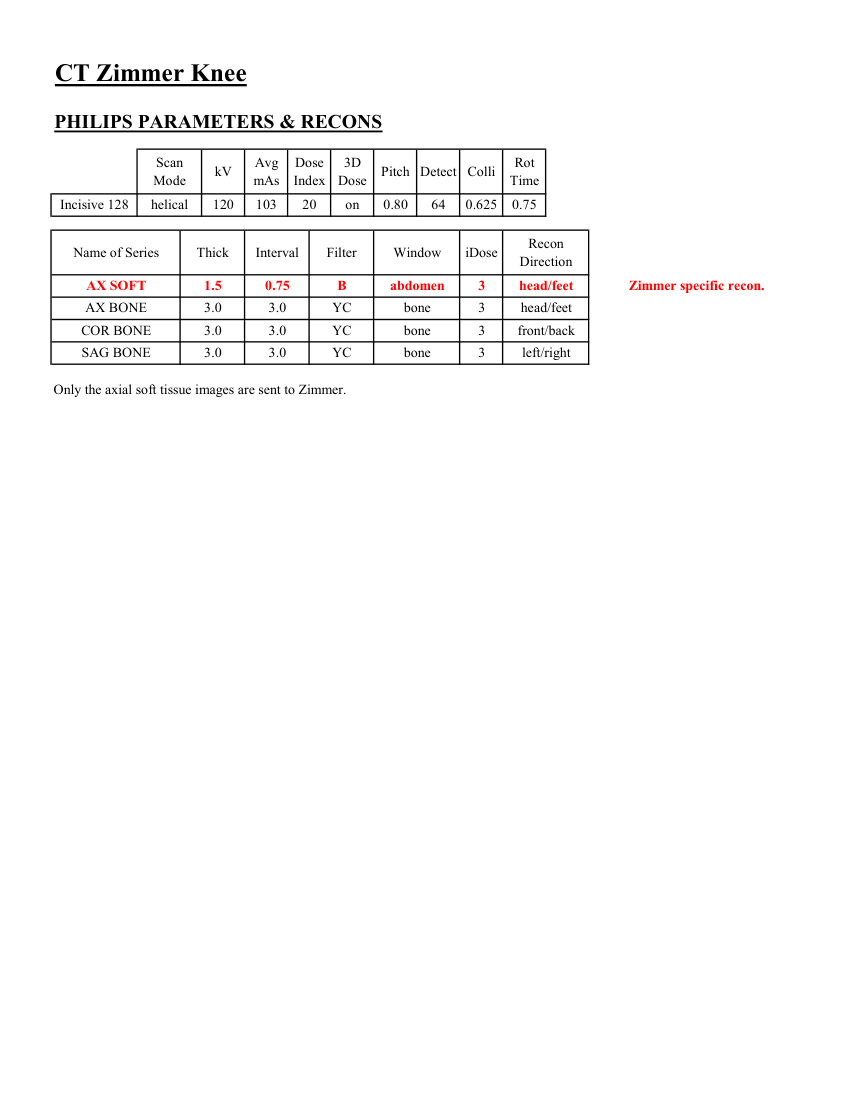
CT Zimmer Knee PHILIPS PARAMETERS & RECONS Scan
Dose
3D
Rot Mode kV Avg
mAs Index Dose Pitch Detect Colli Time Incisive 128 helical 120 103 20 on 0.80 64 0.625 0.75 Name of Series Thick Interval Filter Window iDose Recon
Direction AX SOFT 1.5 0.75 B abdomen 3 head/feet Zimmer specific recon. AX BONE 3.0 3.0 YC bone 3 head/feet COR BONE 3.0 3.0 YC bone 3 front/back SAG BONE 3.0 3.0 YC bone 3 left/right Only the axial soft tissue images are sent to Zimmer.
Dose
3D
Rot Mode kV Avg
mAs Index Dose Pitch Detect Colli Time Incisive 128 helical 120 103 20 on 0.80 64 0.625 0.75 Name of Series Thick Interval Filter Window iDose Recon
Direction AX SOFT 1.5 0.75 B abdomen 3 head/feet Zimmer specific recon. AX BONE 3.0 3.0 YC bone 3 head/feet COR BONE 3.0 3.0 YC bone 3 front/back SAG BONE 3.0 3.0 YC bone 3 left/right Only the axial soft tissue images are sent to Zimmer.
Pagina 4 — text în engleză
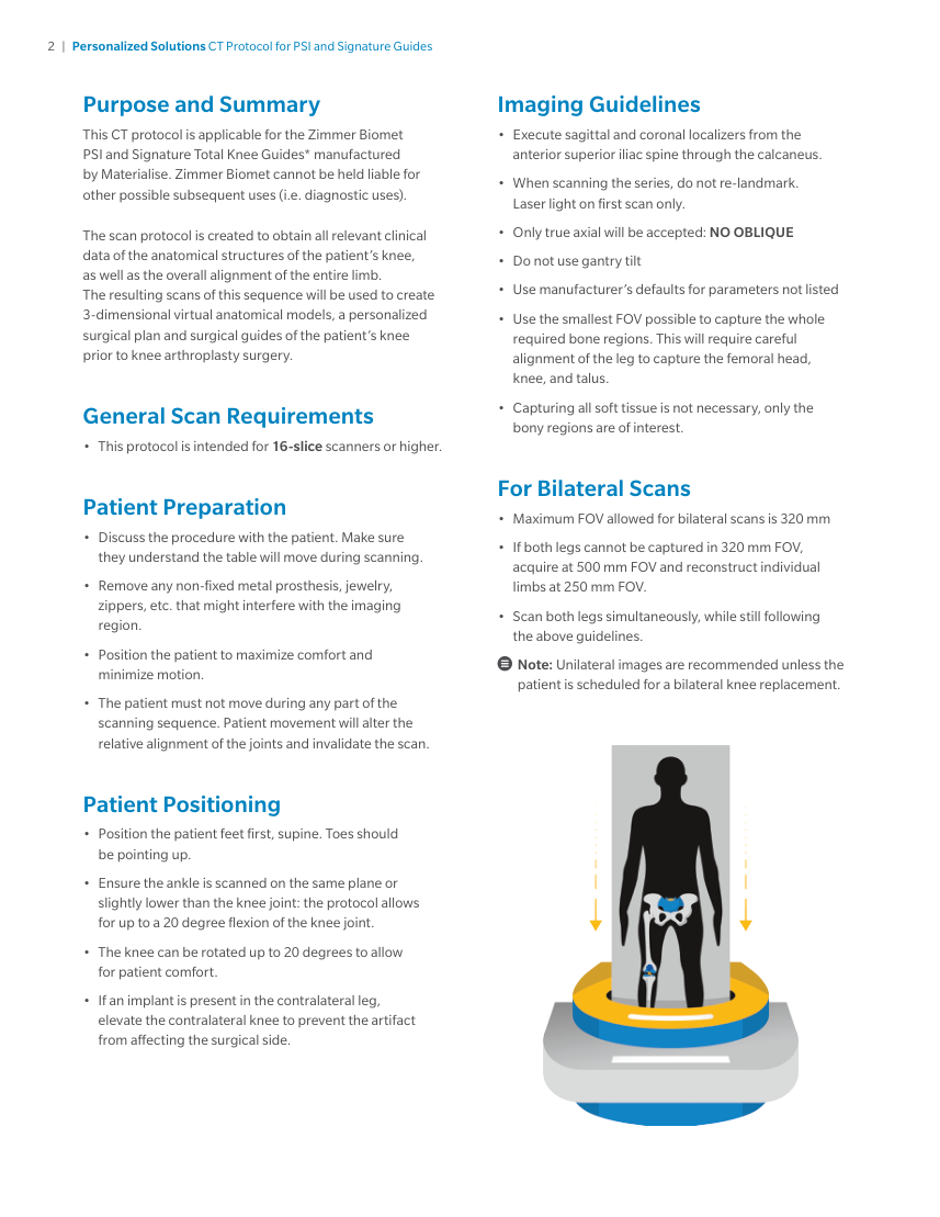
2 | Personalized Solutions CT Protocol for PSI and Signature Guides Purpose and Summary Imaging Guidelines • Execute sagittal and coronal localizers from the anterior superior iliac spine through the calcaneus. • When scanning the series, do not re-landmark. This CT protocol is applicable for the Zimmer Biomet PSI and Signature Total Knee Guides* manufactured by Materialise. Zimmer Biomet cannot be held liable for other possible subsequent uses (i.e. diagnostic uses). Laser light on first scan only. • Only true axial will be accepted: NO OBLIQUE • Do not use gantry tilt • Use manufacturer’s defaults for parameters not listed • Use the smallest FOV possible to capture the whole The scan protocol is created to obtain all relevant clinical data of the anatomical structures of the patient’s knee, as well as the overall alignment of the entire limb. The resulting scans of this sequence will be used to create 3-dimensional virtual anatomical models, a personalized surgical plan and surgical guides of the patient’s knee prior to knee arthroplasty surgery. required bone regions. This will require careful alignment of the leg to capture the femoral head, knee, and talus. • Capturing all soft tissue is not necessary, only the General Scan Requirements bony regions are of interest. • This protocol is intended for 16-slice scanners or higher. For Bilateral Scans Patient Preparation • Maximum FOV allowed for bilateral scans is 320 mm • Discuss the procedure with the patient. Make sure • If both legs cannot be captured in 320 mm FOV, they understand the table will move during scanning. • Remove any non-fixed metal prosthesis, jewelry, acquire at 500 mm FOV and reconstruct individual limbs at 250 mm FOV. • Scan both legs simultaneously, while still following zippers, etc. that might interfere with the imaging region. the above guidelines. • Position the patient to maximize comfort and Note: Unilateral images are recommended unless the minimize motion. patient is scheduled for a bilateral knee replacement. • The patient must not move during any part of the scanning sequence. Patient movement will alter the relative alignment of the joints and invalidate the scan. Patient Positioning • Position the patient feet first, supine. Toes should be pointing up. • Ensure the ankle is scanned on the same plane or slightly lower than the knee joint: the protocol allows for up to a 20 degree flexion of the knee joint. • The knee can be rotated up to 20 degrees to allow for patient comfort. • If an implant is present in the contralateral leg, elevate the contralateral knee to prevent the artifact from affecting the surgical side.
Pagina 5 — text în engleză
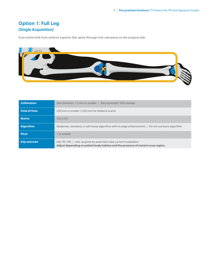
3 | Personalized Solutions CT Protocol for PSI and Signature Guides Option 1: Full Leg (Single Acquisition) Scan entire limb from anterior superior iliac spine through mid-calcaneus on the surgical side. Collimation Slice thickness: 1.5 mm or smaller | Slice increment: 50% overlap Field of View 250 mm or smaller (<320 mm for bilateral scans) Matrix 512 x 512 Algorithm Moderate, standard, or soft tissue algorithm with no edge enhancement | Do not use bone algorithm Pitch 1 or smaller kVp and mAs kVp: 90-140 | mAs: as given by automatic tube current modulation Adjust depending on patient body habitus and the presence of metal in scan region.
Pagina 6 — text în engleză
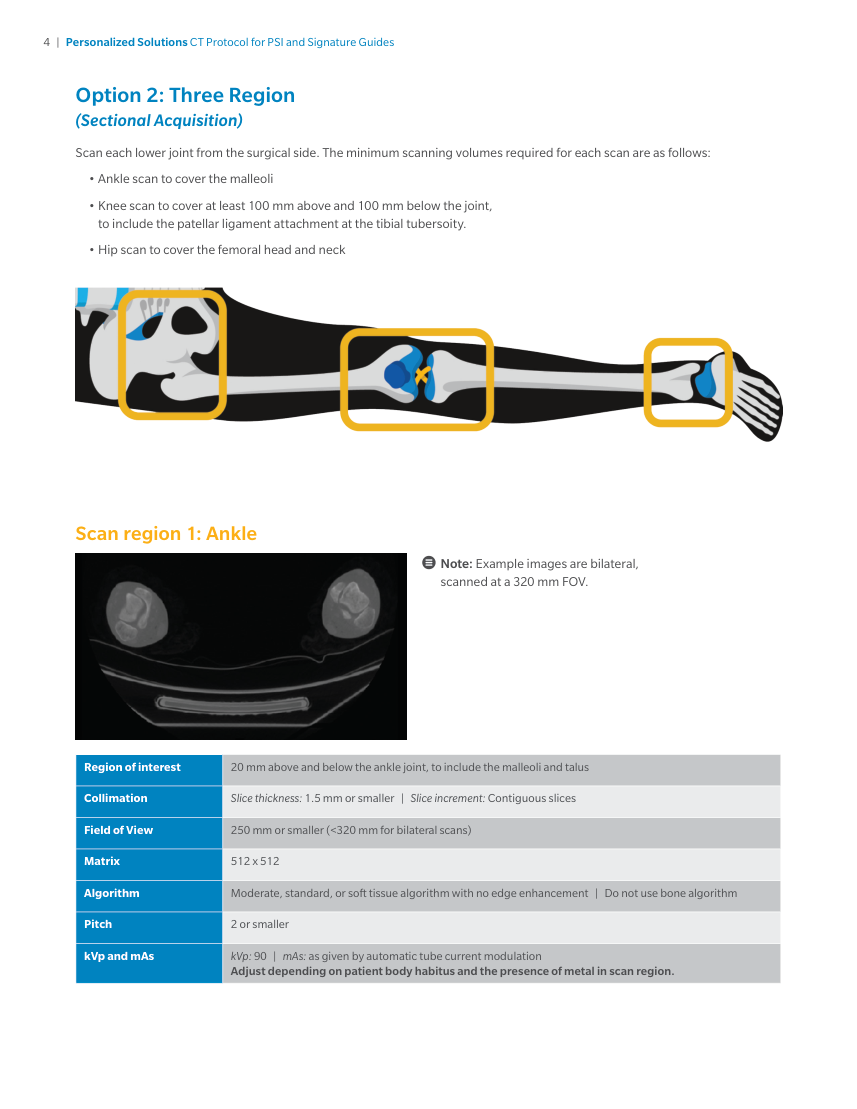
4 | Personalized Solutions CT Protocol for PSI and Signature Guides Option 2: Three Region (Sectional Acquisition) Scan each lower joint from the surgical side. The minimum scanning volumes required for each scan are as follows: • Ankle scan to cover the malleoli • Knee scan to cover at least 100 mm above and 100 mm below the joint, to include the patellar ligament attachment at the tibial tubersoity. • Hip scan to cover the femoral head and neck Scan region 1: Ankle Note: Example images are bilateral, scanned at a 320 mm FOV. Region of interest 20 mm above and below the ankle joint, to include the malleoli and talus Collimation Slice thickness: 1.5 mm or smaller | Slice increment: Contiguous slices Field of View 250 mm or smaller (<320 mm for bilateral scans) Matrix 512 x 512 Algorithm Moderate, standard, or soft tissue algorithm with no edge enhancement | Do not use bone algorithm Pitch 2 or smaller kVp and mAs kVp: 90 | mAs: as given by automatic tube current modulation Adjust depending on patient body habitus and the presence of metal in scan region.
Pagina 7 — text în engleză
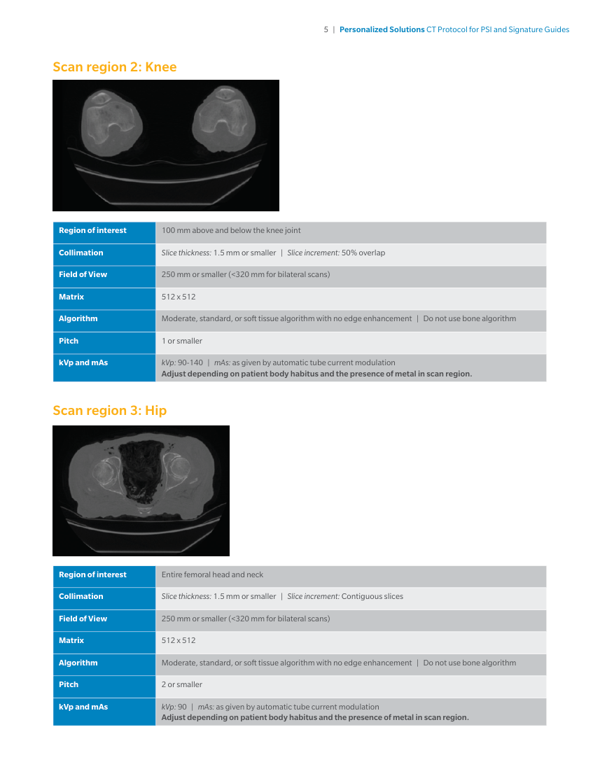
5 | Personalized Solutions CT Protocol for PSI and Signature Guides Scan region 2: Knee Region of interest 100 mm above and below the knee joint Collimation Slice thickness: 1.5 mm or smaller | Slice increment: 50% overlap Field of View 250 mm or smaller (<320 mm for bilateral scans) Matrix 512 x 512 Algorithm Moderate, standard, or soft tissue algorithm with no edge enhancement | Do not use bone algorithm Pitch 1 or smaller kVp and mAs kVp: 90-140 | mAs: as given by automatic tube current modulation Adjust depending on patient body habitus and the presence of metal in scan region. Scan region 3: Hip Region of interest Entire femoral head and neck Collimation Slice thickness: 1.5 mm or smaller | Slice increment: Contiguous slices Field of View 250 mm or smaller (<320 mm for bilateral scans) Matrix 512 x 512 Algorithm Moderate, standard, or soft tissue algorithm with no edge enhancement | Do not use bone algorithm Pitch 2 or smaller kVp and mAs kVp: 90 | mAs: as given by automatic tube current modulation Adjust depending on patient body habitus and the presence of metal in scan region.
Pagina 8 — text în engleză
6 | Personalized Solutions CT Protocol for PSI and Signature Guides Submitting the Images • Only DICOM format will be accepted • No .jpg images or other formats are acceptable • Do not submit reconstructed or reformatted images. • Only original scan data is acceptable. • Uncompressed Dicom data is required for processing. • Lossy and other forms of compression are not recommended • The scanner should be set to DICOM format “raw image,” with no compression. If loading from PACs, import and export the scan as DICOM files with the uncompressed option.