CT — Genunchi Restor3D Conformis (MCB)
Sursă și prelucrare editorială
- Proveniență
- 1 referință declarată
- Fidelitatea preluării
- Nedocumentată
- Revizuire medicală
- Nedocumentată
Referințele declarate nu confirmă singure fidelitatea preluării sau aprobarea protocolului pentru utilizare locală.
Detalii despre surse și verificări
MCB Radiology — 19 Restor3D Conformis Knee
- Ediție / versiune a sursei
- Neprecizată
- Pagină / secțiune
- Pagini: 1–13
- Relația cu protocolul
- Document original importat integral; atribuirea autorilor rămâne cea din PDF.
- Data consultării
- 2026-09-27
Prelucrarea acestei pagini
- Mod de prelucrare
- Titlu și navigare în română; documentul sursă în engleză.; PDF original integral, previzualizare și text extras automat; fără completarea parametrilor clinici lipsă.
- Adaptări față de sursă
- Nedocumentate; absența evidenței nu confirmă o preluare integrală.
- Responsabil pentru prelucrare
- Neprecizat
- Atribuire existentă în pagină
- MCB Radiology (portalul sursă). Această atribuire nu identifică automat autorul sursei sau revizorul medical.
- Versiunea paginii
- mcb-caf28b78a297
- Actualizare declarată a paginii
- 2026-09-27 — distinctă de revizuirea medicală.
Verificarea fidelității: Nedocumentată
Revizuirea medicală: Nedocumentată
Documentul original MCB — Genunchi Restor3D Conformis
Protocol · 13 pagini · consultat la 2026-09-27.
Descarcă PDF-ul integral · Sursa MCB · Catalogul MCB pentru această modalitate
Instrucțiunile, valorile și ilustrațiile din documentul original sunt păstrate în engleză. Titlul și navigarea sunt în română.
Pagina 1
Pagina 2
Pagina 3
Pagina 4
Pagina 5
Pagina 6

Pagina 7
Pagina 8
Pagina 9
Pagina 10
Pagina 11
Pagina 12

Pagina 13
Textul documentului original
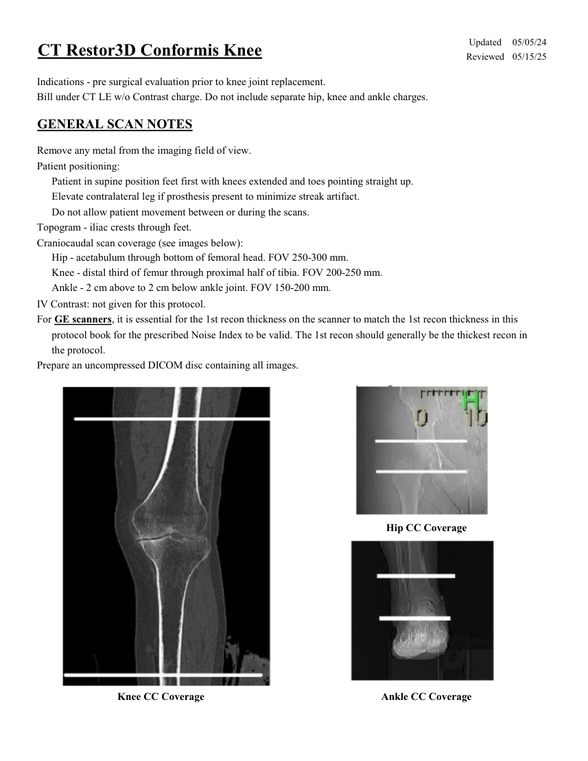
Pagina 1 — text în engleză
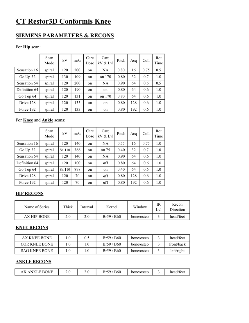
Pagina 2 — text în engleză
Care
Mode kV mAs Care
Dose kV & Lvl Pitch Acq Coll Rot Time Sensation 16 spiral 120 200 on NA 0.80 16 0.75 0.5 Go Up 32 spiral 130 109 on on 170 0.80 32 0.7 1.0 Sensation 64 spiral 120 200 on NA 0.90 64 0.6 0.5 Definition 64 spiral 120 190 on on 0.80 64 0.6 1.0 Go Top 64 spiral 120 131 on on 170 0.80 64 0.6 1.0 Drive 128 spiral 120 133 on on 0.80 128 0.6 1.0 Force 192 spiral 120 133 on on 0.80 192 0.6 1.0 For Knee and Ankle scans: Scan
Care
Mode kV mAs Care
Dose kV & Lvl Pitch Acq Coll Rot Time Sensation 16 spiral 120 140 on NA 0.55 16 0.75 1.0 Go Up 32 spiral on 75 0.40 32 0.7 1.0 Sn 110 366 on Sensation 64 spiral 120 140 on NA 0.90 64 0.6 1.0 0.80 64 0.6 1.0 Definition 64 spiral 120 100 on off Go Top 64 spiral Sn 110 898 on on 64 0.6 1.0 0.40 Drive 128 spiral 0.80 128 0.6 1.0 120 70 on off Force 192 0.80 192 0.6 1.0 spiral 120 70 on off HIP RECONS IR
Recon
Name of Series Thick Interval Kernel Window Lvl Direction AX HIP BONE 2.0 2.0 Br59 / B60 bone/osteo head/feet 3 KNEE RECONS AX KNEE BONE bone/osteo 1.0 0.5 Br59 / B60 3 head/feet COR KNEE BONE 1.0 1.0 Br59 / B60 bone/osteo front/back 3 SAG KNEE BONE 1.0 1.0 Br59 / B60 bone/osteo 3 left/right ANKLE RECONS AX ANKLE BONE 2.0 2.0 Br59 / B60 bone/osteo 3 head/feet
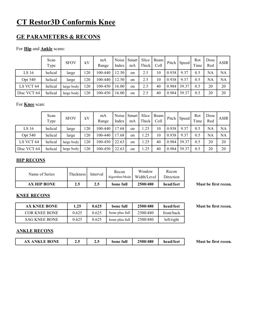
Pagina 3 — text în engleză
Slice
Beam
Dose
Pitch Speed Rot
Type SFOV kV mA
Red ASIR Scan
Range Noise
Index mA Thick Coll Time on 2.5 10 0.938 9.37 0.5 LS 16 helical large 120 100-440 12.50 NA NA Opt 540 helical large 120 100-440 12.50 on 2.5 10 0.938 9.37 0.5 NA NA LS VCT 64 helical large body 120 100-450 16.00 on 2.5 40 0.984 39.37 0.5 20 20 0.984 39.37 0.5 20 20 Disc VCT 64 helical large body 120 100-450 16.00 on 2.5 40 For Knee scan: Scan
Smart
Slice
Beam
Dose
Type SFOV kV mA
Range Noise
Index mA Thick Coll Pitch Speed Rot
Time Red ASIR LS 16 helical large 120 100-440 17.68 on 1.25 10 0.938 9.37 0.5 NA NA NA NA 10 0.938 9.37 0.5 1.25 Opt 540 helical large 120 100-440 17.68 on LS VCT 64 helical large body 120 100-450 22.63 on 1.25 40 0.984 39.37 0.5 20 20 Disc VCT 64 helical large body 120 100-450 22.63 on 1.25 40 0.984 39.37 0.5 20 20 HIP RECONS Window
Recon Recon
Name of Series Thickness Interval Algorithm/Mode Width/Level Direction AX HIP BONE 2.5 2.5 bone full 2500/480 head/feet Must be first recon. KNEE RECONS AX KNEE BONE 1.25 0.625 bone full 2500/480 head/feet Must be first recon. COR KNEE BONE 0.625 0.625 bone plus full 2500/480 front/back SAG KNEE BONE 0.625 0.625 bone plus full 2500/480 left/right ANKLE RECONS AX ANKLE BONE 2.5 2.5 bone full 2500/480 head/feet Must be first recon.
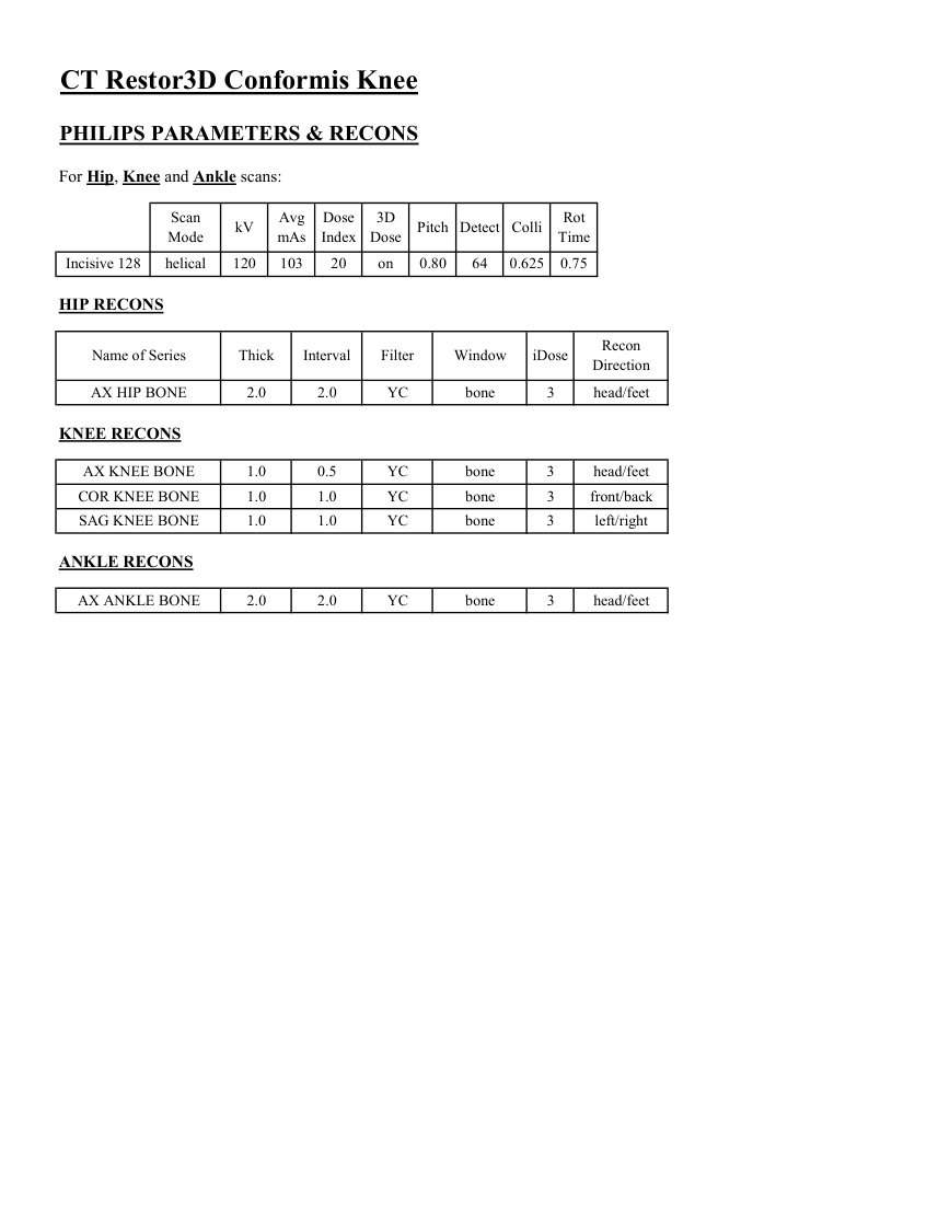
Pagina 4 — text în engleză
Dose
3D
Rot Mode kV Avg
mAs Index Dose Pitch Detect Colli Time Incisive 128 helical 120 103 20 on 0.80 64 0.625 0.75 HIP RECONS Recon
Name of Series Thick Interval Filter Window iDose Direction AX HIP BONE 2.0 2.0 YC bone 3 head/feet KNEE RECONS AX KNEE BONE 1.0 0.5 YC bone 3 head/feet COR KNEE BONE 1.0 1.0 YC bone 3 front/back SAG KNEE BONE 1.0 1.0 YC bone 3 left/right ANKLE RECONS AX ANKLE BONE 2.0 2.0 YC bone 3 head/feet
Pagina 5 — text în engleză
Pagina 6 — text în engleză
Pagina 7 — text în engleză
Pagina 8 — text în engleză
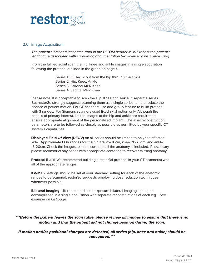
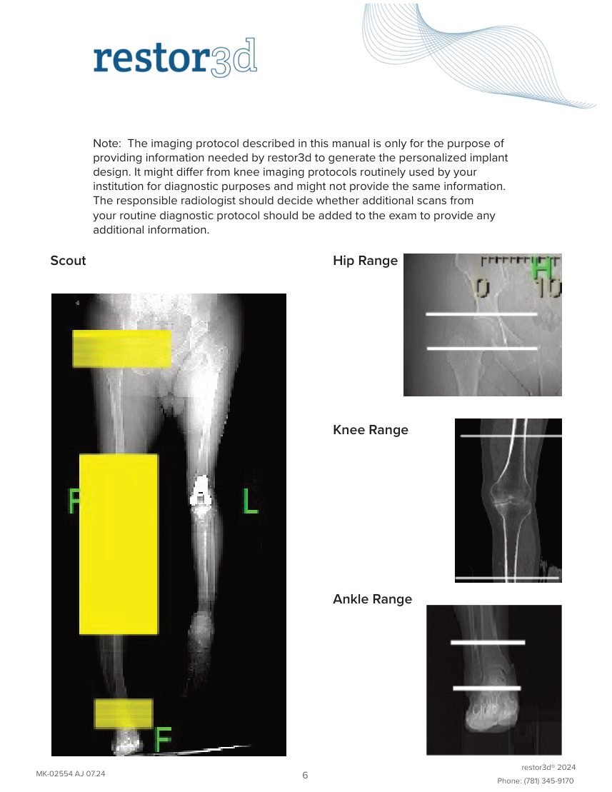
Series 4: Sagittal MPR Knee Please note: It is acceptable to scan the Hip, Knee and Ankle in separate series. But restor3d strongly suggests scanning them as a single series to help reduce the chance of patient motion. For GE scanners use add group feature to build protocol with 3 ranges. For Siemens scanners used fixed axial option only. Although the knee is of primary interest, limited images of the hip and ankle are required to ensure appropriate alignment of the personalized implant. The axial reconstruction parameters are to be followed as closely as possible as permitted by your specific CT system’s capabilities Displayed Field Of View (DFOV) on all series should be limited to only the affected side. Approximate FOV ranges for the hip are 25-30cm, knee 20-25cm, and ankle 15-20cm. Check the images to make sure that all the anatomy is included. If necessary please reconstruct any series with appropriate centering to recover missing anatomy.
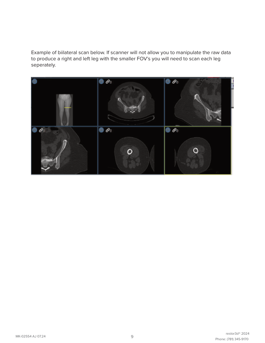
Protocol Build, We recommend building a restor3d protocol in your CT scanner(s) with all of the appropriate ranges. KV/MaS Settings should be set at your standard setting for each of the anatomic ranges to be scanned. restor3d suggests employing dose reduction techniques whenever possible. Bilateral Imaging—To reduce radiation exposure bilateral imaging should be accomplished in a single acquisition with separate reconstructions of each leg. See example on last page. Before the patient leaves the scan table, please review all images to ensure that there is no motion and that the patient did not change position during the scan.
If motion and/or positional changes are detected, all series (hip, knee and ankle) should be reacquired. restor3d® 2024 MK-02554 AJ 07.24 4 Phone: (781) 345-9170
Pagina 9 — text în engleză
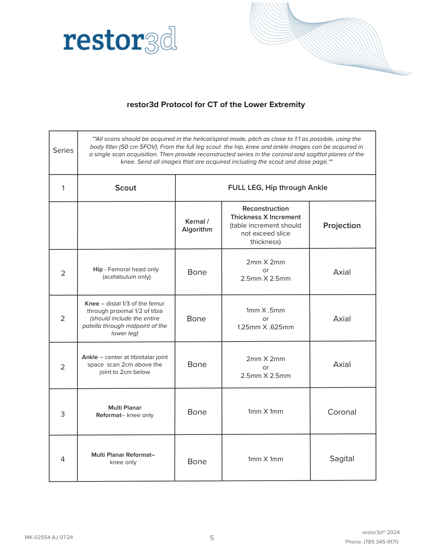
through proximal 1/2 of tibia
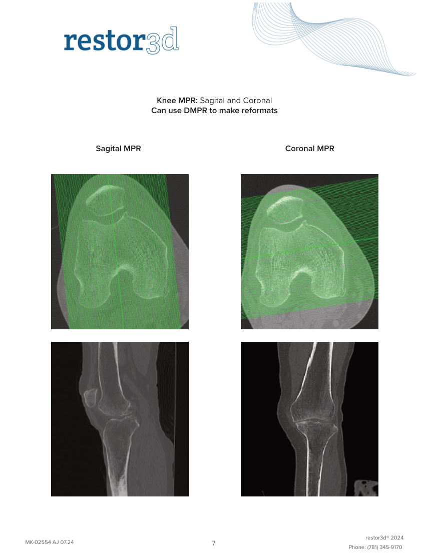
(should include the entire or 2 Bone Axial patella through midpoint of the 1.25mm X .625mm lower leg) Ankle – center at tibiotalar joint 2mm X 2mm space scan 2cm above the Bone Axial or 2 joint to 2cm below 2.5mm X 2.5mm Multi Planar 1mm X 1mm Bone Coronal 3 Reformat– knee only Multi Planar Reformat– 1mm X 1mm Sagital 4 knee only Bone restor3d® 2024 MK-02554 AJ 07.24 5 Phone: (781) 345-9170
Pagina 10 — text în engleză
Pagina 11 — text în engleză
Pagina 12 — text în engleză
secure website. Go to https://widgets.nuancepowershare.com/easyupload/Con formis to upload a scan through our secure .ftp site. 4.2 Secure DICOM transfer via Cloud Sharing Networks: restor3d is able to retrieve images from cloud based image sharing sites. If you are currently using one of these types of applications or are interested in establishing a secure cloud connection please contact restor3d Image Support at 781-345-9170 to discuss establishing a connection. We are a Nuance PowerShare Hub and can be found under Conformis Inc. Just send an invite. 4.3 Priority Shipping: To ensure that the patient’s images are received and reviewed as quickly as possible we strongly recommend the use of an electronic method of DICOM data transfer for restor3d protocol scans. However if you are unable to send studies electronically restor3d scans that have been saved in uncompressed or loss–less compression DICOM format on a disk (CD or DVD) can be shipped to restor3d.
We provide pre-paid envelopes labels. To obtain a supply please email imaging- [email protected] restor3d® 2024 MK-02554 AJ 07.24 8 Phone: (781) 345-9170