CT — Torace: VIDA (MCB)
Sursă și prelucrare editorială
- Proveniență
- 1 referință declarată
- Fidelitatea preluării
- Nedocumentată
- Revizuire medicală
- Nedocumentată
Referințele declarate nu confirmă singure fidelitatea preluării sau aprobarea protocolului pentru utilizare locală.
Detalii despre surse și verificări
MCB Radiology — 10 VIDA
- Ediție / versiune a sursei
- Neprecizată
- Pagină / secțiune
- Pagini: 1–16
- Relația cu protocolul
- Document original importat integral; atribuirea autorilor rămâne cea din PDF.
- Data consultării
- 2026-09-27
Prelucrarea acestei pagini
- Mod de prelucrare
- Titlu și navigare în română; documentul sursă în engleză.; PDF original integral, previzualizare și text extras automat; fără completarea parametrilor clinici lipsă.
- Adaptări față de sursă
- Nedocumentate; absența evidenței nu confirmă o preluare integrală.
- Responsabil pentru prelucrare
- Neprecizat
- Atribuire existentă în pagină
- MCB Radiology (portalul sursă). Această atribuire nu identifică automat autorul sursei sau revizorul medical.
- Versiunea paginii
- mcb-1e3d1cd813b6
- Actualizare declarată a paginii
- 2026-09-27 — distinctă de revizuirea medicală.
Verificarea fidelității: Nedocumentată
Revizuirea medicală: Nedocumentată
Documentul original MCB — Torace: VIDA
Protocol · 16 pagini · consultat la 2026-09-27.
Descarcă PDF-ul integral · Sursa MCB · Catalogul MCB pentru această modalitate
Instrucțiunile, valorile și ilustrațiile din documentul original sunt păstrate în engleză. Titlul și navigarea sunt în română.
Pagina 1
Pagina 2
Pagina 3
Pagina 4
Pagina 5
Pagina 6
Pagina 7
Pagina 8
Pagina 9
Pagina 10
Pagina 11
Pagina 12
Pagina 13
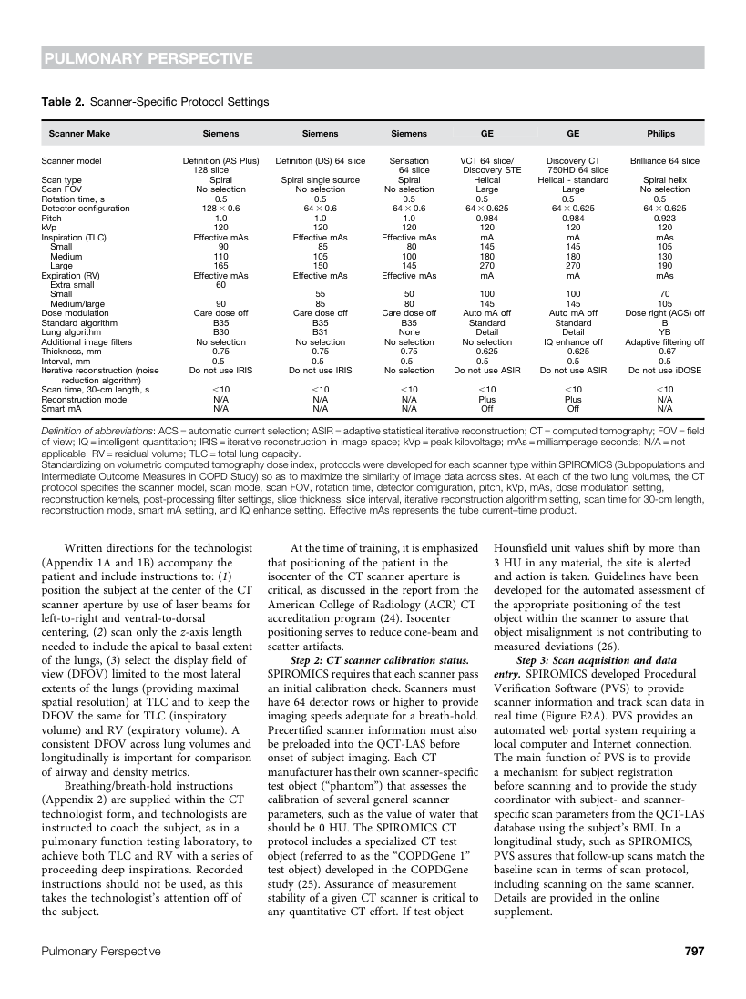
Pagina 14
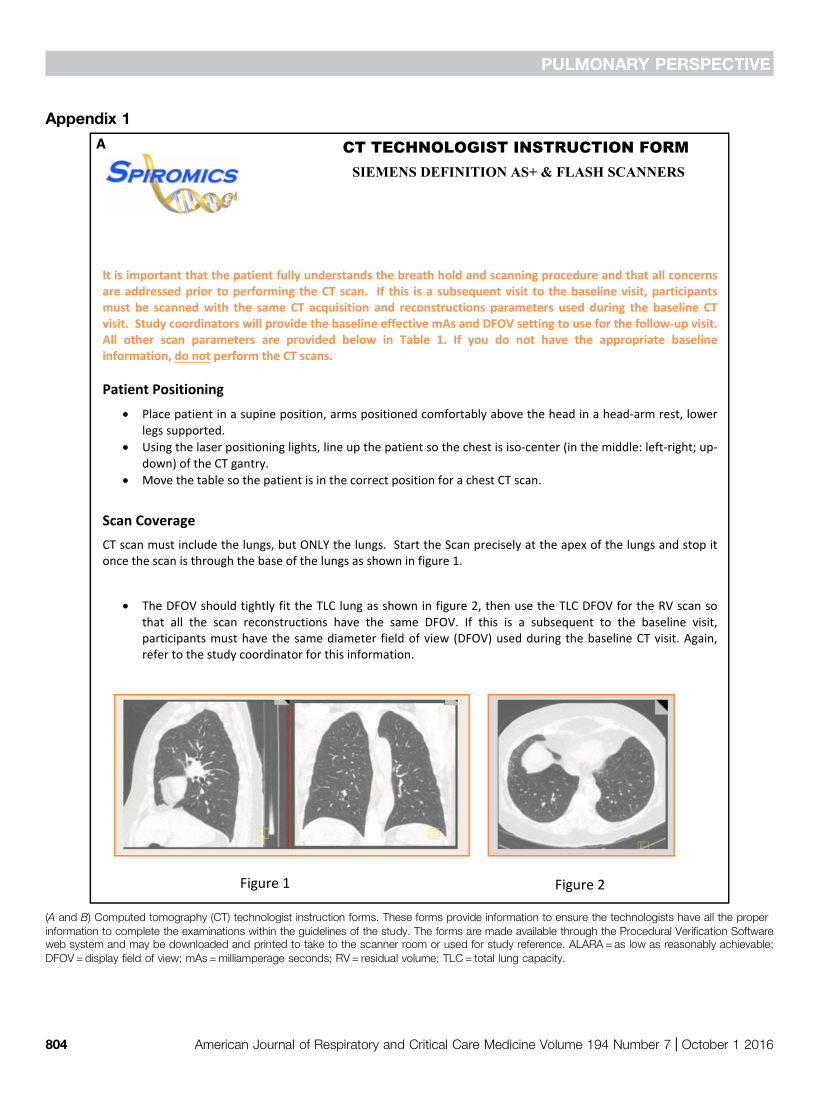
Pagina 15
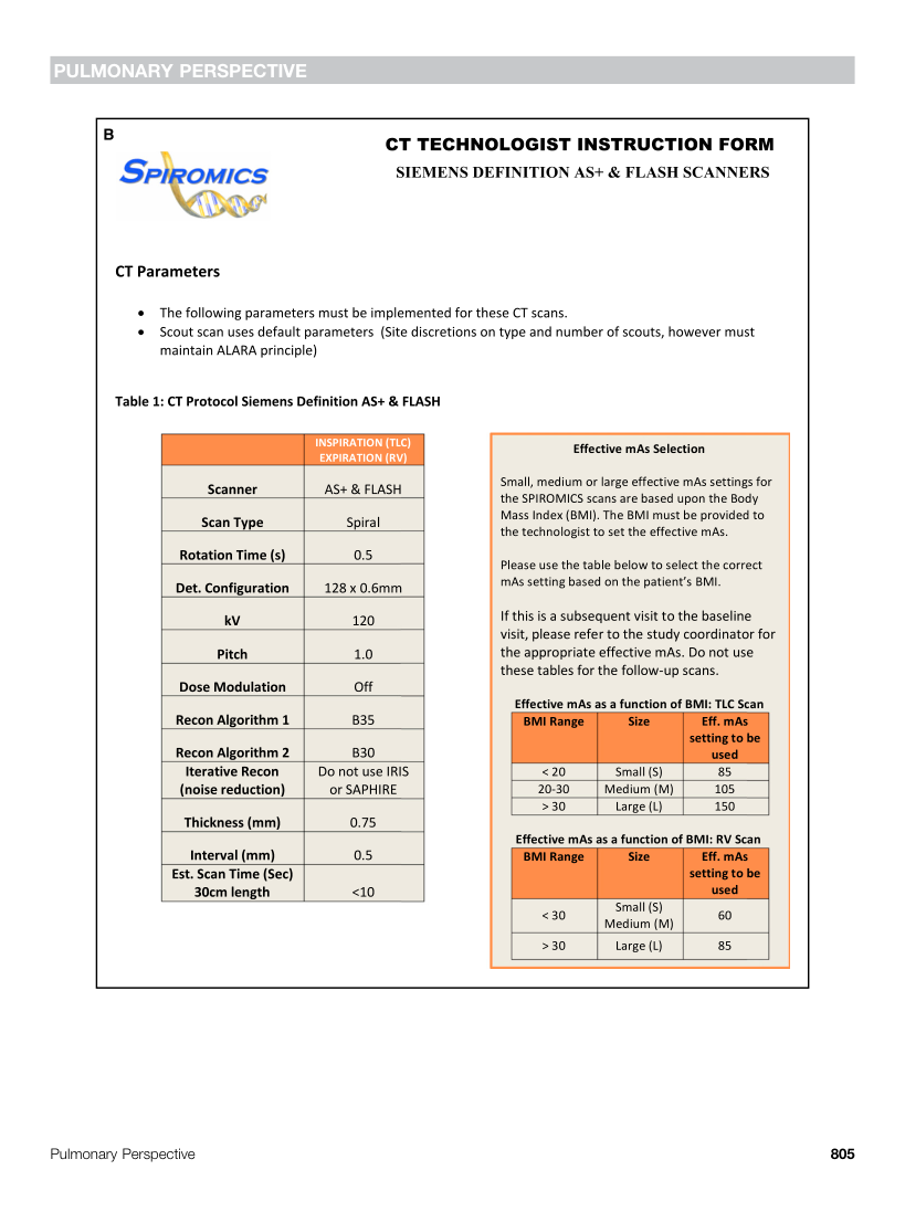
Pagina 16
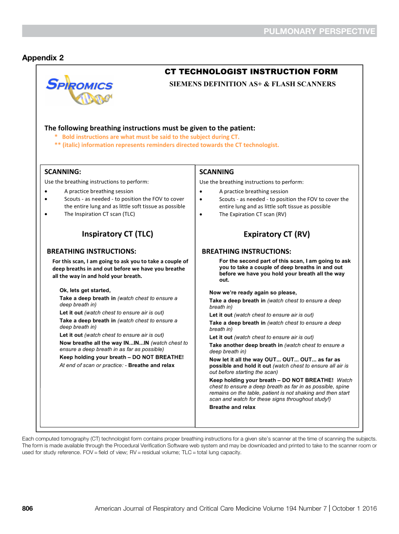
Textul documentului original
Pagina 1 — text în engleză
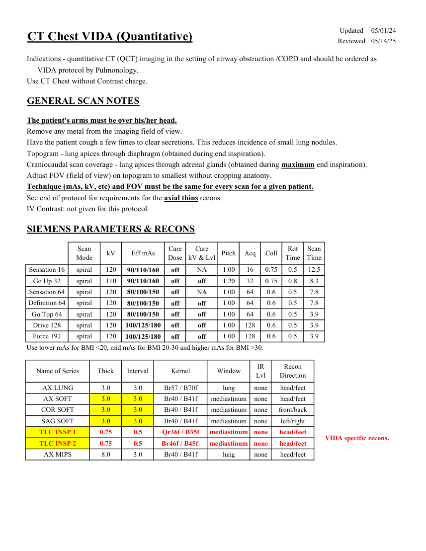
Care
Eff mAs Scan
Mode kV Care
Dose Pitch Acq Coll Rot kV & Lvl Time Time NA 16 0.5 0.75 12.5 Sensation 16 spiral 120 90/110/160 off 1.00 0.8 8.3 Go Up 32 spiral 110 off 1.20 32 0.75 90/110/160 off Sensation 64 spiral 64 0.6 0.5 NA 1.00 7.8 120 off 80/100/150 Definition 64 spiral 64 0.6 0.5 7.8 120 off 1.00 80/100/150 off Go Top 64 spiral 120 1.00 64 0.6 0.5 3.9 80/100/150 off off 1.00 128 0.6 0.5 3.9 100/125/180 Drive 128 spiral 120 off off 3.9 0.6 0.5 Force 192 spiral 120 off 100/125/180 1.00 128 off Use lower mAs for BMI <20, mid mAs for BMI 20-30 and higher mAs for BMI >30. Recon
Name of Series Thick Interval Kernel Window IR
Lvl Direction AX LUNG 3.0 3.0 Br57 / B70f lung none head/feet AX SOFT 3.0 3.0 Br40 / B41f mediastinum none head/feet COR SOFT 3.0 3.0 Br40 / B41f mediastinum none front/back SAG SOFT 3.0 3.0 Br40 / B41f mediastinum none left/right TLC INSP 1 0.75 0.5 mediastinum Qr36f / B35f none head/feet VIDA specific recons. TLC INSP 2 0.75 0.5 Br46f / B45f mediastinum none head/feet AX MIPS 8.0 3.0 Br40 / B41f lung none head/feet
Pagina 2 — text în engleză
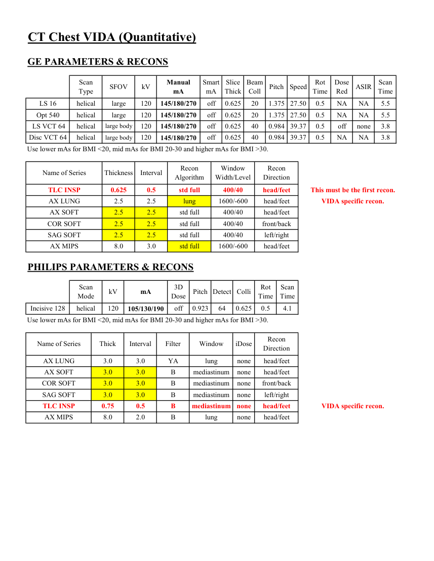
Slice
Beam
Dose
Scan
Manual
ASIR Type SFOV kV Smart
mA Thick Pitch Speed Rot
Coll Time Red Time mA LS 16 helical large 120 NA 20 0.625 1.375 27.50 off 0.5 NA 5.5 145/180/270 Opt 540 helical large 120 20 0.625 1.375 off 27.50 0.5 NA NA 5.5 145/180/270 LS VCT 64 helical large body 120 off off 40 0.625 0.984 39.37 0.5 none 3.8 145/180/270 Disc VCT 64 helical large body 120 40 0.625 off 0.984 39.37 0.5 NA NA 3.8 145/180/270 Use lower mAs for BMI <20, mid mAs for BMI 20-30 and higher mAs for BMI >30. Window
Recon
Name of Series Thickness Interval Recon
Algorithm Width/Level Direction TLC INSP 0.625 0.5 std full 400/40 head/feet This must be the first recon. AX LUNG 2.5 2.5 lung 1600/-600 head/feet VIDA specific recon. AX SOFT 2.5 2.5 std full 400/40 head/feet COR SOFT 2.5 2.5 std full 400/40 front/back SAG SOFT 2.5 2.5 std full 400/40 left/right AX MIPS 8.0 3.0 std full 1600/-600 head/feet PHILIPS PARAMETERS & RECONS Scan
3D
Rot Scan
mA Mode kV Dose Pitch Detect Colli Time Time Incisive 128 helical 120 off 0.923 64 0.625 0.5 4.1 105/130/190 Use lower mAs for BMI <20, mid mAs for BMI 20-30 and higher mAs for BMI >30. Recon
Name of Series Thick Interval Filter Window iDose Direction AX LUNG 3.0 3.0 YA lung none head/feet AX SOFT 3.0 3.0 B mediastinum none head/feet COR SOFT 3.0 3.0 B mediastinum none front/back SAG SOFT 3.0 3.0 B mediastinum none left/right TLC INSP B mediastinum none 0.75 0.5 head/feet VIDA specific recon. AX MIPS 8.0 2.0 B lung none head/feet
Pagina 3 — text în engleză
Pagina 4 — text în engleză
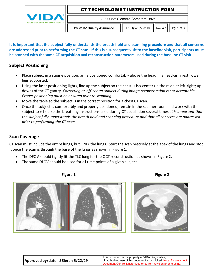
CT-90053: Siemens Somatom Drive
Issued by: Quality Assurance
Eff. Date: 05/22/19 Rev A.1
Pg. 1 of 3
It is important that the subject fully understands the breath hold and scanning procedure and that all concerns are addressed prior to performing the CT scan. If this is a subsequent visit to the baseline visit, participants must be scanned with the same CT acquisition and reconstruction parameters used during the baseline CT visit.
Subject Positioning • Place subject in a supine position, arms positioned comfortably above the head in a head-arm rest, lower legs supported. • Using the laser positioning lights, line up the subject so the chest is iso-center (in the middle: left-right; up- down) of the CT gantry. Correcting an off-center subject during image reconstruction is not acceptable. Proper positioning must be ensured prior to scanning. • Move the table so the subject is in the correct position for a chest CT scan. • Once the subject is comfortably and properly positioned, remain in the scanner room and work with the subject to rehearse the breathing instructions used during CT acquisition several times. It is important that the subject fully understands the breath hold and scanning procedure and that all concerns are addressed prior to performing the CT scan.
Scan Coverage CT scan must include the entire lungs, but ONLY the lungs. Start the scan precisely at the apex of the lungs and stop it once the scan is through the base of the lungs as shown in Figure 1.
• The DFOV should tightly fit the TLC lung for the QCT reconstruction as shown in Figure 2. • The same DFOV should be used for all time points of a given subject.
Figure 1 Figure 2
Approved by/date: J Sieren 5/22/19 This document is the property of VIDA Diagnostics, Inc.
Unauthorized use of this document is prohibited. Note: Always check Document Control Master List for current revision prior to using.
Pagina 5 — text în engleză
CT-90053: Siemens Somatom Drive
Issued by: Quality Assurance
Eff. Date: 05/22/19 Rev A.1
Pg. 2 of 3
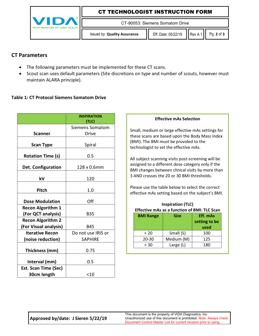
CT Parameters • The following parameters must be implemented for these CT scans.
• Scout scan uses default parameters (Site discretions on type and number of scouts, however must maintain ALARA principle).
Table 1: CT Protocol Siemens Somatom Drive
INSPIRATION Effective mAs Selection (TLC)
Siemens Somatom Scanner Drive Scan Type Spiral Rotation Time (s) 0.5 Det. Configuration 128 x 0.6mm kV 120 Pitch 1.0 Small, medium or large effective mAs settings for these scans are based upon the Body Mass Index (BMI). The BMI must be provided to the technologist to set the effective mAs.
All subject scanning visits post-screening will be assigned to a different dose category only if the BMI changes between clinical visits by more than 3 AND crosses the 20 or 30 BMI thresholds.
Please use the table below to select the correct effective mAs setting based on the subject’s BMI.
Dose Modulation Off Inspiration (TLC) Recon Algorithm 1
Effective mAs as a function of BMI: TLC Scan (For QCT analysis) B35 BMI Range Size Eff. mAs Recon Algorithm 2
setting to be (For Visual analysis) B45 used < 20 Small (S) 100 Iterative Recon Do not use IRIS or 20-30 Medium (M) 125 (noise reduction) SAPHIRE > 30 Large (L) 180
Thickness (mm) 0.75 Interval (mm) 0.5 Est. Scan Time (Sec)
30cm length <10
Approved by/date: J Sieren 5/22/19 This document is the property of VIDA Diagnostics, Inc.
Unauthorized use of this document is prohibited. Note: Always check Document Control Master List for current revision prior to using.
Pagina 6 — text în engleză
CT-90053: Siemens Somatom Drive
Issued by: Quality Assurance
Eff. Date: 05/22/19 Rev A.1
Pg. 3 of 3
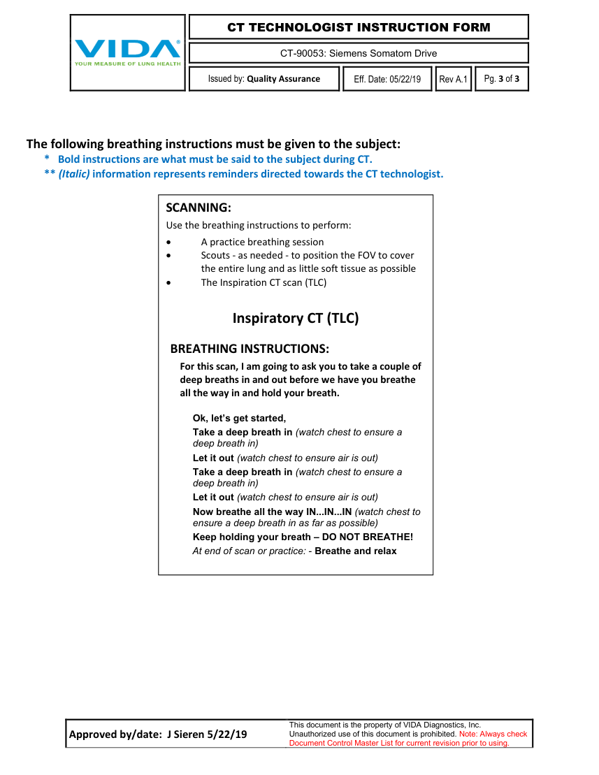
The following breathing instructions must be given to the subject: * Bold instructions are what must be said to the subject during CT. ** (Italic) information represents reminders directed towards the CT technologist.
SCANNING: Use the breathing instructions to perform: • A practice breathing session • Scouts - as needed - to position the FOV to cover the entire lung and as little soft tissue as possible • The Inspiration CT scan (TLC)
Inspiratory CT (TLC)
BREATHING INSTRUCTIONS: For this scan, I am going to ask you to take a couple of deep breaths in and out before we have you breathe all the way in and hold your breath.
Ok, let’s get started, Take a deep breath in (watch chest to ensure a deep breath in) Let it out (watch chest to ensure air is out) Take a deep breath in (watch chest to ensure a deep breath in) Let it out (watch chest to ensure air is out) Now breathe all the way IN...IN...IN (watch chest to ensure a deep breath in as far as possible) Keep holding your breath – DO NOT BREATHE! At end of scan or practice: - Breathe and relax
Approved by/date: J Sieren 5/22/19 This document is the property of VIDA Diagnostics, Inc.
Unauthorized use of this document is prohibited. Note: Always check Document Control Master List for current revision prior to using.
Pagina 7 — text în engleză
CT-90059: Siemens Somatom Go. Up 32
Issued by: Imaging Services
Eff. Date: 6/23/2020
Rev A
Pg. 1 of 3
It is important that the subject fully understands the breath hold and scanning procedure and that all concerns are addressed prior to performing the CT scan. If this is a subsequent visit to the baseline visit, participants must be scanned with the same CT acquisition and reconstruction parameters used during the baseline CT visit.
Subject Positioning • Place subject in a supine position, arms positioned comfortably above the head in a head-arm rest, lower legs supported. • Using the laser positioning lights, line up the subject so the chest is iso-center (in the middle: left-right; up-down) of the CT gantry. Correcting an off-center subject during image reconstruction is not acceptable. Proper positioning must be ensured prior to scanning. • Move the table so the subject is in the correct position for a chest CT scan. • Once the subject is comfortably and properly positioned, remain in the scanner room and work with the subject to rehearse the breathing instructions used during CT acquisition several times. It is important that the subject fully understands the breath hold and scanning procedure and that all concerns are addressed prior to performing the CT scan.
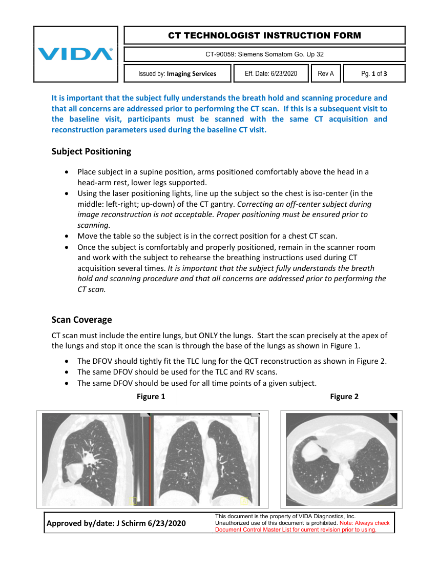
Scan Coverage CT scan must include the entire lungs, but ONLY the lungs. Start the scan precisely at the apex of the lungs and stop it once the scan is through the base of the lungs as shown in Figure 1.
• The DFOV should tightly fit the TLC lung for the QCT reconstruction as shown in Figure 2. • The same DFOV should be used for the TLC and RV scans. • The same DFOV should be used for all time points of a given subject.
Figure 1 Figure 2
Approved by/date: J Schirm 6/23/2020 This document is the property of VIDA Diagnostics, Inc.
Unauthorized use of this document is prohibited. Note: Always check Document Control Master List for current revision prior to using.
Pagina 8 — text în engleză
CT-90059: Siemens Somatom Go. Up 32
Issued by: Imaging Services
Eff. Date: 6/23/2020
Rev A
Pg. 2 of 3
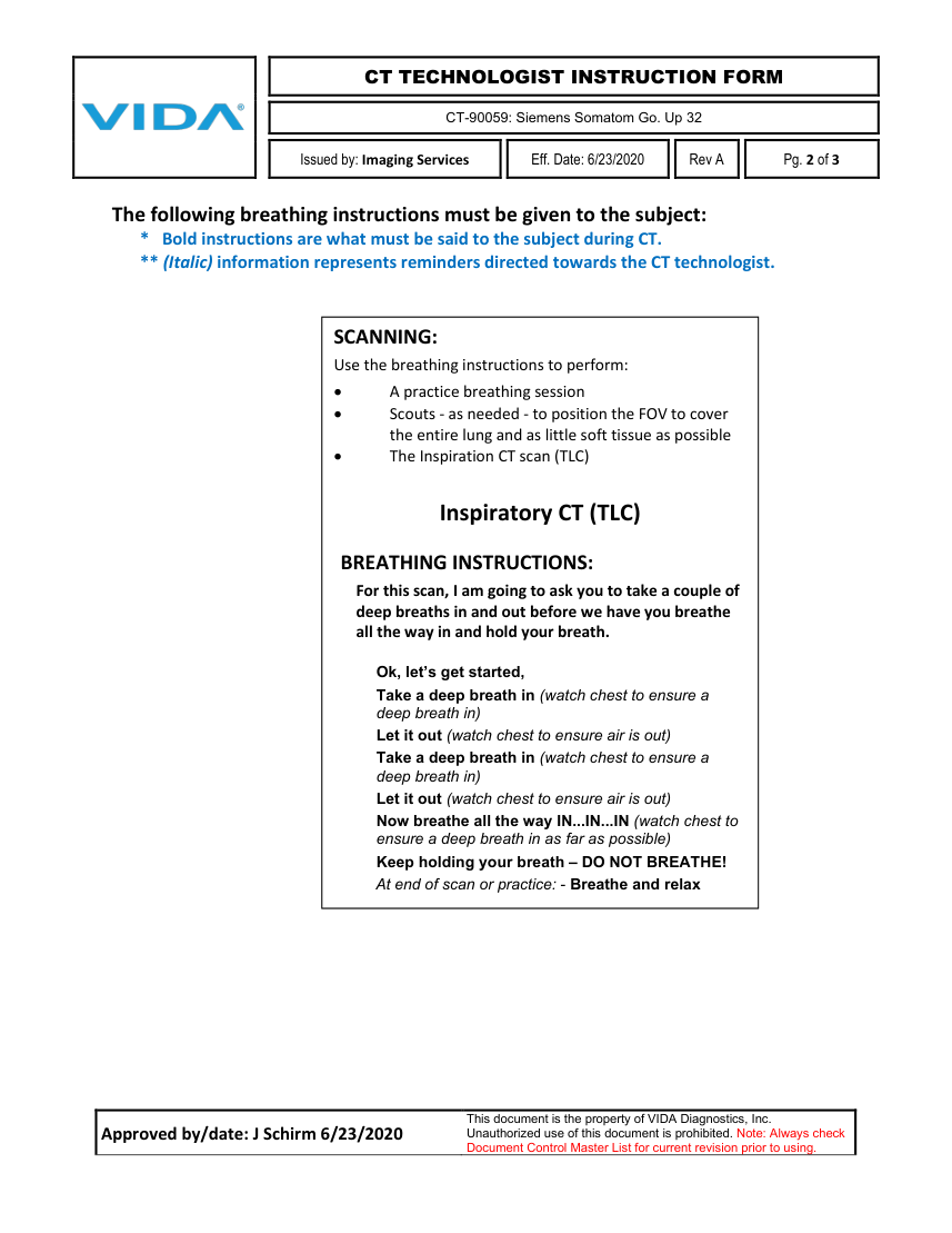
The following breathing instructions must be given to the subject: * Bold instructions are what must be said to the subject during CT. ** (Italic) information represents reminders directed towards the CT technologist.
SCANNING: Use the breathing instructions to perform: • A practice breathing session • Scouts - as needed - to position the FOV to cover the entire lung and as little soft tissue as possible • The Inspiration CT scan (TLC)
Inspiratory CT (TLC)
BREATHING INSTRUCTIONS: For this scan, I am going to ask you to take a couple of deep breaths in and out before we have you breathe all the way in and hold your breath.
Ok, let’s get started, Take a deep breath in (watch chest to ensure a deep breath in) Let it out (watch chest to ensure air is out) Take a deep breath in (watch chest to ensure a deep breath in) Let it out (watch chest to ensure air is out) Now breathe all the way IN...IN...IN (watch chest to ensure a deep breath in as far as possible) Keep holding your breath – DO NOT BREATHE! At end of scan or practice: - Breathe and relax
Approved by/date: J Schirm 6/23/2020 This document is the property of VIDA Diagnostics, Inc.
Unauthorized use of this document is prohibited. Note: Always check Document Control Master List for current revision prior to using.
Pagina 9 — text în engleză
CT-90059: Siemens Somatom Go. Up 32
Issued by: Imaging Services
Eff. Date: 6/23/2020
Rev A
Pg. 3 of 3
CT Parameters • The following parameters must be implemented for these CT scans.
• Scout scan uses default parameters (Site discretions on type and number of scouts, however must maintain ALARA principle).
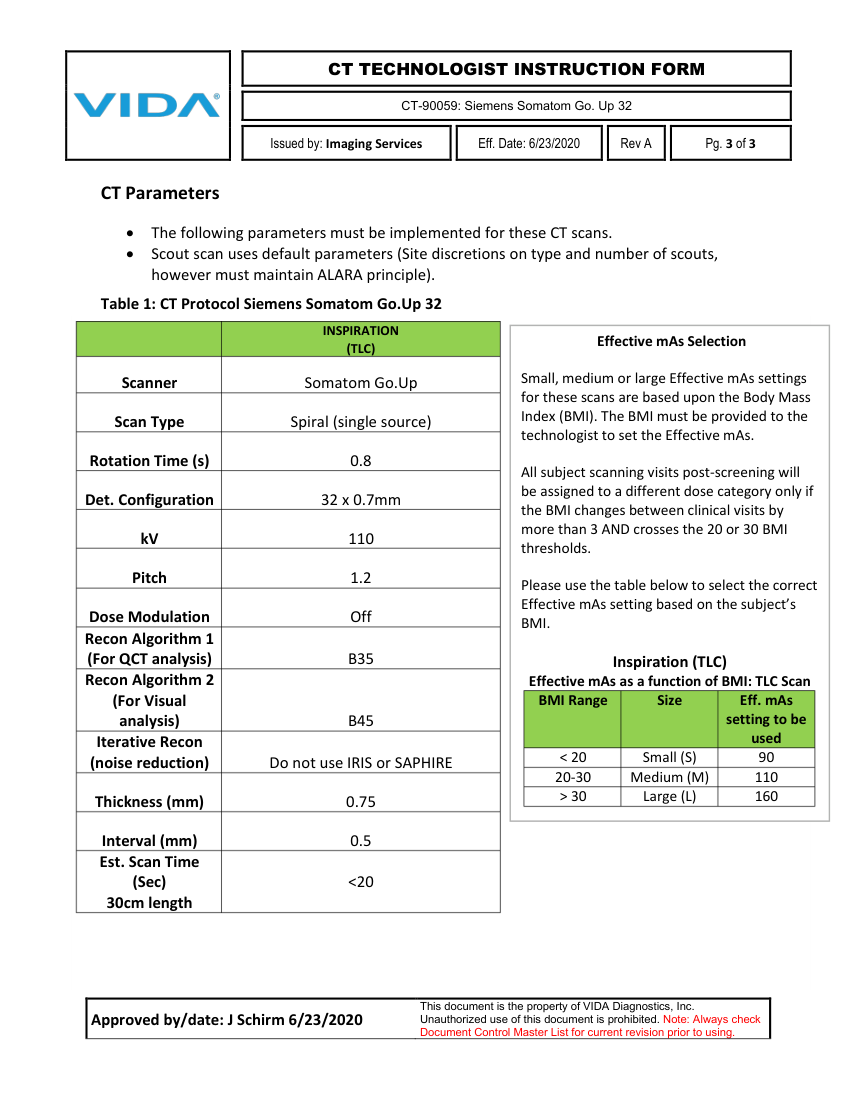
Table 1: CT Protocol Siemens Somatom Go.Up 32
INSPIRATION Effective mAs Selection (TLC)
Scanner Somatom Go.Up
Scan Type Spiral (single source) Rotation Time (s) 0.8 Det. Configuration 32 x 0.7mm kV 110 Pitch 1.2 Dose Modulation Off Recon Algorithm 1
Small, medium or large Effective mAs settings for these scans are based upon the Body Mass Index (BMI). The BMI must be provided to the technologist to set the Effective mAs.
All subject scanning visits post-screening will be assigned to a different dose category only if the BMI changes between clinical visits by more than 3 AND crosses the 20 or 30 BMI thresholds.
Please use the table below to select the correct Effective mAs setting based on the subject’s BMI.
(For QCT analysis) B35 Inspiration (TLC) Recon Algorithm 2 Effective mAs as a function of BMI: TLC Scan BMI Range Size Eff. mAs (For Visual setting to be analysis) B45 used Iterative Recon < 20 Small (S) 90 (noise reduction) Do not use IRIS or SAPHIRE 20-30 Medium (M) 110 > 30 Large (L) 160 Thickness (mm) 0.75
Interval (mm) 0.5 Est. Scan Time
(Sec) <20 30cm length
Approved by/date: J Schirm 6/23/2020 This document is the property of VIDA Diagnostics, Inc.
Unauthorized use of this document is prohibited. Note: Always check Document Control Master List for current revision prior to using.
Pagina 10 — text în engleză
CT-90043: SIEMENS SENSATION 64
Issued by: Quality Assurance
Eff. Date: 6/9/2014
Rev D
Pg. 1 of 3
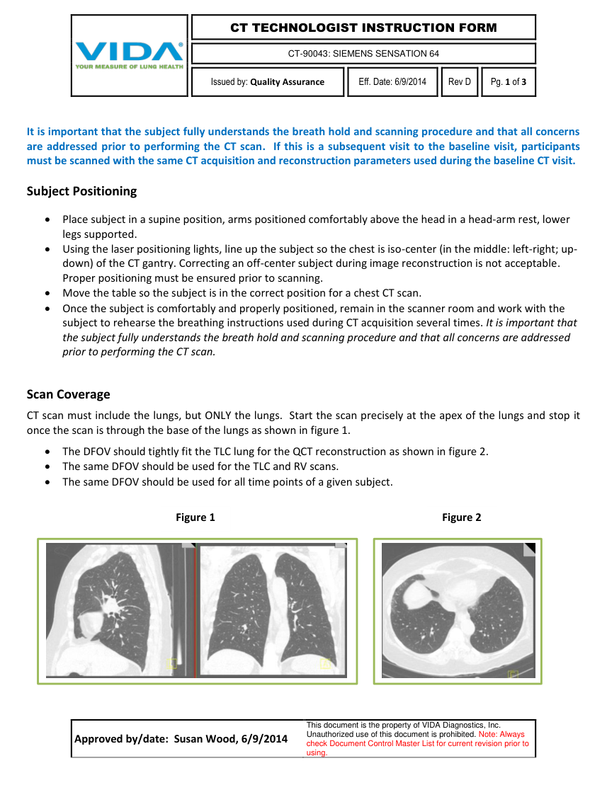
It is important that the subject fully understands the breath hold and scanning procedure and that all concerns are addressed prior to performing the CT scan. If this is a subsequent visit to the baseline visit, participants must be scanned with the same CT acquisition and reconstruction parameters used during the baseline CT visit.
Subject Positioning Place subject in a supine position, arms positioned comfortably above the head in a head-arm rest, lower legs supported. Using the laser positioning lights, line up the subject so the chest is iso-center (in the middle: left-right; up- down) of the CT gantry. Correcting an off-center subject during image reconstruction is not acceptable. Proper positioning must be ensured prior to scanning. Move the table so the subject is in the correct position for a chest CT scan. Once the subject is comfortably and properly positioned, remain in the scanner room and work with the subject to rehearse the breathing instructions used during CT acquisition several times. It is important that the subject fully understands the breath hold and scanning procedure and that all concerns are addressed prior to performing the CT scan.
Scan Coverage CT scan must include the lungs, but ONLY the lungs. Start the scan precisely at the apex of the lungs and stop it once the scan is through the base of the lungs as shown in figure 1.
The DFOV should tightly fit the TLC lung for the QCT reconstruction as shown in figure 2. The same DFOV should be used for the TLC and RV scans. The same DFOV should be used for all time points of a given subject.
Figure 1 Figure 2
Approved by/date: Susan Wood, 6/9/2014 This document is the property of VIDA Diagnostics, Inc.
Unauthorized use of this document is prohibited. Note: Always check Document Control Master List for current revision prior to using.
Pagina 11 — text în engleză
CT-90043: SIEMENS SENSATION 64
Issued by: Quality Assurance
Eff. Date: 6/9/2014
Rev D
Pg. 2 of 3
CT Parameters The following parameters must be implemented for these CT scans.
Scout scan uses default parameters (Site discretions on type and number of scouts, however must maintain ALARA principle).
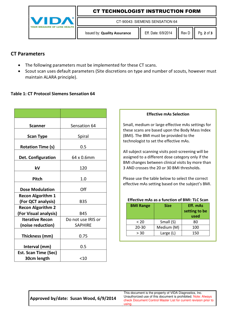
Table 1: CT Protocol Siemens Sensation 64
Effective mAs Selection
Scanner Sensation 64 Scan Type Spiral Rotation Time (s) 0.5 Det. Configuration 64 x 0.6mm kV 120 Pitch 1.0 Dose Modulation Off Small, medium or large effective mAs settings for these scans are based upon the Body Mass Index (BMI). The BMI must be provided to the technologist to set the effective mAs.
All subject scanning visits post-screening will be assigned to a different dose category only if the BMI changes between clinical visits by more than 3 AND crosses the 20 or 30 BMI thresholds.
Please use the table below to select the correct effective mAs setting based on the subject’s BMI.
Recon Algorithm 1
Effective mAs as a function of BMI: TLC Scan (For QCT analysis) B35 BMI Range Size Eff. mAs Recon Algorithm 2
setting to be (For Visual analysis) B45 used Iterative Recon Do not use IRIS or < 20 Small (S) 80 (noise reduction) SAPHIRE 20-30 Medium (M) 100 > 30 Large (L) 150 Thickness (mm) 0.75
Interval (mm) 0.5 Est. Scan Time (Sec)
30cm length <10
Approved by/date: Susan Wood, 6/9/2014 This document is the property of VIDA Diagnostics, Inc.
Unauthorized use of this document is prohibited. Note: Always check Document Control Master List for current revision prior to using.
Pagina 12 — text în engleză
CT-90043: SIEMENS SENSATION 64
Issued by: Quality Assurance
Eff. Date: 6/9/2014
Rev D
Pg. 3 of 3
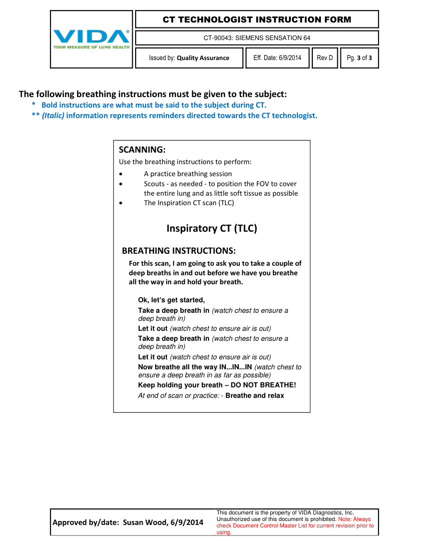
The following breathing instructions must be given to the subject: * Bold instructions are what must be said to the subject during CT. ** (Italic) information represents reminders directed towards the CT technologist.
SCANNING: Use the breathing instructions to perform: A practice breathing session Scouts - as needed - to position the FOV to cover the entire lung and as little soft tissue as possible The Inspiration CT scan (TLC)
Inspiratory CT (TLC)
BREATHING INSTRUCTIONS: For this scan, I am going to ask you to take a couple of deep breaths in and out before we have you breathe all the way in and hold your breath.
Ok, let’s get started, Take a deep breath in (watch chest to ensure a deep breath in) Let it out (watch chest to ensure air is out) Take a deep breath in (watch chest to ensure a deep breath in) Let it out (watch chest to ensure air is out) Now breathe all the way IN...IN...IN (watch chest to ensure a deep breath in as far as possible) Keep holding your breath – DO NOT BREATHE! At end of scan or practice: - Breathe and relax
Approved by/date: Susan Wood, 6/9/2014 This document is the property of VIDA Diagnostics, Inc.
Unauthorized use of this document is prohibited. Note: Always check Document Control Master List for current revision prior to using.