CT — Torace: Veran (MCB)
Sursă și prelucrare editorială
- Proveniență
- 1 referință declarată
- Fidelitatea preluării
- Nedocumentată
- Revizuire medicală
- Nedocumentată
Referințele declarate nu confirmă singure fidelitatea preluării sau aprobarea protocolului pentru utilizare locală.
Detalii despre surse și verificări
MCB Radiology — 7 Veran
- Ediție / versiune a sursei
- Neprecizată
- Pagină / secțiune
- Pagini: 1–7
- Relația cu protocolul
- Document original importat integral; atribuirea autorilor rămâne cea din PDF.
- Data consultării
- 2026-09-27
Prelucrarea acestei pagini
- Mod de prelucrare
- Titlu și navigare în română; documentul sursă în engleză.; PDF original integral, previzualizare și text extras automat; fără completarea parametrilor clinici lipsă.
- Adaptări față de sursă
- Nedocumentate; absența evidenței nu confirmă o preluare integrală.
- Responsabil pentru prelucrare
- Neprecizat
- Atribuire existentă în pagină
- MCB Radiology (portalul sursă). Această atribuire nu identifică automat autorul sursei sau revizorul medical.
- Versiunea paginii
- mcb-83f6defe8956
- Actualizare declarată a paginii
- 2026-09-27 — distinctă de revizuirea medicală.
Verificarea fidelității: Nedocumentată
Revizuirea medicală: Nedocumentată
Documentul original MCB — Torace: Veran
Protocol · 7 pagini · consultat la 2026-09-27.
Descarcă PDF-ul integral · Sursa MCB · Catalogul MCB pentru această modalitate
Instrucțiunile, valorile și ilustrațiile din documentul original sunt păstrate în engleză. Titlul și navigarea sunt în română.
Pagina 1
Pagina 2
Pagina 3
Pagina 4
Pagina 5
Pagina 6
Pagina 7
Textul documentului original
Pagina 1 — text în engleză
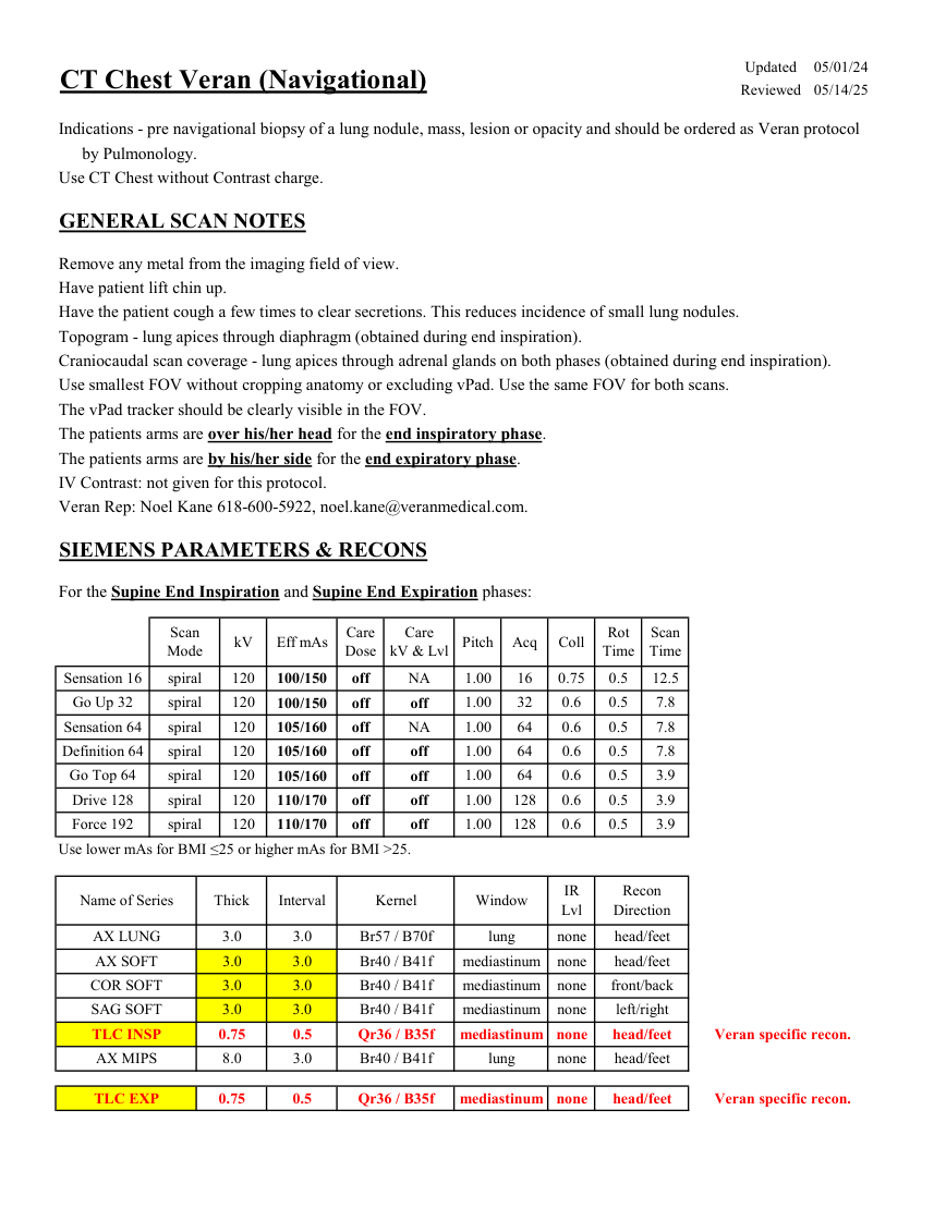
Care
Scan
Pitch Acq Coll Rot Mode kV Eff mAs Care
Dose kV & Lvl Time Time 1.00 0.5 0.75 16 Sensation 16 spiral 120 NA 12.5 100/150 off 0.5 7.8 Go Up 32 spiral 120 off 1.00 32 0.6 100/150 off Sensation 64 spiral 120 NA 0.5 7.8 105/160 off 1.00 64 0.6 0.5 7.8 Definition 64 spiral 120 off 1.00 105/160 64 0.6 off Go Top 64 spiral 120 3.9 105/160 off 1.00 64 0.6 0.5 off 3.9 Drive 128 spiral 120 off 1.00 128 0.6 0.5 110/170 off Force 192 spiral 3.9 120 off 1.00 128 0.6 0.5 110/170 off Use lower mAs for BMI ≤25 or higher mAs for BMI >25. IR
Name of Series Thick Interval Window Recon
Kernel Lvl Direction AX LUNG 3.0 3.0 Br57 / B70f lung none head/feet AX SOFT 3.0 3.0 mediastinum head/feet Br40 / B41f none COR SOFT 3.0 3.0 none Br40 / B41f mediastinum front/back SAG SOFT 3.0 3.0 mediastinum Br40 / B41f none left/right 0.75 0.5 mediastinum head/feet none TLC INSP Qr36 / B35f Veran specific recon. AX MIPS 8.0 3.0 lung head/feet none Br40 / B41f TLC EXP 0.75 0.5 mediastinum Veran specific recon. Qr36 / B35f none head/feet
Pagina 2 — text în engleză
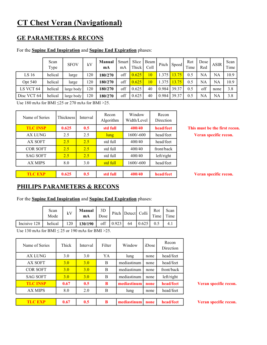
Smart
Beam
Dose
Scan
Manual
SFOV kV Slice
Pitch Speed Rot
ASIR Type mA Thick Coll Time Red Time mA 0.625 13.75 0.5 LS 16 helical large 120 NA 10 1.375 off NA 10.9 180/270 Opt 540 helical large 120 NA 10 0.625 1.375 off 13.75 0.5 NA 10.9 180/270 LS VCT 64 helical large body 120 off off 0.625 40 0.984 39.37 0.5 none 3.8 180/270 off NA 3.8 Disc VCT 64 helical large body 120 NA 40 0.984 39.37 0.625 180/270 0.5 Use 180 mAs for BMI ≤25 or 270 mAs for BMI >25. Window
Recon
Name of Series Thickness Interval Recon
Algorithm Width/Level Direction This must be the first recon. TLC INSP 0.625 0.5 std full 400/40 head/feet AX LUNG 2.5 2.5 lung 1600/-600 head/feet Veran specific recon. AX SOFT 2.5 2.5 std full 400/40 head/feet COR SOFT 2.5 2.5 std full 400/40 front/back SAG SOFT 2.5 2.5 std full 400/40 left/right AX MIPS 8.0 3.0 std full 1600/-600 head/feet TLC EXP 0.625 0.5 std full 400/40 head/feet Veran specific recon. PHILIPS PARAMETERS & RECONS For the Supine End Inspiration and Supine End Expiration phases: Scan
Rot Scan
Manual
Mode kV 3D
Dose Pitch Detect Colli Time Time mA Incisive 128 helical 120 off 0.923 64 0.625 0.5 4.1 130/190 Use 130 mAs for BMI ≤ 25 or 190 mAs for BMI >25. Name of Series Thick Interval Filter Window iDose Recon
Direction AX LUNG 3.0 3.0 YA lung none head/feet AX SOFT 3.0 3.0 B mediastinum none head/feet COR SOFT 3.0 3.0 B mediastinum none front/back SAG SOFT 3.0 3.0 B mediastinum none left/right TLC INSP 0.67 0.5 B mediastinum none head/feet Veran specific recon. AX MIPS 8.0 2.0 B lung none head/feet head/feet TLC EXP 0.67 0.5 B mediastinum none Veran specific recon.
Pagina 3 — text în engleză
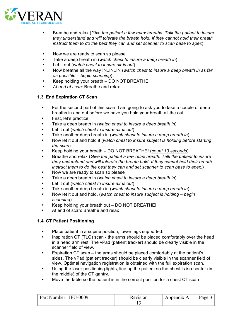
• Now we are ready to scan so please • Take a deep breath in (watch chest to insure a deep breath in) • Let it out (watch chest to insure air is out) • Now breathe all the way IN..IN..IN (watch chest to insure a deep breath in as far as possible – begin scanning) • Keep holding your breath – DO NOT BREATHE! • At end of scan: Breathe and relax
1.3 End Expiration CT Scan
• For the second part of this scan, I am going to ask you to take a couple of deep breaths in and out before we have you hold your breath all the out. • First, let’s practice • Take a deep breath in (watch chest to insure a deep breath in) • Let it out (watch chest to insure air is out)
• Take another deep breath in (watch chest to insure a deep breath in) • Now let it out and hold it (watch chest to insure subject is holding before starting the scan) • Keep holding your breath – DO NOT BREATHE! (count 10 seconds) • Breathe and relax (Give the patient a few relax breath. Talk the patient to insure they understand and will tolerate the breath hold. If they cannot hold their breath instruct them to do the best they can and set scanner to scan base to apex.) • Now we are ready to scan so please • Take a deep breath in (watch chest to insure a deep breath in) • Let it out (watch chest to insure air is out) • Take another deep breath in (watch chest to insure a deep breath in) • Now let it out and hold. (watch chest to insure subject is holding – begin scanning) • Keep holding your breath out – DO NOT BREATHE! • At end of scan: Breathe and relax
1.4 CT Patient Positioning
• Place patient in a supine position, lower legs supported. • Inspiration CT (TLC) scan - the arms should be placed comfortably over the head in a head arm rest. The vPad (patient tracker) should be clearly visible in the scanner field of view. • Expiration CT scan – the arms should be placed comfortably at the patient’s sides. The vPad (patient tracker) should be clearly visible in the scanner field of view. Optimal navigation registration is obtained with the full expiration scan. • Using the laser positioning lights, line up the patient so the chest is iso-center (in the middle) of the CT gantry. • Move the table so the patient is in the correct position for a chest CT scan
Part Number: IFU-0009 Revision
Appendix A Page 3 13
Pagina 4 — text în engleză
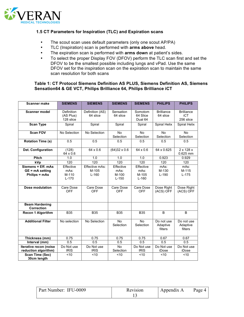
• The scout scan uses default parameters (only one scout AP/PA) • TLC (Inspiration) scan is performed with arms above head. • The expiration scan is performed with arms down at patient’s sides. • To select the proper Display FOV (DFOV) perform the TLC scan first and set the DFOV to be the smallest possible including lungs and vPad. Use the same DFOV set for the inspiration scan on the expiration scan to maintain the same scan resolution for both scans
Table 1: CT Protocol Siemens Definition AS PLUS, Siemens Definition AS, Siemens Sensation64 & GE VCT, Philips Brilliance 64, Philips Brilliance iCT
Scanner make SIEMENS SIEMENS SIEMENS SIEMENS PHILIPS PHILIPS
Definition (AS) Sensation Somotom Brilliance Brilliance Scanner model Definition (AS Plus) 64 slice 64 slice 64 slice iCT 128 slice 64 Slice Dual 64 256 slice Scan Type Spiral Spiral Spiral Spiral Spiral Helix Spiral Helix Scan FOV No Selection No Selection No No No No Selection Selection Selection Selection Rotation Time (s) 0.5 0.5 0.5 0.5 0.5 0.5 Det. Configuration (128) 64 x 0.6 (64)32 x 0.6 64 x 0.6 64 x 0.625 2 x 128 x 64 x 0.6 0.625 mm Pitch 1.0 1.0 1.0 1.0 0.923 0.929 kVp 120 120 120 120 120 120 Siemens = Eff. mAs Effective Effective mAs: Effective Effective mAs: mAs: GE = mA setting mAs: M-105 mAs: mAs: M-130 M-115 Philips = mAs M-110 L-160 M-100 M-105 L-190 L-175
L-170 L-150 L-160 Dose modulation Care Dose Care Dose Care Dose Care Dose OFF OFF OFF OFF Dose Right (ACS) OFF Dose Right (ACS) OFF Beam Hardening
Correction Recon 1 Algorithm B35 B35 B35 B35 B B Additional Filter No selection No Selection No No Do not use Do not use Selection Selection Adaptive Adaptive filters filters Thickness (mm) 0.75 0.75 0.75 0.75 0.67 0.67 Interval (mm) 0.5 0.5 0.5 0.5 0.5 0.5 Iterative recon (noise Do Not use Do Not use No Do Not use Do Not use Do Not use reduction algorithm) IRIS IRIS Selection IRIS iDose iDose Scan Time (Sec) <10 <10 <10 <10 <10 <10 30cm length
Part Number: IFU-0009 Revision
Appendix A Page 4 13
Pagina 5 — text în engleză
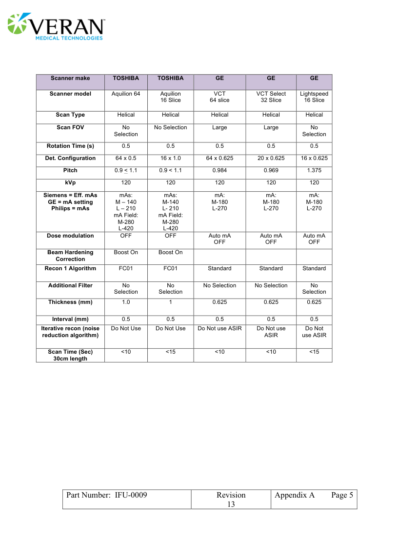
Scanner make TOSHIBA TOSHIBA GE GE GE VCT VCT Select Lightspeed Scanner model Aquilion 64
Aquilion 16 Slice 16 Slice 64 slice 32 Slice Scan Type Helical Helical Helical Helical Helical Scan FOV No No Selection Large Large No Selection Selection Rotation Time (s) 0.5 0.5 0.5 0.5 0.5 Det. Configuration 64 x 0.5 16 x 1.0 64 x 0.625 20 x 0.625 16 x 0.625 Pitch 0.9 < 1.1 0.9 < 1.1 0.984 0.969 1.375 kVp 120 120 120 120 120 Siemens = Eff. mAs mAs: mAs: mA: mA: mA: GE = mA setting M – 140 M-180 M-180 M-180 Philips = mAs L – 210 M-140 L- 210 L-270 L-270 L-270 mA Field: mA Field:
M-280 M-280 L-420 L-420 Auto mA Auto mA Dose modulation OFF OFF Auto mA OFF OFF OFF Beam Hardening Boost On Boost On
Correction Recon 1 Algorithm FC01 FC01 Standard Standard Standard No Additional Filter No No Selection No Selection No Selection Selection Selection Thickness (mm) 1.0 1 0.625 0.625 0.625 Interval (mm) 0.5 0.5 0.5 0.5 0.5 Iterative recon (noise Do Not Do Not Use Do Not Use Do Not use ASIR Do Not use use ASIR reduction algorithm) ASIR Scan Time (Sec) <10 <15 <10 <10 <15 30cm length
Part Number: IFU-0009 Revision
Appendix A Page 5 13
Pagina 6 — text în engleză
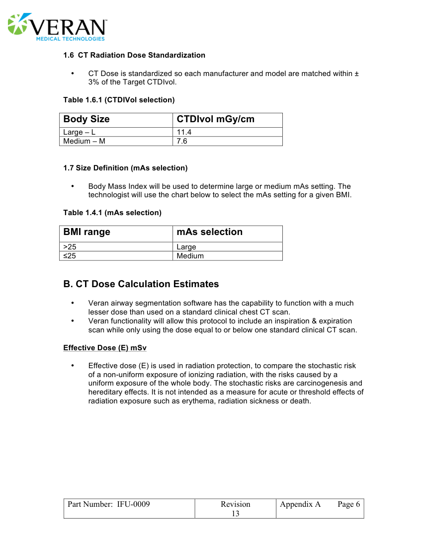
• CT Dose is standardized so each manufacturer and model are matched within ± 3% of the Target CTDIvol.
Table 1.6.1 (CTDIVol selection)
Body Size CTDIvol mGy/cm Large – L 11.4 Medium – M 7.6
1.7 Size Definition (mAs selection)
• Body Mass Index will be used to determine large or medium mAs setting. The technologist will use the chart below to select the mAs setting for a given BMI.
Table 1.4.1 (mAs selection)
BMI range mAs selection >25 Large ≤25 Medium
B. CT Dose Calculation Estimates
• Veran airway segmentation software has the capability to function with a much lesser dose than used on a standard clinical chest CT scan. • Veran functionality will allow this protocol to include an inspiration & expiration scan while only using the dose equal to or below one standard clinical CT scan.
Effective Dose (E) mSv
• Effective dose (E) is used in radiation protection, to compare the stochastic risk of a non-uniform exposure of ionizing radiation, with the risks caused by a uniform exposure of the whole body. The stochastic risks are carcinogenesis and hereditary effects. It is not intended as a measure for acute or threshold effects of radiation exposure such as erythema, radiation sickness or death.
Part Number: IFU-0009 Revision
Appendix A Page 6 13
Pagina 7 — text în engleză
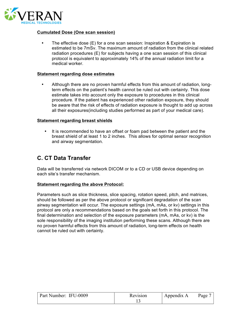
• The effective dose (E) for a one scan session: Inspiration & Expiration is estimated to be 7mSv. The maximum amount of radiation from the clinical related radiation procedures (E) for subjects having a one scan session of this clinical protocol is equivalent to approximately 14% of the annual radiation limit for a medical worker.
Statement regarding dose estimates
• Although there are no proven harmful effects from this amount of radiation, long- term effects on the patient’s health cannot be ruled out with certainty. This dose estimate takes into account only the exposure to procedures in this clinical procedure. If the patient has experienced other radiation exposure, they should be aware that the risk of effects of radiation exposure is thought to add up across all their exposures(including studies performed as part of your medical care).
Statement regarding breast shields
• It is recommended to have an offset or foam pad between the patient and the breast shield of at least 1 to 2 inches. This allows for optimal sensor recognition and airway segmentation.
C. CT Data Transfer
Data will be transferred via network DICOM or to a CD or USB device depending on each site’s transfer mechanism.
Statement regarding the above Protocol:
Parameters such as slice thickness, slice spacing, rotation speed, pitch, and matrices, should be followed as per the above protocol or significant degradation of the scan airway segmentation will occur. The exposure settings (mA, mAs, or kv) settings in this protocol are only a recommendations based on the goals set forth in this protocol. The final determination and selection of the exposure parameters (mA, mAs, or kv) is the sole responsibility of the imaging institution performing these scans. Although there are no proven harmful effects from this amount of radiation, long-term effects on health cannot be ruled out with certainty.
Part Number: IFU-0009 Revision
Appendix A Page 7 13