CT — Torace: Teton (MCB)
Sursă și prelucrare editorială
- Proveniență
- 1 referință declarată
- Fidelitatea preluării
- Nedocumentată
- Revizuire medicală
- Nedocumentată
Referințele declarate nu confirmă singure fidelitatea preluării sau aprobarea protocolului pentru utilizare locală.
Detalii despre surse și verificări
MCB Radiology — 5 Teton
- Ediție / versiune a sursei
- Neprecizată
- Pagină / secțiune
- Pagini: 1–7
- Relația cu protocolul
- Document original importat integral; atribuirea autorilor rămâne cea din PDF.
- Data consultării
- 2026-09-27
Prelucrarea acestei pagini
- Mod de prelucrare
- Titlu și navigare în română; documentul sursă în engleză.; PDF original integral, previzualizare și text extras automat; fără completarea parametrilor clinici lipsă.
- Adaptări față de sursă
- Nedocumentate; absența evidenței nu confirmă o preluare integrală.
- Responsabil pentru prelucrare
- Neprecizat
- Atribuire existentă în pagină
- MCB Radiology (portalul sursă). Această atribuire nu identifică automat autorul sursei sau revizorul medical.
- Versiunea paginii
- mcb-fb7c512c937b
- Actualizare declarată a paginii
- 2026-09-27 — distinctă de revizuirea medicală.
Verificarea fidelității: Nedocumentată
Revizuirea medicală: Nedocumentată
Documentul original MCB — Torace: Teton
Protocol · 7 pagini · consultat la 2026-09-27.
Descarcă PDF-ul integral · Sursa MCB · Catalogul MCB pentru această modalitate
Instrucțiunile, valorile și ilustrațiile din documentul original sunt păstrate în engleză. Titlul și navigarea sunt în română.
Pagina 1
Pagina 2
Pagina 3
Pagina 4
Pagina 5
Pagina 6

Pagina 7
Textul documentului original
Pagina 1 — text în engleză
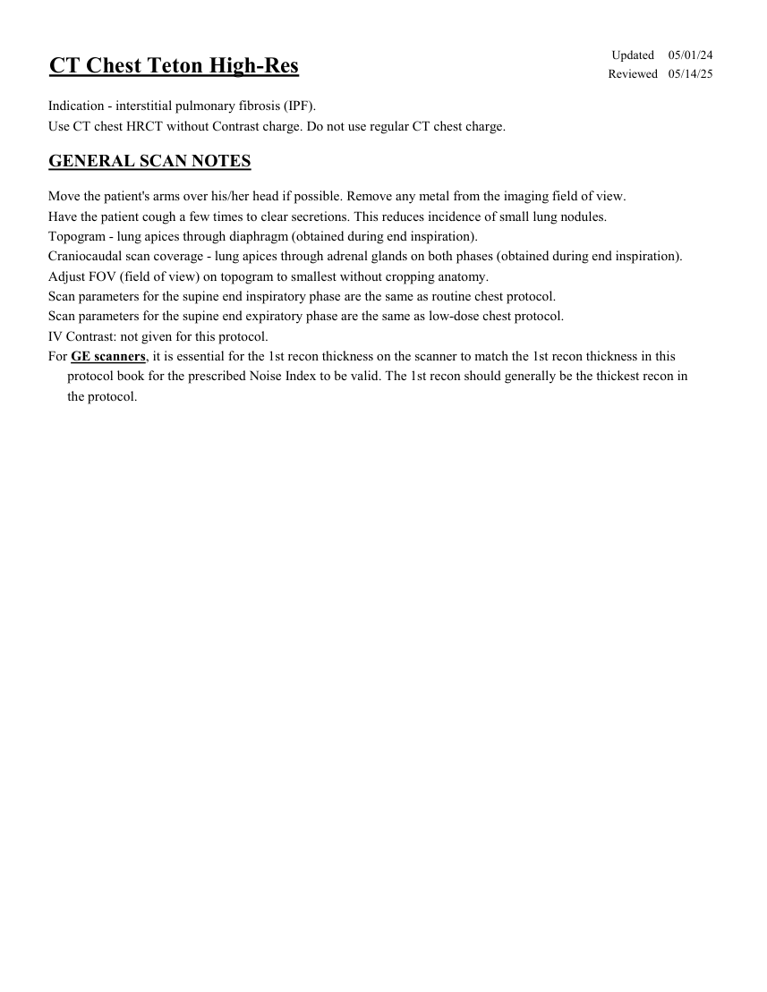
Pagina 2 — text în engleză
Care
Scan
Acq Coll Rot Mode kV mAs Care
Dose kV & Lvl Pitch Time Time Sensation 16 spiral 120 100 on NA 1.15 16 0.75 0.5 10.9 Go Up 32 spiral 130 51 on on 80 1.50 32 0.7 0.8 7.1 100 on Sensation 64 spiral 120 NA 1.40 64 0.6 0.5 5.6 Definition 64 spiral 120 110 on on 1.20 64 0.6 0.5 6.5 Go Top 64 spiral 120 62 on on 80 1.20 64 0.6 0.33 2.1 Drive 128 spiral 120 66 on on 1.20 128 0.6 0.5 3.3 Force 192 spiral 110 51 on on 1.20 192 0.5 0.5 2.6 Recon
Name of Series Thick Interval Kernel Window IR
Lvl Direction AX INSP LUNG 3.0 3.0 Br57 / B70f lung 3 head/feet AX INSP SOFT 3.0 3.0 Br40 / B41f mediastinum 3 head/feet COR INSP SOFT 3.0 3.0 Br40 / B41f mediastinum 3 front/back SAG INSP SOFT 3.0 3.0 Br40 / B41f mediastinum 3 left/right AX INSP HRCT 1.0 0.8 Bl57 / B80f lung 3 head/feet Teton specific recon. COR INSP HRCT 1.0 5.0 Br57 / B70f lung 3 front/back AX INSP MIPS 8.0 3.0 Br40 / B41f lung 3 head/feet For the Supine End Expiration phase: Scan
Care
Rot Scan
Acq Coll Mode kV mAs Care
Dose kV & Lvl Pitch Time Time NA 1.15 16 0.75 0.5 10.9 Sensation 16 spiral 120 60 on on Go Up 32 spiral 130 on 80 1.50 32 0.7 0.8 7.1 31 NA 1.40 64 0.6 0.5 5.6 Sensation 64 spiral 120 60 on on 1.20 64 0.6 0.5 6.5 Definition 64 spiral 120 66 on on 80 1.20 64 0.6 0.33 2.1 Go Top 64 spiral 120 37 on 0.5 on 1.20 128 0.6 3.3 Drive 128 spiral 120 40 on on 1.20 192 0.5 0.5 2.6 Force 192 spiral 110 31 on Recon
Name of Series Thick Interval Kernel Window IR
Lvl Direction lung AX EXP HRCT 1.0 0.8 Bl57 / B80 3 head/feet Teton specific recon.
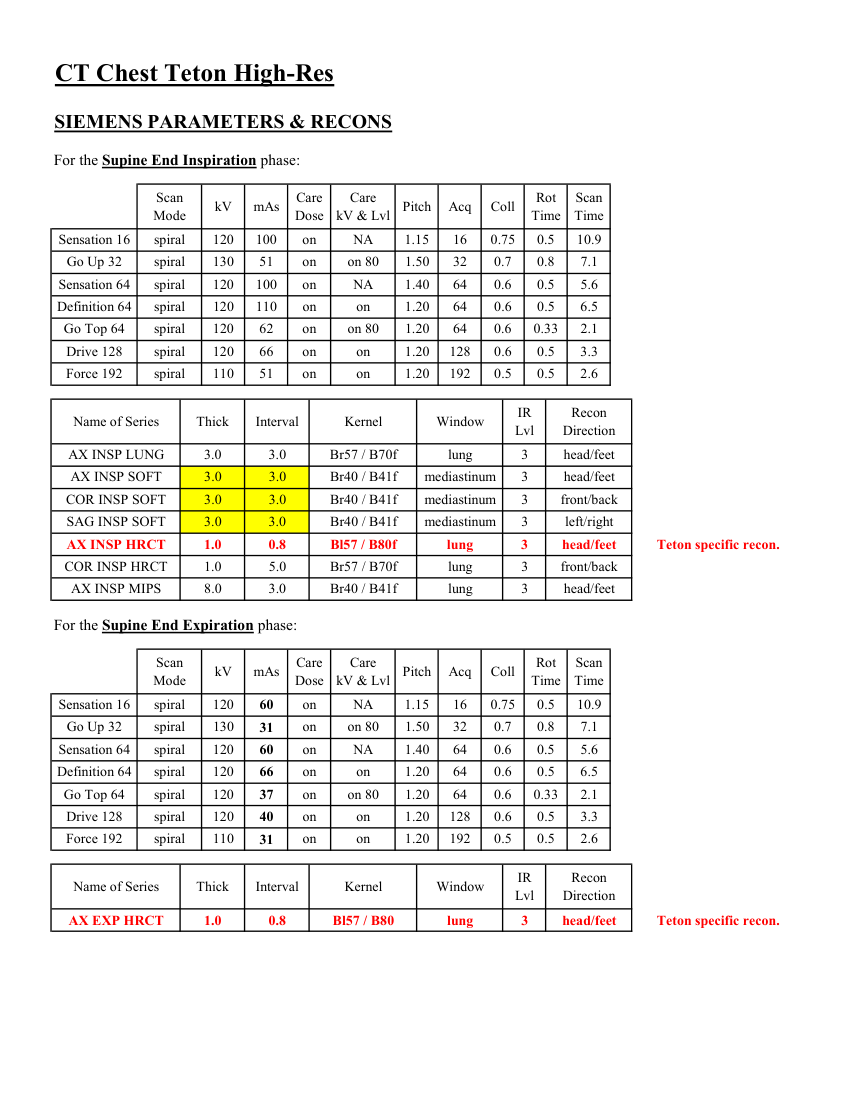
Pagina 3 — text în engleză
mA
Smart
Slice
Beam
Rot
Dose
Scan
Pitch Speed Type SFOV kV Range Noise
Index mA Thick Coll Time Red ASIR Time LS 16 helical large 120 100-440 16.36 on 2.5 20 1.375 27.50 0.5 NA NA 5.5 Opt 540 helical large 120 100-440 16.36 on 2.5 20 1.375 27.50 0.5 NA NA 5.5 LS VCT 64 helical large body 120 100-650 18.38 on 2.5 40 1.375 55.00 0.4 50 50 2.2 Disc VCT 64 helical large body 120 100-650 18.38 on 2.5 40 1.375 55.00 0.4 NA NA 2.2 Recon
Name of Series Interval Recon
Thickness Window
Algorithm Width/Level Direction head/feet AX INSP LUNG 2.5 2.5 lung 1600/-600 Must be first recon. AX INSP SOFT 2.5 2.5 std full 400/40 head/feet COR INSP SOFT 2.5 2.5 std full 400/40 front/back SAG INSP SOFT 2.5 2.5 std full 400/40 left/right AX INSP HRCT 1.25 1.0 bone plus full 1600/-600 head/feet Teton specific recon. COR INSP HRCT 1.25 5.0 bone plus full 1600/-600 front/back AX INSP MIPS 8.0 3.0 std full 1600/-600 head/feet For the Supine End Expiration phase: Scan
Smart
Scan
Beam
Dose
Speed Rot
Thick Coll Pitch Slice
Time Red ASIR Type SFOV kV mA
Range Noise
Index mA Time LS 16 helical large 120 100-300 1.375 27.50 0.5 NA NA 5.5 36.20 on 1.25 20 on 1.25 20 1.375 27.50 0.5 NA NA 5.5 Opt 540 helical large 120 100-300 36.20 40 0.984 39.375 0.5 30 70 3.8 LS VCT 64 helical large body 120 50-300 36.01 on 1.25 on 1.25 40 0.984 39.375 0.5 NA NA 3.8 Disc VCT 64 helical large body 120 50-300 36.01 Window
Recon
Name of Series Thickness Interval Recon
Algorithm Width/Level Direction AX EXP HRCT 1.25 1.0 bone plus full 1600/-600 Teton specific recon. head/feet
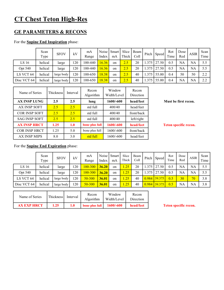
Pagina 4 — text în engleză
Avg
Dose
3D
Rot Scan
Mode kV mAs Index Dose Pitch Time Detect Colli Time 0.75 Incisive 128 helical 120 92 19 on 1.00 64 0.625 5.6 Name of Series Thick Interval Filter Window iDose Recon
Direction AX INSP LUNG 3.0 3.0 lung 3 head/feet YA AX INSP SOFT 3.0 3.0 B mediastinum 3 head/feet COR INSP SOFT 3.0 3.0 B mediastinum 3 front/back mediastinum 3 left/right SAG INSP SOFT 3.0 3.0 B AX INSP HRCT 1.0 0.8 YA lung 3 head/feet Teton specific recon. COR INSP HRCT 1.0 5.0 YA lung 3 front/back AX INSP MIPS 8.0 2.0 B lung 3 head/feet For the Supine End Expiration phase: Scan
Dose
3D
Pitch Detect Colli Rot Mode kV Avg
mAs Index Dose Time 64 0.625 0.75 Incisive 128 helical 120 55 19 on 1.00 Name of Series Thick Interval Filter Window iDose Recon
Direction AX EXP HRCT 1.0 0.8 YA lung 3 head/feet Teton specific recon.
Pagina 5 — text în engleză
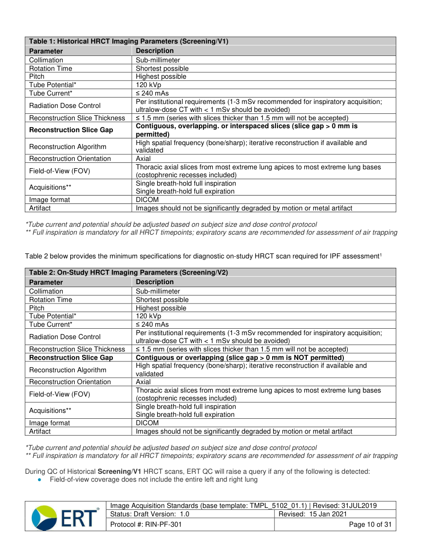
Table 1: Historical HRCT Imaging Parameters (Screening/V1) Parameter Description Collimation Sub-millimeter Rotation Time Shortest possible Pitch Highest possible Tube Potential 120 kVp Tube Current ≤ 240 mAs Radiation Dose Control Per institutional requirements (1-3 mSv recommended for inspiratory acquisition; ultralow-dose CT with < 1 mSv should be avoided) Reconstruction Slice Thickness ≤ 1.5 mm (series with slices thicker than 1.5 mm will not be accepted) Reconstruction Slice Gap Contiguous, overlapping. or interspaced slices (slice gap > 0 mm is permitted) Reconstruction Algorithm High spatial frequency (bone/sharp); iterative reconstruction if available and validated Reconstruction Orientation Axial Field-of-View (FOV) Thoracic axial slices from most extreme lung apices to most extreme lung bases (costophrenic recesses included) Acquisitions** Single breath-hold full inspiration Single breath-hold full expiration Image format DICOM
Artifact Images should not be significantly degraded by motion or metal artifact
*Tube current and potential should be adjusted based on subject size and dose control protocol ** Full inspiration is mandatory for all HRCT timepoints; expiratory scans are recommended for assessment of air trapping
Table 2 below provides the minimum specifications for diagnostic on-study HRCT scan required for IPF assessment1
Table 2: On-Study HRCT Imaging Parameters (Screening/V2) Parameter Description Collimation Sub-millimeter Rotation Time Shortest possible Pitch Highest possible Tube Potential 120 kVp Tube Current ≤ 240 mAs Radiation Dose Control Per institutional requirements (1-3 mSv recommended for inspiratory acquisition; ultralow-dose CT with < 1 mSv should be avoided) Reconstruction Slice Thickness ≤ 1.5 mm (series with slices thicker than 1.5 mm will not be accepted) Reconstruction Slice Gap Contiguous or overlapping (slice gap > 0 mm is NOT permitted) Reconstruction Algorithm High spatial frequency (bone/sharp); iterative reconstruction if available and validated Reconstruction Orientation Axial Field-of-View (FOV) Thoracic axial slices from most extreme lung apices to most extreme lung bases (costophrenic recesses included) Acquisitions** Single breath-hold full inspiration Single breath-hold full expiration Image format DICOM
Artifact Images should not be significantly degraded by motion or metal artifact
*Tube current and potential should be adjusted based on subject size and dose control protocol ** Full inspiration is mandatory for all HRCT timepoints; expiratory scans are recommended for assessment of air trapping
During QC of Historical Screening/V1 HRCT scans, ERT QC will raise a query if any of the following is detected: ● Field-of-view coverage does not include the entire left and right lung
Image Acquisition Standards (base template: TMPL_5102_01.1) | Revised: 31JUL2019 Status: Draft Version: 1.0 Revised: 15 Jan 2021
Protocol #: RIN-PF-301 Page 10 of 31
Pagina 6 — text în engleză
Appendix 1: The Image Management Solution (IMS)
1.0 IMS Access and Training
1.1 Site User Access and Training
ERT will provide web-based training sessions for sites detailing the processes required to create subjects, add timepoints for subjects, and upload timepoint images in the IMS. Detailed instructions, workflows, and descriptions of IMS eCRF content are also provided in the Appendix of this document.
Following the training and completion of training documentation, ERT will email trained site users with a link to ERT’s Global Single Sign-on (GSSO) page and associated registration instructions. If the site user has not previously registered for an ERT study, he/she will be required to self-register. Note sites must use the Google Chrome internet browser to access and use the ERT IMS platform (Internet Explorer/Microsoft Edge do not support the IMS functionality).
Site users should only use their designated accounts to access the study. If the person responsible for transferring imaging via the IMS is absent for an extended period, a new user at the site should be identified with a training request submitted to ERT for the user. Additionally, if a site user is no longer employed at the site or participating in the study, the site should notify ERT to inactivate the user’s access to the study. Sites should contact the ERT Customer Care for any technical issues pertaining to the use of the IMS. Detailed instructions for image upload will be provided to the site in the IMS Image Transfer Instructions document.
1.2 Sponsor User Access and Training ERT will provide a representative(s) from United Therapeutics access to the “data review” user role in the IMS. This is a read-only user role for United Therapeutics to review the information completed by the site user(s), QC user(s), and/or central reviewer(s). The data review user(s) will be able to: ● View all subject timepoints and their current workflow status ● View submitted images for timepoints within the IMS (post-QC) ● View reader annotations (read-only) ● View completed eCRFs/Reports: ○ Timepoint Submission ○ Reader Assessments ○ Timepoint QC ● Download Reports: ○ Site Compliance Report - provides an overview of site submission information, current workflow states, and number of submission cycles for each timepoint ○ QC Compliance Report - provides an evaluation of QC performance for each timepoint (outstanding query information and turnaround time) ○ Image Analysis Compliance Report - provides an evaluation of a central reviewer’s performance for timepoint (read status and turnaround time) ○ User Access and Activity Report - provides a summary of the users that have access to the study, their training dates, activation status, and date/time of last access to the study ○ Imaging Study Tracker Report - provides an overall status for each timepoint created in the study and any associated queries ○ Eligibility Reports
The data review user(s) must complete an ERT IMS training session before ERT will grant the user access to the study.
Image Acquisition Standards (base template: TMPL_5102_01.1) | Revised: 31JUL2019 Status: Draft Version: 1.0 Revised: 15 Jan 2021
Protocol #: RIN-PF-301 Page 16 of 31
Pagina 7 — text în engleză
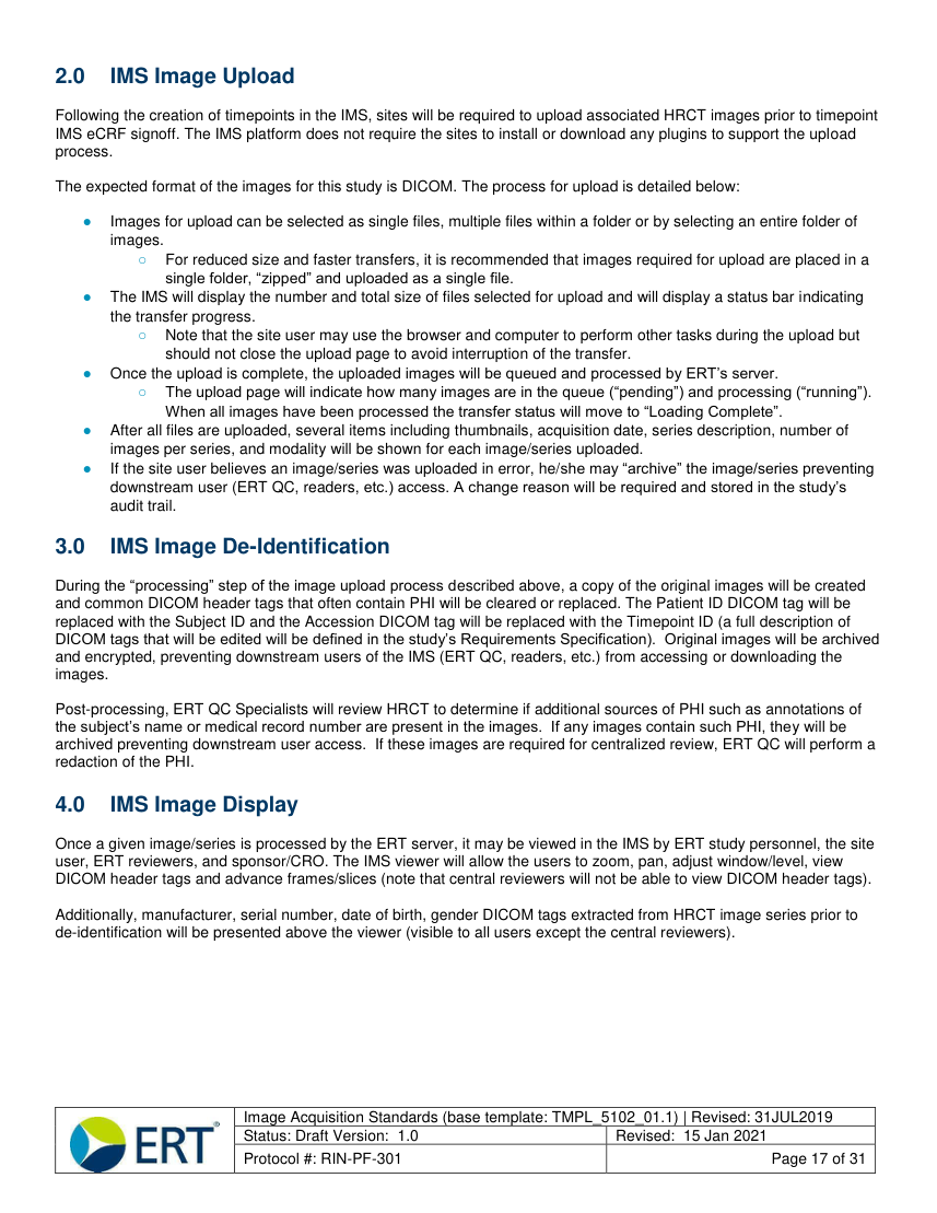
2.0 IMS Image Upload Following the creation of timepoints in the IMS, sites will be required to upload associated HRCT images prior to timepoint IMS eCRF signoff. The IMS platform does not require the sites to install or download any plugins to support the upload process.
The expected format of the images for this study is DICOM. The process for upload is detailed below:
● Images for upload can be selected as single files, multiple files within a folder or by selecting an entire folder of images.
○ For reduced size and faster transfers, it is recommended that images required for upload are placed in a single folder, “zipped” and uploaded as a single file.
● The IMS will display the number and total size of files selected for upload and will display a status bar indicating the transfer progress.
○ Note that the site user may use the browser and computer to perform other tasks during the upload but should not close the upload page to avoid interruption of the transfer.
● Once the upload is complete, the uploaded images will be queued and processed by ERT’s server.
○ The upload page will indicate how many images are in the queue (“pending”) and processing (“running”).
When all images have been processed the transfer status will move to “Loading Complete”.
● After all files are uploaded, several items including thumbnails, acquisition date, series description, number of images per series, and modality will be shown for each image/series uploaded.
● If the site user believes an image/series was uploaded in error, he/she may “archive” the image/series preventing downstream user (ERT QC, readers, etc.) access. A change reason will be required and stored in the study’s audit trail.
3.0 IMS Image De-Identification
During the “processing” step of the image upload process described above, a copy of the original images will be created and common DICOM header tags that often contain PHI will be cleared or replaced. The Patient ID DICOM tag will be replaced with the Subject ID and the Accession DICOM tag will be replaced with the Timepoint ID (a full description of DICOM tags that will be edited will be defined in the study’s Requirements Specification). Original images will be archived and encrypted, preventing downstream users of the IMS (ERT QC, readers, etc.) from accessing or downloading the images.
Post-processing, ERT QC Specialists will review HRCT to determine if additional sources of PHI such as annotations of the subject’s name or medical record number are present in the images. If any images contain such PHI, they will be archived preventing downstream user access. If these images are required for centralized review, ERT QC will perform a redaction of the PHI.
4.0 IMS Image Display Once a given image/series is processed by the ERT server, it may be viewed in the IMS by ERT study personnel, the site user, ERT reviewers, and sponsor/CRO. The IMS viewer will allow the users to zoom, pan, adjust window/level, view DICOM header tags and advance frames/slices (note that central reviewers will not be able to view DICOM header tags).
Additionally, manufacturer, serial number, date of birth, gender DICOM tags extracted from HRCT image series prior to de-identification will be presented above the viewer (visible to all users except the central reviewers).
Image Acquisition Standards (base template: TMPL_5102_01.1) | Revised: 31JUL2019 Status: Draft Version: 1.0 Revised: 15 Jan 2021
Protocol #: RIN-PF-301 Page 17 of 31