CT — Torace: Olympus (MCB)
Sursă și prelucrare editorială
- Proveniență
- 1 referință declarată
- Fidelitatea preluării
- Nedocumentată
- Revizuire medicală
- Nedocumentată
Referințele declarate nu confirmă singure fidelitatea preluării sau aprobarea protocolului pentru utilizare locală.
Detalii despre surse și verificări
MCB Radiology — 9 Olympus
- Ediție / versiune a sursei
- Neprecizată
- Pagină / secțiune
- Pagini: 1–7
- Relația cu protocolul
- Document original importat integral; atribuirea autorilor rămâne cea din PDF.
- Data consultării
- 2026-09-27
Prelucrarea acestei pagini
- Mod de prelucrare
- Titlu și navigare în română; documentul sursă în engleză.; PDF original integral, previzualizare și text extras automat; fără completarea parametrilor clinici lipsă.
- Adaptări față de sursă
- Nedocumentate; absența evidenței nu confirmă o preluare integrală.
- Responsabil pentru prelucrare
- Neprecizat
- Atribuire existentă în pagină
- MCB Radiology (portalul sursă). Această atribuire nu identifică automat autorul sursei sau revizorul medical.
- Versiunea paginii
- mcb-f742ab1d2293
- Actualizare declarată a paginii
- 2026-09-27 — distinctă de revizuirea medicală.
Verificarea fidelității: Nedocumentată
Revizuirea medicală: Nedocumentată
Documentul original MCB — Torace: Olympus
Protocol · 7 pagini · consultat la 2026-09-27.
Descarcă PDF-ul integral · Sursa MCB · Catalogul MCB pentru această modalitate
Instrucțiunile, valorile și ilustrațiile din documentul original sunt păstrate în engleză. Titlul și navigarea sunt în română.
Pagina 1
Pagina 2
Pagina 3
Pagina 4
Pagina 5
Pagina 6
Pagina 7
Textul documentului original
Pagina 1 — text în engleză
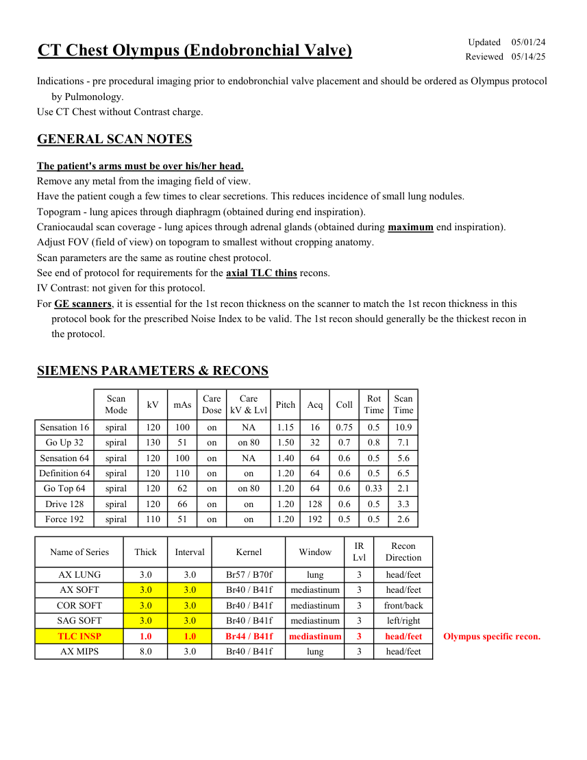
Care
Scan
Mode kV mAs Care
Dose kV & Lvl Pitch Acq Coll Rot Time Time Sensation 16 spiral 120 100 on NA 1.15 16 0.75 0.5 10.9 Go Up 32 spiral 130 51 on on 80 1.50 32 0.7 0.8 7.1 Sensation 64 spiral 120 100 on NA 1.40 64 0.6 0.5 5.6 Definition 64 spiral 120 110 on on 1.20 64 0.6 0.5 6.5 Go Top 64 spiral 120 62 on on 80 1.20 64 0.6 0.33 2.1 Drive 128 spiral 120 66 on on 1.20 128 0.6 0.5 3.3 Force 192 spiral 110 51 on on 1.20 192 0.5 0.5 2.6 Recon
Name of Series Thick Interval Kernel Window IR
Lvl Direction AX LUNG 3.0 3.0 Br57 / B70f lung 3 head/feet AX SOFT 3.0 3.0 Br40 / B41f mediastinum 3 head/feet COR SOFT 3.0 3.0 Br40 / B41f mediastinum 3 front/back SAG SOFT 3.0 3.0 Br40 / B41f mediastinum 3 left/right TLC INSP 1.0 1.0 Br44 / B41f 3 mediastinum head/feet Olympus specific recon. AX MIPS 8.0 3.0 Br40 / B41f lung 3 head/feet
Pagina 2 — text în engleză
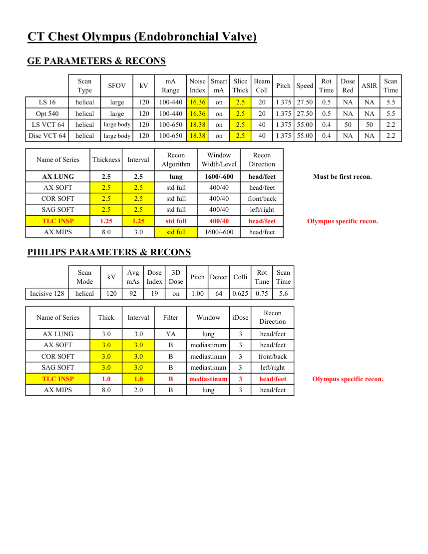
Smart
Slice
Beam
Rot
Dose
Scan
Pitch Speed Type SFOV kV mA
Range Noise
Index mA Thick Coll Time Red ASIR Time LS 16 helical large 120 100-440 16.36 on 2.5 20 1.375 27.50 0.5 NA NA 5.5 Opt 540 helical large 120 100-440 16.36 on 2.5 20 1.375 27.50 0.5 NA NA 5.5 LS VCT 64 helical large body 120 100-650 18.38 on 2.5 40 1.375 55.00 0.4 50 50 2.2 Disc VCT 64 helical large body 120 100-650 18.38 on 2.5 40 1.375 55.00 0.4 NA NA 2.2 Window
Recon
Name of Series Thickness Interval Recon
Algorithm Width/Level Direction AX LUNG 2.5 2.5 lung 1600/-600 head/feet Must be first recon. AX SOFT 2.5 2.5 std full 400/40 head/feet COR SOFT 2.5 2.5 std full 400/40 front/back SAG SOFT 2.5 2.5 std full 400/40 left/right TLC INSP 1.25 1.25 std full 400/40 head/feet Olympus specific recon. AX MIPS 8.0 3.0 std full 1600/-600 head/feet PHILIPS PARAMETERS & RECONS Scan
Dose
3D
Scan
Mode kV Avg
mAs Index Dose Pitch Detect Colli Rot Time Time 0.75 Incisive 128 helical 120 92 19 on 1.00 64 0.625 5.6 Name of Series Thick Interval Filter Window iDose Recon
Direction AX LUNG 3.0 3.0 YA lung 3 head/feet AX SOFT 3.0 3.0 B mediastinum 3 head/feet COR SOFT 3.0 3.0 B mediastinum 3 front/back SAG SOFT 3.0 3.0 B mediastinum 3 left/right TLC INSP 1.0 1.0 B mediastinum 3 head/feet Olympus specific recon. AX MIPS 8.0 2.0 B lung 3 head/feet
Pagina 3 — text în engleză
Pagina 4 — text în engleză
Pagina 5 — text în engleză
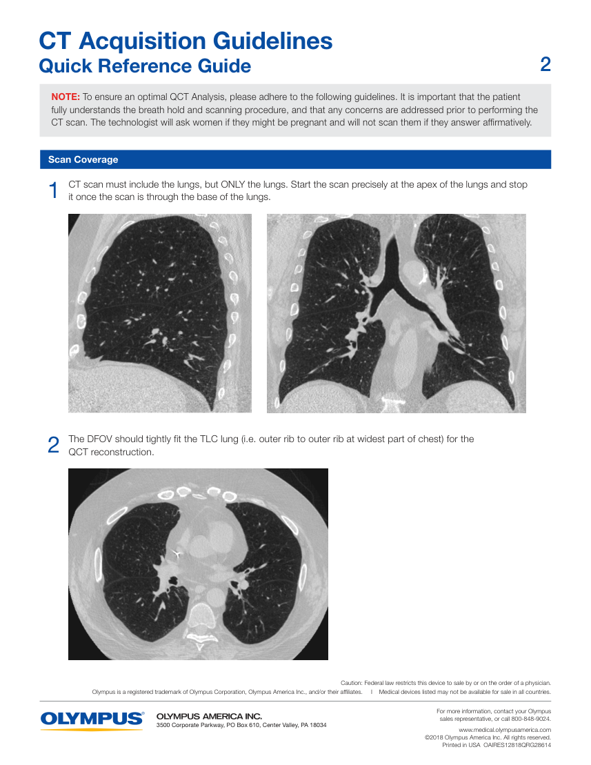
sales representative, or call 800-848-9024. 3500 Corporate Parkway, PO Box 610, Center Valley, PA 18034 www.medical.olympusamerica.com ©2018 Olympus America Inc. All rights reserved. Printed in USA OAIRES12818QRG28614
Pagina 6 — text în engleză
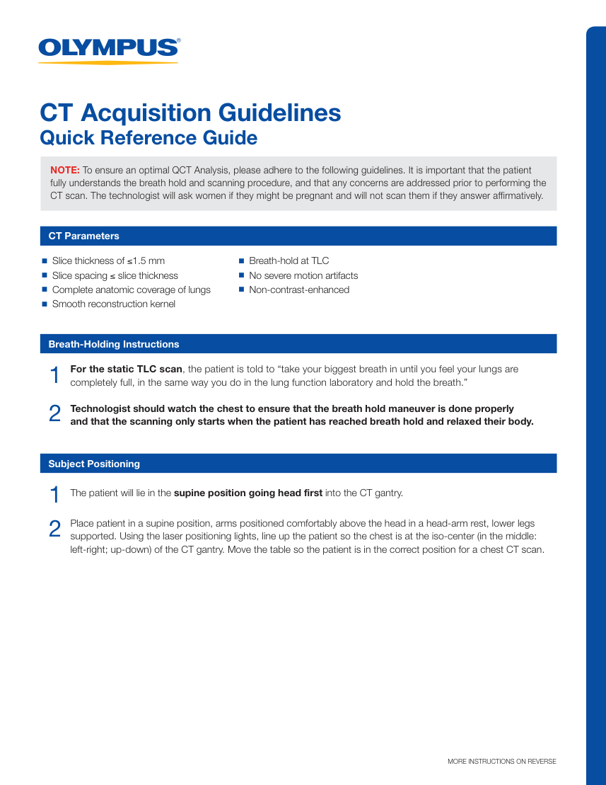
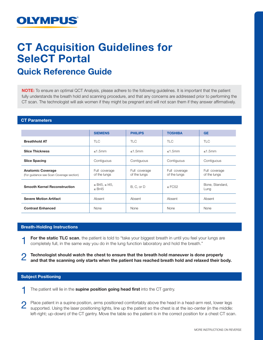
SeleCT Portal Quick Reference Guide NOTE: To ensure an optimal QCT Analysis, please adhere to the following guidelines. It is important that the patient
fully understands the breath hold and scanning procedure, and that any concerns are addressed prior to performing the
CT scan. The technologist will ask women if they might be pregnant and will not scan them if they answer affirmatively. CT Parameters SIEMENS PHILIPS TOSHIBA GE Breathhold AT TLC TLC TLC TLC Slice Thickness ≤1.5mm ≤1.5mm ≤1.5mm ≤1.5mm Slice Spacing Contiguous Contiguous Contiguous Contiguous Anatomic Coverage
(For guidance see Scan Coverage section) Full coverage
of the lungs Full coverage
of the lungs Full coverage
of the lungs Full coverage
of the lungs Smooth Kernel Reconstruction ≤ B45, ≤ I45, ≤ Br45 B, C, or D ≤ FC52 Bone, Standard,
Lung Severe Motion Artifact Absent Absent Absent Absent Contrast Enhanced None None None None Breath-Holding Instructions For the static TLC scan, the patient is told to “take your biggest breath in until you feel your lungs are
completely full, in the same way you do in the lung function laboratory and hold the breath.” 1 2 Technologist should watch the chest to ensure that the breath hold maneuver is done properly
and that the scanning only starts when the patient has reached breath hold and relaxed their body. Subject Positioning The patient will lie in the supine position going head first into the CT gantry. 1 2 Place patient in a supine position, arms positioned comfortably above the head in a head-arm rest, lower legs supported. Using the laser positioning lights, line up the patient so the chest is at the iso-center (in the middle: left-right; up-down) of the CT gantry. Move the table so the patient is in the correct position for a chest CT scan. MORE INSTRUCTIONS ON REVERSE
Pagina 7 — text în engleză
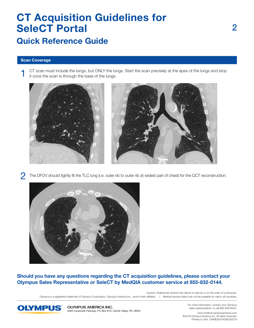
SeleCT Portal Quick Reference Guide Scan Coverage CT scan must include the lungs, but ONLY the lungs. Start the scan precisely at the apex of the lungs and stop it once the scan is through the base of the lungs. 1 The DFOV should tightly fit the TLC lung (i.e. outer rib to outer rib at widest part of chest) for the QCT reconstruction. 2 Should you have any questions regarding the CT acquisition guidelines, please contact your Olympus Sales Representative or SeleCT by MedQIA customer service at 855-832-0144. Caution: Federal law restricts this device to sale by or on the order of a physician. Olympus is a registered trademark of Olympus Corporation, Olympus America Inc., and/or their affiliates. I Medical devices listed may not be available for sale in all countries. For more information, contact your Olympus
sales representative, or call 800-848-9024. 3500 Corporate Parkway, PO Box 610, Center Valley, PA 18034 www.medical.olympusamerica.com ©2019 Olympus America Inc. All rights reserved. Printed in USA OAIRES0419QRG30379