CT — Esofagografie (MCB)
Sursă și prelucrare editorială
- Proveniență
- 1 referință declarată
- Fidelitatea preluării
- Nedocumentată
- Revizuire medicală
- Nedocumentată
Referințele declarate nu confirmă singure fidelitatea preluării sau aprobarea protocolului pentru utilizare locală.
Detalii despre surse și verificări
MCB Radiology — 12 Esophagram
- Ediție / versiune a sursei
- Neprecizată
- Pagină / secțiune
- Pagini: 1–2
- Relația cu protocolul
- Document original importat integral; atribuirea autorilor rămâne cea din PDF.
- Data consultării
- 2026-09-27
Prelucrarea acestei pagini
- Mod de prelucrare
- Titlu și navigare în română; documentul sursă în engleză.; PDF original integral, previzualizare și text extras automat; fără completarea parametrilor clinici lipsă.
- Adaptări față de sursă
- Nedocumentate; absența evidenței nu confirmă o preluare integrală.
- Responsabil pentru prelucrare
- Neprecizat
- Atribuire existentă în pagină
- MCB Radiology (portalul sursă). Această atribuire nu identifică automat autorul sursei sau revizorul medical.
- Versiunea paginii
- mcb-2678a9986b12
- Actualizare declarată a paginii
- 2026-09-27 — distinctă de revizuirea medicală.
Verificarea fidelității: Nedocumentată
Revizuirea medicală: Nedocumentată
Documentul original MCB — Esofagografie
Protocol · 2 pagini · consultat la 2026-09-27.
Descarcă PDF-ul integral · Sursa MCB · Catalogul MCB pentru această modalitate
Instrucțiunile, valorile și ilustrațiile din documentul original sunt păstrate în engleză. Titlul și navigarea sunt în română.
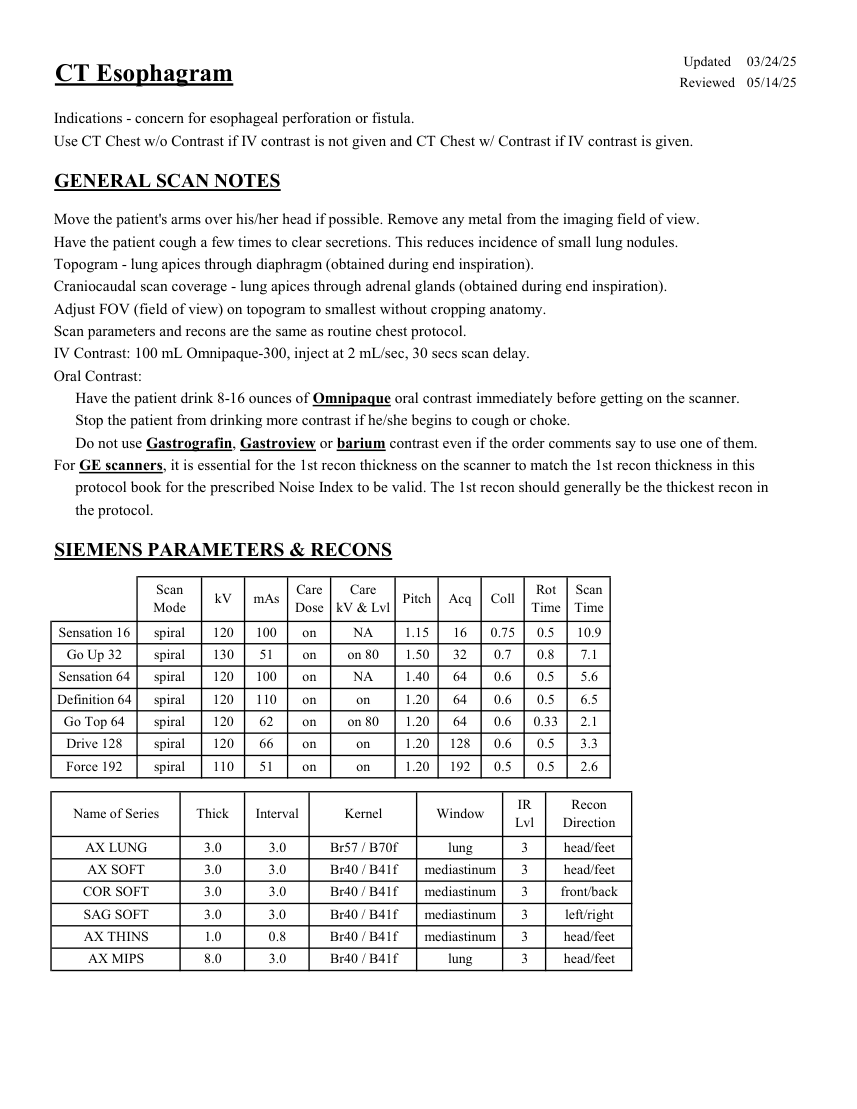
Pagina 1
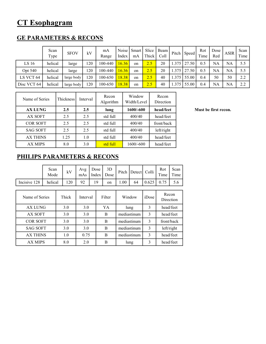
Pagina 2
Textul documentului original
Pagina 1 — text în engleză
Updated 03/24/25 CT Esophagram Reviewed 05/14/25 Indications - concern for esophageal perforation or fistula. Use CT Chest w/o Contrast if IV contrast is not given and CT Chest w/ Contrast if IV contrast is given. GENERAL SCAN NOTES Move the patient's arms over his/her head if possible. Remove any metal from the imaging field of view. Have the patient cough a few times to clear secretions. This reduces incidence of small lung nodules. Topogram - lung apices through diaphragm (obtained during end inspiration). Craniocaudal scan coverage - lung apices through adrenal glands (obtained during end inspiration). Adjust FOV (field of view) on topogram to smallest without cropping anatomy. Scan parameters and recons are the same as routine chest protocol. IV Contrast: 100 mL Omnipaque-300, inject at 2 mL/sec, 30 secs scan delay. Oral Contrast: Have the patient drink 8-16 ounces of Omnipaque oral contrast immediately before getting on the scanner. Stop the patient from drinking more contrast if he/she begins to cough or choke. Do not use Gastrografin, Gastroview or barium contrast even if the order comments say to use one of them. For GE scanners, it is essential for the 1st recon thickness on the scanner to match the 1st recon thickness in this protocol book for the prescribed Noise Index to be valid. The 1st recon should generally be the thickest recon in the protocol. SIEMENS PARAMETERS & RECONS Scan
Care
Scan
Mode kV mAs Care
Dose kV & Lvl Pitch Acq Coll Rot Time Time Sensation 16 spiral 120 100 on NA 1.15 16 0.75 0.5 10.9 Go Up 32 spiral 130 51 on on 80 1.50 32 0.7 0.8 7.1 Sensation 64 spiral 120 100 on NA 1.40 64 0.6 0.5 5.6 Definition 64 spiral 120 110 on on 1.20 0.5 64 0.6 6.5 Go Top 64 spiral 120 62 on on 80 1.20 64 0.6 0.33 2.1 Drive 128 spiral 120 66 on on 1.20 128 0.6 0.5 3.3 on 1.20 192 Force 192 spiral 110 51 on 0.5 0.5 2.6 Recon
Name of Series Thick Interval Kernel Window IR
Lvl Direction AX LUNG 3.0 3.0 Br57 / B70f lung 3 head/feet AX SOFT 3.0 3.0 Br40 / B41f mediastinum 3 head/feet COR SOFT 3.0 3.0 Br40 / B41f mediastinum 3 front/back SAG SOFT 3.0 3.0 Br40 / B41f mediastinum 3 left/right AX THINS 1.0 0.8 Br40 / B41f mediastinum 3 head/feet head/feet AX MIPS 8.0 3.0 Br40 / B41f lung 3
Care
Scan
Mode kV mAs Care
Dose kV & Lvl Pitch Acq Coll Rot Time Time Sensation 16 spiral 120 100 on NA 1.15 16 0.75 0.5 10.9 Go Up 32 spiral 130 51 on on 80 1.50 32 0.7 0.8 7.1 Sensation 64 spiral 120 100 on NA 1.40 64 0.6 0.5 5.6 Definition 64 spiral 120 110 on on 1.20 0.5 64 0.6 6.5 Go Top 64 spiral 120 62 on on 80 1.20 64 0.6 0.33 2.1 Drive 128 spiral 120 66 on on 1.20 128 0.6 0.5 3.3 on 1.20 192 Force 192 spiral 110 51 on 0.5 0.5 2.6 Recon
Name of Series Thick Interval Kernel Window IR
Lvl Direction AX LUNG 3.0 3.0 Br57 / B70f lung 3 head/feet AX SOFT 3.0 3.0 Br40 / B41f mediastinum 3 head/feet COR SOFT 3.0 3.0 Br40 / B41f mediastinum 3 front/back SAG SOFT 3.0 3.0 Br40 / B41f mediastinum 3 left/right AX THINS 1.0 0.8 Br40 / B41f mediastinum 3 head/feet head/feet AX MIPS 8.0 3.0 Br40 / B41f lung 3
Pagina 2 — text în engleză
CT Esophagram GE PARAMETERS & RECONS Scan
Smart
Slice
Beam
Rot
Dose
Scan
Pitch Speed Type SFOV kV mA
Range Noise
Index mA Thick Coll Time Red ASIR Time LS 16 helical large 120 100-440 16.36 on 2.5 20 1.375 27.50 0.5 NA NA 5.5 Opt 540 helical large 120 100-440 16.36 on 2.5 20 1.375 27.50 0.5 NA NA 5.5 LS VCT 64 helical large body 120 100-650 18.38 on 2.5 40 1.375 55.00 0.4 50 50 2.2 Disc VCT 64 helical large body 120 100-650 18.38 on 2.5 40 1.375 55.00 0.4 NA NA 2.2 Window
Recon
Name of Series Thickness Interval Recon
Algorithm Width/Level Direction AX LUNG 2.5 2.5 lung 1600/-600 head/feet Must be first recon. AX SOFT 2.5 2.5 std full 400/40 head/feet COR SOFT 2.5 2.5 std full 400/40 front/back SAG SOFT 2.5 2.5 std full 400/40 left/right AX THINS 1.25 1.0 std full 400/40 head/feet AX MIPS 8.0 3.0 std full 1600/-600 head/feet PHILIPS PARAMETERS & RECONS Scan
Dose
3D
Rot Scan
Detect Colli Mode kV Avg
mAs Index Dose Pitch Time Time 0.75 Incisive 128 helical 120 92 19 on 1.00 64 0.625 5.6 Recon
Name of Series Thick Interval Filter Window iDose Direction AX LUNG 3.0 3.0 YA lung 3 head/feet AX SOFT 3.0 3.0 B mediastinum 3 head/feet COR SOFT 3.0 3.0 B mediastinum 3 front/back SAG SOFT 3.0 3.0 B mediastinum 3 left/right AX THINS 1.0 0.75 B mediastinum 3 head/feet head/feet AX MIPS 8.0 2.0 B lung 3
Smart
Slice
Beam
Rot
Dose
Scan
Pitch Speed Type SFOV kV mA
Range Noise
Index mA Thick Coll Time Red ASIR Time LS 16 helical large 120 100-440 16.36 on 2.5 20 1.375 27.50 0.5 NA NA 5.5 Opt 540 helical large 120 100-440 16.36 on 2.5 20 1.375 27.50 0.5 NA NA 5.5 LS VCT 64 helical large body 120 100-650 18.38 on 2.5 40 1.375 55.00 0.4 50 50 2.2 Disc VCT 64 helical large body 120 100-650 18.38 on 2.5 40 1.375 55.00 0.4 NA NA 2.2 Window
Recon
Name of Series Thickness Interval Recon
Algorithm Width/Level Direction AX LUNG 2.5 2.5 lung 1600/-600 head/feet Must be first recon. AX SOFT 2.5 2.5 std full 400/40 head/feet COR SOFT 2.5 2.5 std full 400/40 front/back SAG SOFT 2.5 2.5 std full 400/40 left/right AX THINS 1.25 1.0 std full 400/40 head/feet AX MIPS 8.0 3.0 std full 1600/-600 head/feet PHILIPS PARAMETERS & RECONS Scan
Dose
3D
Rot Scan
Detect Colli Mode kV Avg
mAs Index Dose Pitch Time Time 0.75 Incisive 128 helical 120 92 19 on 1.00 64 0.625 5.6 Recon
Name of Series Thick Interval Filter Window iDose Direction AX LUNG 3.0 3.0 YA lung 3 head/feet AX SOFT 3.0 3.0 B mediastinum 3 head/feet COR SOFT 3.0 3.0 B mediastinum 3 front/back SAG SOFT 3.0 3.0 B mediastinum 3 left/right AX THINS 1.0 0.75 B mediastinum 3 head/feet head/feet AX MIPS 8.0 2.0 B lung 3