CT — Parametri aparat: GE LS VCT 64 Protocols (MCB)
Sursă și prelucrare editorială
- Proveniență
- 1 referință declarată
- Fidelitatea preluării
- Nedocumentată
- Revizuire medicală
- Nedocumentată
Referințele declarate nu confirmă singure fidelitatea preluării sau aprobarea protocolului pentru utilizare locală.
Detalii despre surse și verificări
MCB Radiology — GE LS VCT 64 Protocols
- Ediție / versiune a sursei
- Neprecizată
- Pagină / secțiune
- Pagini: 1–356
- Relația cu protocolul
- Document original importat integral; atribuirea autorilor rămâne cea din PDF.
- Data consultării
- 2026-09-27
Prelucrarea acestei pagini
- Mod de prelucrare
- Titlu și navigare în română; documentul sursă în engleză.; PDF original integral, previzualizare și text extras automat; fără completarea parametrilor clinici lipsă.
- Adaptări față de sursă
- Nedocumentate; absența evidenței nu confirmă o preluare integrală.
- Responsabil pentru prelucrare
- Neprecizat
- Atribuire existentă în pagină
- MCB Radiology (portalul sursă). Această atribuire nu identifică automat autorul sursei sau revizorul medical.
- Versiunea paginii
- mcb-c78dd8718351
- Actualizare declarată a paginii
- 2026-09-27 — distinctă de revizuirea medicală.
Verificarea fidelității: Nedocumentată
Revizuirea medicală: Nedocumentată
Documentul original MCB — Parametri aparat: GE LS VCT 64 Protocols
Manual de aparat · 356 pagini · consultat la 2026-09-27.
Descarcă PDF-ul integral · Sursa MCB · Catalogul MCB pentru această modalitate
Instrucțiunile, valorile și ilustrațiile din documentul original sunt păstrate în engleză. Titlul și navigarea sunt în română.
Previzualizarea de mai jos prezintă primele 3 pagini. PDF-ul descărcabil conține toate cele 356 de pagini.
Pagina 1
Pagina 2

Pagina 3
Textul documentului original
Pagina 1 — text în engleză
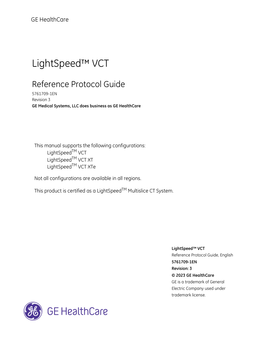
GE HealthCare LightSpeed™ VCT Reference Protocol Guide 5761709-1EN Revision 3 GE Medical Systems, LLC does business as GE HealthCare This manual supports the following configurations: LightSpeedTM VCT LightSpeedTM VCT XT LightSpeedTM VCT XTe Not all configurations are available in all regions. This product is certified as a LightSpeedTM Multislice CT System. LightSpeed™ VCT Reference Protocol Guide, English 5761709-1EN Revision: 3 © 2023 GE HealthCare GE is a trademark of General Electric Company used under trademark license.
Pagina 2 — text în engleză
Reference Protocol Guide 5761709-1EN r3 Mar. 2023
Pagina 3 — text în engleză
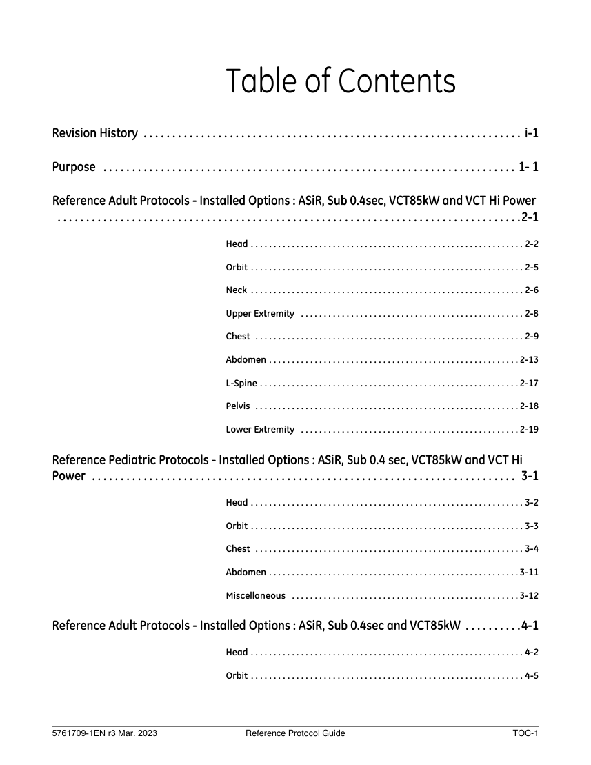
Table of Contents Revision History . . . . . . . . . . . . . . . . . . . . . . . . . . . . . . . . . . . . . . . . . . . . . . . . . . . . . . . . . . . . . . . . . . i-1 Purpose . . . . . . . . . . . . . . . . . . . . . . . . . . . . . . . . . . . . . . . . . . . . . . . . . . . . . . . . . . . . . . . . . . . . . . . . 1- 1 Reference Adult Protocols - Installed Options : ASiR, Sub 0.4sec, VCT85kW and VCT Hi Power
. . . . . . . . . . . . . . . . . . . . . . . . . . . . . . . . . . . . . . . . . . . . . . . . . . . . . . . . . . . . . . . . . . . . . . . . . . . . . . . . .2-1 Head . . . . . . . . . . . . . . . . . . . . . . . . . . . . . . . . . . . . . . . . . . . . . . . . . . . . . . . . . . . . 2-2 Orbit . . . . . . . . . . . . . . . . . . . . . . . . . . . . . . . . . . . . . . . . . . . . . . . . . . . . . . . . . . . . 2-5 Neck . . . . . . . . . . . . . . . . . . . . . . . . . . . . . . . . . . . . . . . . . . . . . . . . . . . . . . . . . . . . 2-6 Upper Extremity . . . . . . . . . . . . . . . . . . . . . . . . . . . . . . . . . . . . . . . . . . . . . . . . . 2-8 Chest . . . . . . . . . . . . . . . . . . . . . . . . . . . . . . . . . . . . . . . . . . . . . . . . . . . . . . . . . . . 2-9 Abdomen . . . . . . . . . . . . . . . . . . . . . . . . . . . . . . . . . . . . . . . . . . . . . . . . . . . . . . . 2-13 L-Spine . . . . . . . . . . . . . . . . . . . . . . . . . . . . . . . . . . . . . . . . . . . . . . . . . . . . . . . . . 2-17 Pelvis . . . . . . . . . . . . . . . . . . . . . . . . . . . . . . . . . . . . . . . . . . . . . . . . . . . . . . . . . . 2-18 Lower Extremity . . . . . . . . . . . . . . . . . . . . . . . . . . . . . . . . . . . . . . . . . . . . . . . . 2-19 Reference Pediatric Protocols - Installed Options : ASiR, Sub 0.4 sec, VCT85kW and VCT Hi Power . . . . . . . . . . . . . . . . . . . . . . . . . . . . . . . . . . . . . . . . . . . . . . . . . . . . . . . . . . . . . . . . . . . . . . . . . . 3-1 Head . . . . . . . . . . . . . . . . . . . . . . . . . . . . . . . . . . . . . . . . . . . . . . . . . . . . . . . . . . . . 3-2 Orbit . . . . . . . . . . . . . . . . . . . . . . . . . . . . . . . . . . . . . . . . . . . . . . . . . . . . . . . . . . . . 3-3 Chest . . . . . . . . . . . . . . . . . . . . . . . . . . . . . . . . . . . . . . . . . . . . . . . . . . . . . . . . . . . 3-4 Abdomen . . . . . . . . . . . . . . . . . . . . . . . . . . . . . . . . . . . . . . . . . . . . . . . . . . . . . . . 3-11 Miscellaneous . . . . . . . . . . . . . . . . . . . . . . . . . . . . . . . . . . . . . . . . . . . . . . . . . . 3-12 Reference Adult Protocols - Installed Options : ASiR, Sub 0.4sec and VCT85kW . . . . . . . . . .4-1 Head . . . . . . . . . . . . . . . . . . . . . . . . . . . . . . . . . . . . . . . . . . . . . . . . . . . . . . . . . . . . 4-2 Orbit . . . . . . . . . . . . . . . . . . . . . . . . . . . . . . . . . . . . . . . . . . . . . . . . . . . . . . . . . . . . 4-5 5761709-1EN r3 Mar. 2023 Reference Protocol Guide TOC-1
. . . . . . . . . . . . . . . . . . . . . . . . . . . . . . . . . . . . . . . . . . . . . . . . . . . . . . . . . . . . . . . . . . . . . . . . . . . . . . . . .2-1 Head . . . . . . . . . . . . . . . . . . . . . . . . . . . . . . . . . . . . . . . . . . . . . . . . . . . . . . . . . . . . 2-2 Orbit . . . . . . . . . . . . . . . . . . . . . . . . . . . . . . . . . . . . . . . . . . . . . . . . . . . . . . . . . . . . 2-5 Neck . . . . . . . . . . . . . . . . . . . . . . . . . . . . . . . . . . . . . . . . . . . . . . . . . . . . . . . . . . . . 2-6 Upper Extremity . . . . . . . . . . . . . . . . . . . . . . . . . . . . . . . . . . . . . . . . . . . . . . . . . 2-8 Chest . . . . . . . . . . . . . . . . . . . . . . . . . . . . . . . . . . . . . . . . . . . . . . . . . . . . . . . . . . . 2-9 Abdomen . . . . . . . . . . . . . . . . . . . . . . . . . . . . . . . . . . . . . . . . . . . . . . . . . . . . . . . 2-13 L-Spine . . . . . . . . . . . . . . . . . . . . . . . . . . . . . . . . . . . . . . . . . . . . . . . . . . . . . . . . . 2-17 Pelvis . . . . . . . . . . . . . . . . . . . . . . . . . . . . . . . . . . . . . . . . . . . . . . . . . . . . . . . . . . 2-18 Lower Extremity . . . . . . . . . . . . . . . . . . . . . . . . . . . . . . . . . . . . . . . . . . . . . . . . 2-19 Reference Pediatric Protocols - Installed Options : ASiR, Sub 0.4 sec, VCT85kW and VCT Hi Power . . . . . . . . . . . . . . . . . . . . . . . . . . . . . . . . . . . . . . . . . . . . . . . . . . . . . . . . . . . . . . . . . . . . . . . . . . 3-1 Head . . . . . . . . . . . . . . . . . . . . . . . . . . . . . . . . . . . . . . . . . . . . . . . . . . . . . . . . . . . . 3-2 Orbit . . . . . . . . . . . . . . . . . . . . . . . . . . . . . . . . . . . . . . . . . . . . . . . . . . . . . . . . . . . . 3-3 Chest . . . . . . . . . . . . . . . . . . . . . . . . . . . . . . . . . . . . . . . . . . . . . . . . . . . . . . . . . . . 3-4 Abdomen . . . . . . . . . . . . . . . . . . . . . . . . . . . . . . . . . . . . . . . . . . . . . . . . . . . . . . . 3-11 Miscellaneous . . . . . . . . . . . . . . . . . . . . . . . . . . . . . . . . . . . . . . . . . . . . . . . . . . 3-12 Reference Adult Protocols - Installed Options : ASiR, Sub 0.4sec and VCT85kW . . . . . . . . . .4-1 Head . . . . . . . . . . . . . . . . . . . . . . . . . . . . . . . . . . . . . . . . . . . . . . . . . . . . . . . . . . . . 4-2 Orbit . . . . . . . . . . . . . . . . . . . . . . . . . . . . . . . . . . . . . . . . . . . . . . . . . . . . . . . . . . . . 4-5 5761709-1EN r3 Mar. 2023 Reference Protocol Guide TOC-1
Pagina 4 — text în engleză
Neck . . . . . . . . . . . . . . . . . . . . . . . . . . . . . . . . . . . . . . . . . . . . . . . . . . . . . . . . . . . . 4-6 Upper Extremity . . . . . . . . . . . . . . . . . . . . . . . . . . . . . . . . . . . . . . . . . . . . . . . . . 4-8 Chest . . . . . . . . . . . . . . . . . . . . . . . . . . . . . . . . . . . . . . . . . . . . . . . . . . . . . . . . . . . 4-9 Abdomen . . . . . . . . . . . . . . . . . . . . . . . . . . . . . . . . . . . . . . . . . . . . . . . . . . . . . . . 4-13 L-Spine . . . . . . . . . . . . . . . . . . . . . . . . . . . . . . . . . . . . . . . . . . . . . . . . . . . . . . . . . 4-17 Pelvis . . . . . . . . . . . . . . . . . . . . . . . . . . . . . . . . . . . . . . . . . . . . . . . . . . . . . . . . . . 4-18 Lower Extremity . . . . . . . . . . . . . . . . . . . . . . . . . . . . . . . . . . . . . . . . . . . . . . . . 4-19 Reference Pediatric Protocols - Installed Options : ASiR, Sub 0.4 sec and VCT85kW . . . . . 5-1 Head . . . . . . . . . . . . . . . . . . . . . . . . . . . . . . . . . . . . . . . . . . . . . . . . . . . . . . . . . . . . 5-2 Orbit . . . . . . . . . . . . . . . . . . . . . . . . . . . . . . . . . . . . . . . . . . . . . . . . . . . . . . . . . . . . 5-3 Chest . . . . . . . . . . . . . . . . . . . . . . . . . . . . . . . . . . . . . . . . . . . . . . . . . . . . . . . . . . . 5-4 Abdomen . . . . . . . . . . . . . . . . . . . . . . . . . . . . . . . . . . . . . . . . . . . . . . . . . . . . . . . 5-11 Miscellaneous . . . . . . . . . . . . . . . . . . . . . . . . . . . . . . . . . . . . . . . . . . . . . . . . . . 5-12 Reference Adult Protocols - Installed Options : ASiR and Sub 0.4sec . . . . . . . . . . . . . . . . . . . 6-1 Head . . . . . . . . . . . . . . . . . . . . . . . . . . . . . . . . . . . . . . . . . . . . . . . . . . . . . . . . . . . . 6-2 Orbit . . . . . . . . . . . . . . . . . . . . . . . . . . . . . . . . . . . . . . . . . . . . . . . . . . . . . . . . . . . . 6-5 Neck . . . . . . . . . . . . . . . . . . . . . . . . . . . . . . . . . . . . . . . . . . . . . . . . . . . . . . . . . . . . 6-6 Upper Extremity . . . . . . . . . . . . . . . . . . . . . . . . . . . . . . . . . . . . . . . . . . . . . . . . . 6-8 Chest . . . . . . . . . . . . . . . . . . . . . . . . . . . . . . . . . . . . . . . . . . . . . . . . . . . . . . . . . . . 6-9 Abdomen . . . . . . . . . . . . . . . . . . . . . . . . . . . . . . . . . . . . . . . . . . . . . . . . . . . . . . . 6-13 L-Spine . . . . . . . . . . . . . . . . . . . . . . . . . . . . . . . . . . . . . . . . . . . . . . . . . . . . . . . . . 6-17 Pelvis . . . . . . . . . . . . . . . . . . . . . . . . . . . . . . . . . . . . . . . . . . . . . . . . . . . . . . . . . . 6-18 Lower Extremity . . . . . . . . . . . . . . . . . . . . . . . . . . . . . . . . . . . . . . . . . . . . . . . . 6-19 Reference Pediatric Protocols - Installed Options : ASiR and Sub 0.4 sec . . . . . . . . . . . . . . . 7-1 Head . . . . . . . . . . . . . . . . . . . . . . . . . . . . . . . . . . . . . . . . . . . . . . . . . . . . . . . . . . . . 7-2 Orbit . . . . . . . . . . . . . . . . . . . . . . . . . . . . . . . . . . . . . . . . . . . . . . . . . . . . . . . . . . . . 7-3 5761709-1EN r3 Mar. 2023 Reference Protocol Guide TOC-2
Pagina 5 — text în engleză
Chest . . . . . . . . . . . . . . . . . . . . . . . . . . . . . . . . . . . . . . . . . . . . . . . . . . . . . . . . . . . 7-4 Abdomen . . . . . . . . . . . . . . . . . . . . . . . . . . . . . . . . . . . . . . . . . . . . . . . . . . . . . . . 7-11 Miscellaneous . . . . . . . . . . . . . . . . . . . . . . . . . . . . . . . . . . . . . . . . . . . . . . . . . . 7-12 Reference Adult Protocols - Installed Options : Sub 0.4sec, VCT85kW and VCT Hi Power . 8-1 Head . . . . . . . . . . . . . . . . . . . . . . . . . . . . . . . . . . . . . . . . . . . . . . . . . . . . . . . . . . . . 8-2 Orbit . . . . . . . . . . . . . . . . . . . . . . . . . . . . . . . . . . . . . . . . . . . . . . . . . . . . . . . . . . . . 8-5 Neck . . . . . . . . . . . . . . . . . . . . . . . . . . . . . . . . . . . . . . . . . . . . . . . . . . . . . . . . . . . . 8-6 Upper Extremity . . . . . . . . . . . . . . . . . . . . . . . . . . . . . . . . . . . . . . . . . . . . . . . . . 8-8 Chest . . . . . . . . . . . . . . . . . . . . . . . . . . . . . . . . . . . . . . . . . . . . . . . . . . . . . . . . . . . 8-9 Abdomen . . . . . . . . . . . . . . . . . . . . . . . . . . . . . . . . . . . . . . . . . . . . . . . . . . . . . . . 8-13 L-Spine . . . . . . . . . . . . . . . . . . . . . . . . . . . . . . . . . . . . . . . . . . . . . . . . . . . . . . . . . 8-17 Pelvis . . . . . . . . . . . . . . . . . . . . . . . . . . . . . . . . . . . . . . . . . . . . . . . . . . . . . . . . . . 8-18 Lower Extremity . . . . . . . . . . . . . . . . . . . . . . . . . . . . . . . . . . . . . . . . . . . . . . . . 8-19 Reference Pediatric Protocols - Installed Options : Sub 0.4 sec, VCT85kW and VCT Hi Power . . . . . . . . . . . . . . . . . . . . . . . . . . . . . . . . . . . . . . . . . . . . . . . . . . . . . . . . . . . . . . . . . . . . . . . . . . . . . . . . . .9-1 Head . . . . . . . . . . . . . . . . . . . . . . . . . . . . . . . . . . . . . . . . . . . . . . . . . . . . . . . . . . . . 9-2 Orbit . . . . . . . . . . . . . . . . . . . . . . . . . . . . . . . . . . . . . . . . . . . . . . . . . . . . . . . . . . . . 9-3 Chest . . . . . . . . . . . . . . . . . . . . . . . . . . . . . . . . . . . . . . . . . . . . . . . . . . . . . . . . . . . 9-4 Abdomen . . . . . . . . . . . . . . . . . . . . . . . . . . . . . . . . . . . . . . . . . . . . . . . . . . . . . . . 9-11 Miscellaneous . . . . . . . . . . . . . . . . . . . . . . . . . . . . . . . . . . . . . . . . . . . . . . . . . . 9-12 Reference Adult Protocols - Installed Options : Sub 0.4sec and VCT85kW . . . . . . . . . . . . . 10-1 Head . . . . . . . . . . . . . . . . . . . . . . . . . . . . . . . . . . . . . . . . . . . . . . . . . . . . . . . . . . . 10-2 Orbit . . . . . . . . . . . . . . . . . . . . . . . . . . . . . . . . . . . . . . . . . . . . . . . . . . . . . . . . . . . 10-5 Neck . . . . . . . . . . . . . . . . . . . . . . . . . . . . . . . . . . . . . . . . . . . . . . . . . . . . . . . . . . . 10-6 Upper Extremity . . . . . . . . . . . . . . . . . . . . . . . . . . . . . . . . . . . . . . . . . . . . . . . . 10-8 Chest . . . . . . . . . . . . . . . . . . . . . . . . . . . . . . . . . . . . . . . . . . . . . . . . . . . . . . . . . . 10-9 5761709-1EN r3 Mar. 2023 Reference Protocol Guide TOC-3
Pagina 6 — text în engleză
Abdomen . . . . . . . . . . . . . . . . . . . . . . . . . . . . . . . . . . . . . . . . . . . . . . . . . . . . . .10-13 L-Spine . . . . . . . . . . . . . . . . . . . . . . . . . . . . . . . . . . . . . . . . . . . . . . . . . . . . . . . .10-17 Pelvis . . . . . . . . . . . . . . . . . . . . . . . . . . . . . . . . . . . . . . . . . . . . . . . . . . . . . . . . .10-18 Lower Extremity . . . . . . . . . . . . . . . . . . . . . . . . . . . . . . . . . . . . . . . . . . . . . . .10-19 Reference Pediatric Protocols - Installed Options : Sub 0.4 sec and VCT85kW . . . . . . . . . 11-1 Head . . . . . . . . . . . . . . . . . . . . . . . . . . . . . . . . . . . . . . . . . . . . . . . . . . . . . . . . . . . 11-2 Orbit . . . . . . . . . . . . . . . . . . . . . . . . . . . . . . . . . . . . . . . . . . . . . . . . . . . . . . . . . . . 11-3 Chest . . . . . . . . . . . . . . . . . . . . . . . . . . . . . . . . . . . . . . . . . . . . . . . . . . . . . . . . . . 11-4 Abdomen . . . . . . . . . . . . . . . . . . . . . . . . . . . . . . . . . . . . . . . . . . . . . . . . . . . . . .11-11 Miscellaneous . . . . . . . . . . . . . . . . . . . . . . . . . . . . . . . . . . . . . . . . . . . . . . . . .11-12 Reference Adult Protocols - Installed Options : Sub 0.4sec . . . . . . . . . . . . . . . . . . . . . . . . . . 12-1 Head . . . . . . . . . . . . . . . . . . . . . . . . . . . . . . . . . . . . . . . . . . . . . . . . . . . . . . . . . . . 12-2 Orbit . . . . . . . . . . . . . . . . . . . . . . . . . . . . . . . . . . . . . . . . . . . . . . . . . . . . . . . . . . . 12-5 Neck . . . . . . . . . . . . . . . . . . . . . . . . . . . . . . . . . . . . . . . . . . . . . . . . . . . . . . . . . . . 12-6 Upper Extremity . . . . . . . . . . . . . . . . . . . . . . . . . . . . . . . . . . . . . . . . . . . . . . . . 12-8 Chest . . . . . . . . . . . . . . . . . . . . . . . . . . . . . . . . . . . . . . . . . . . . . . . . . . . . . . . . . . 12-9 Abdomen . . . . . . . . . . . . . . . . . . . . . . . . . . . . . . . . . . . . . . . . . . . . . . . . . . . . . .12-13 L-Spine . . . . . . . . . . . . . . . . . . . . . . . . . . . . . . . . . . . . . . . . . . . . . . . . . . . . . . . .12-17 Pelvis . . . . . . . . . . . . . . . . . . . . . . . . . . . . . . . . . . . . . . . . . . . . . . . . . . . . . . . . .12-18 Lower Extremity . . . . . . . . . . . . . . . . . . . . . . . . . . . . . . . . . . . . . . . . . . . . . . .12-19 Reference Pediatric Protocols - Installed Options : Sub 0.4 sec . . . . . . . . . . . . . . . . . . . . . . 13-1 Head . . . . . . . . . . . . . . . . . . . . . . . . . . . . . . . . . . . . . . . . . . . . . . . . . . . . . . . . . . . 13-2 Orbit . . . . . . . . . . . . . . . . . . . . . . . . . . . . . . . . . . . . . . . . . . . . . . . . . . . . . . . . . . . 13-3 Chest . . . . . . . . . . . . . . . . . . . . . . . . . . . . . . . . . . . . . . . . . . . . . . . . . . . . . . . . . . 13-4 Abdomen . . . . . . . . . . . . . . . . . . . . . . . . . . . . . . . . . . . . . . . . . . . . . . . . . . . . . .13-11 Miscellaneous . . . . . . . . . . . . . . . . . . . . . . . . . . . . . . . . . . . . . . . . . . . . . . . . .13-12 TOC-4 Reference Protocol Guide 5761709-1EN r3 Mar. 2023
Pagina 7 — text în engleză
Reference Adult Protocols - Installed Options : ASiR, VCT85kW and VCT Hi Power . . . . . 14-1 Head . . . . . . . . . . . . . . . . . . . . . . . . . . . . . . . . . . . . . . . . . . . . . . . . . . . . . . . . . . . 14-2 Orbit . . . . . . . . . . . . . . . . . . . . . . . . . . . . . . . . . . . . . . . . . . . . . . . . . . . . . . . . . . . 14-5 Neck . . . . . . . . . . . . . . . . . . . . . . . . . . . . . . . . . . . . . . . . . . . . . . . . . . . . . . . . . . . 14-6 Upper Extremity . . . . . . . . . . . . . . . . . . . . . . . . . . . . . . . . . . . . . . . . . . . . . . . . 14-8 Chest . . . . . . . . . . . . . . . . . . . . . . . . . . . . . . . . . . . . . . . . . . . . . . . . . . . . . . . . . . 14-9 Abdomen . . . . . . . . . . . . . . . . . . . . . . . . . . . . . . . . . . . . . . . . . . . . . . . . . . . . . .14-12 L-Spine . . . . . . . . . . . . . . . . . . . . . . . . . . . . . . . . . . . . . . . . . . . . . . . . . . . . . . . .14-16 Pelvis . . . . . . . . . . . . . . . . . . . . . . . . . . . . . . . . . . . . . . . . . . . . . . . . . . . . . . . . .14-17 Lower Extremity . . . . . . . . . . . . . . . . . . . . . . . . . . . . . . . . . . . . . . . . . . . . . . .14-18 Reference Pediatric Protocols - Installed Options : ASiR, VCT85kW and VCT Hi Power . . 15-1 Head . . . . . . . . . . . . . . . . . . . . . . . . . . . . . . . . . . . . . . . . . . . . . . . . . . . . . . . . . . . 15-2 Orbit . . . . . . . . . . . . . . . . . . . . . . . . . . . . . . . . . . . . . . . . . . . . . . . . . . . . . . . . . . . 15-3 Chest . . . . . . . . . . . . . . . . . . . . . . . . . . . . . . . . . . . . . . . . . . . . . . . . . . . . . . . . . . 15-4 Abdomen . . . . . . . . . . . . . . . . . . . . . . . . . . . . . . . . . . . . . . . . . . . . . . . . . . . . . .15-11 Miscellaneous . . . . . . . . . . . . . . . . . . . . . . . . . . . . . . . . . . . . . . . . . . . . . . . . .15-12 Reference Adult Protocols - Installed Options : ASiR . . . . . . . . . . . . . . . . . . . . . . . . . . . . . . . . 16-1 Head . . . . . . . . . . . . . . . . . . . . . . . . . . . . . . . . . . . . . . . . . . . . . . . . . . . . . . . . . . . 16-2 Orbit . . . . . . . . . . . . . . . . . . . . . . . . . . . . . . . . . . . . . . . . . . . . . . . . . . . . . . . . . . . 16-5 Neck . . . . . . . . . . . . . . . . . . . . . . . . . . . . . . . . . . . . . . . . . . . . . . . . . . . . . . . . . . . 16-6 Upper Extremity . . . . . . . . . . . . . . . . . . . . . . . . . . . . . . . . . . . . . . . . . . . . . . . . 16-8 Chest . . . . . . . . . . . . . . . . . . . . . . . . . . . . . . . . . . . . . . . . . . . . . . . . . . . . . . . . . . 16-9 Abdomen . . . . . . . . . . . . . . . . . . . . . . . . . . . . . . . . . . . . . . . . . . . . . . . . . . . . . .16-12 L-Spine . . . . . . . . . . . . . . . . . . . . . . . . . . . . . . . . . . . . . . . . . . . . . . . . . . . . . . . .16-16 Pelvis . . . . . . . . . . . . . . . . . . . . . . . . . . . . . . . . . . . . . . . . . . . . . . . . . . . . . . . . .16-17 Lower Extremity . . . . . . . . . . . . . . . . . . . . . . . . . . . . . . . . . . . . . . . . . . . . . . .16-18 5761709-1EN r3 Mar. 2023 Reference Protocol Guide TOC-5
Pagina 8 — text în engleză
Reference Pediatric Protocols - Installed Options : ASiR . . . . . . . . . . . . . . . . . . . . . . . . . . . . . 17-1 Head . . . . . . . . . . . . . . . . . . . . . . . . . . . . . . . . . . . . . . . . . . . . . . . . . . . . . . . . . . . 17-2 Orbit . . . . . . . . . . . . . . . . . . . . . . . . . . . . . . . . . . . . . . . . . . . . . . . . . . . . . . . . . . . 17-3 Chest . . . . . . . . . . . . . . . . . . . . . . . . . . . . . . . . . . . . . . . . . . . . . . . . . . . . . . . . . . 17-4 Abdomen . . . . . . . . . . . . . . . . . . . . . . . . . . . . . . . . . . . . . . . . . . . . . . . . . . . . . .17-11 Miscellaneous . . . . . . . . . . . . . . . . . . . . . . . . . . . . . . . . . . . . . . . . . . . . . . . . .17-12 Reference Adult Protocols - Installed Options : VCT85kW and/or VCT Hi Power . . . . . . . . 18-1 Head . . . . . . . . . . . . . . . . . . . . . . . . . . . . . . . . . . . . . . . . . . . . . . . . . . . . . . . . . . . 18-2 Orbit . . . . . . . . . . . . . . . . . . . . . . . . . . . . . . . . . . . . . . . . . . . . . . . . . . . . . . . . . . . 18-5 Neck . . . . . . . . . . . . . . . . . . . . . . . . . . . . . . . . . . . . . . . . . . . . . . . . . . . . . . . . . . . 18-6 Upper Extremity . . . . . . . . . . . . . . . . . . . . . . . . . . . . . . . . . . . . . . . . . . . . . . . . 18-8 Chest . . . . . . . . . . . . . . . . . . . . . . . . . . . . . . . . . . . . . . . . . . . . . . . . . . . . . . . . . . 18-9 Abdomen . . . . . . . . . . . . . . . . . . . . . . . . . . . . . . . . . . . . . . . . . . . . . . . . . . . . . .18-12 L-Spine . . . . . . . . . . . . . . . . . . . . . . . . . . . . . . . . . . . . . . . . . . . . . . . . . . . . . . . .18-16 Pelvis . . . . . . . . . . . . . . . . . . . . . . . . . . . . . . . . . . . . . . . . . . . . . . . . . . . . . . . . .18-17 Lower Extremity . . . . . . . . . . . . . . . . . . . . . . . . . . . . . . . . . . . . . . . . . . . . . . .18-18 Reference Pediatric Protocols - Installed Options : VCT85kW and/or VCT Hi Power . . . . 19-1 Head . . . . . . . . . . . . . . . . . . . . . . . . . . . . . . . . . . . . . . . . . . . . . . . . . . . . . . . . . . . 19-2 Orbit . . . . . . . . . . . . . . . . . . . . . . . . . . . . . . . . . . . . . . . . . . . . . . . . . . . . . . . . . . . 19-3 Chest . . . . . . . . . . . . . . . . . . . . . . . . . . . . . . . . . . . . . . . . . . . . . . . . . . . . . . . . . . 19-4 Abdomen . . . . . . . . . . . . . . . . . . . . . . . . . . . . . . . . . . . . . . . . . . . . . . . . . . . . . .19-11 Miscellaneous . . . . . . . . . . . . . . . . . . . . . . . . . . . . . . . . . . . . . . . . . . . . . . . . .19-12 Reference Adult Protocols - Installed Options : 72kW . . . . . . . . . . . . . . . . . . . . . . . . . . . . . . . 20-1 Head . . . . . . . . . . . . . . . . . . . . . . . . . . . . . . . . . . . . . . . . . . . . . . . . . . . . . . . . . . . 20-2 Orbit . . . . . . . . . . . . . . . . . . . . . . . . . . . . . . . . . . . . . . . . . . . . . . . . . . . . . . . . . . . 20-5 Neck . . . . . . . . . . . . . . . . . . . . . . . . . . . . . . . . . . . . . . . . . . . . . . . . . . . . . . . . . . . 20-6 TOC-6 Reference Protocol Guide 5761709-1EN r3 Mar. 2023
Pagina 9 — text în engleză
Upper Extremity . . . . . . . . . . . . . . . . . . . . . . . . . . . . . . . . . . . . . . . . . . . . . . . . 20-8 Chest . . . . . . . . . . . . . . . . . . . . . . . . . . . . . . . . . . . . . . . . . . . . . . . . . . . . . . . . . . 20-9 Abdomen . . . . . . . . . . . . . . . . . . . . . . . . . . . . . . . . . . . . . . . . . . . . . . . . . . . . . .20-12 L-Spine . . . . . . . . . . . . . . . . . . . . . . . . . . . . . . . . . . . . . . . . . . . . . . . . . . . . . . . .20-16 Pelvis . . . . . . . . . . . . . . . . . . . . . . . . . . . . . . . . . . . . . . . . . . . . . . . . . . . . . . . . .20-17 Lower Extremity . . . . . . . . . . . . . . . . . . . . . . . . . . . . . . . . . . . . . . . . . . . . . . .20-18 Reference Pediatric Protocols - Installed Options : 72kW . . . . . . . . . . . . . . . . . . . . . . . . . . . . 21-1 Head . . . . . . . . . . . . . . . . . . . . . . . . . . . . . . . . . . . . . . . . . . . . . . . . . . . . . . . . . . . 21-2 Orbit . . . . . . . . . . . . . . . . . . . . . . . . . . . . . . . . . . . . . . . . . . . . . . . . . . . . . . . . . . . 21-3 Chest . . . . . . . . . . . . . . . . . . . . . . . . . . . . . . . . . . . . . . . . . . . . . . . . . . . . . . . . . . 21-4 Abdomen . . . . . . . . . . . . . . . . . . . . . . . . . . . . . . . . . . . . . . . . . . . . . . . . . . . . . .21-11 Miscellaneous . . . . . . . . . . . . . . . . . . . . . . . . . . . . . . . . . . . . . . . . . . . . . . . . .21-12 Lexicon . . . . . . . . . . . . . . . . . . . . . . . . . . . . . . . . . . . . . . . . . . . . . . . . . . . . . . . . . . . . . . . . . . . . . . . . 22-1 5761709-1EN r3 Mar. 2023 Reference Protocol Guide TOC-7
Pagina 10 — text în engleză
TOC-8 Reference Protocol Guide 5761709-1EN r3 Mar. 2023
Pagina 11 — text în engleză
Revision History i-1
Pagina 12 — text în engleză
Revision History REV DATE REASON FOR CHANGE 1 Nov. 2016 Initial Release Dec. 2016 2 Typo 3 Mar. 2023 Typo i-2
Pagina 13 — text în engleză
Purpose 1-1
Pagina 14 — text în engleză
Purpose This document provides a listing of the parameters provided in the reference protocols contained under the GE tab selector. Each table lists the Protocol Number, Protocol Name, Post Processing software associated with, Scan Type, SFOV, Pitch/Table Speed/Row, Gantry Rotation Time, Slice Thickness, Beam Collimation, kV, mA/Min-Max/Noise Index/Average mA, Recon Algorithm Type used, CTDIvol, DLP (mGy-cm), Scan Length, and Phantom Type used and Description. If the ASiR option is installed on the system, there will be a % Dose Reduction column and % ASiR column listed in the table indicating the % Dose Reduction and % ASiR value in the reference protocol. For protocols with manual mA, the mA listed is the actual mA used for the scan. The Reference mA is calculated as: Manual mA mode allows you to scan without enabling Auto mA mode. When building protocols, make sure the mA value field contains a reasonable mA value in the event that AutomA is turned off. In the GE reference protocols, the Average (Avg) mA provided in the tables within this document are what is used in calculating the CTDI and DLP for protocols using AutomA or SmartmA. The Avg mA value provided in the table is an estimate based on an average patient size (32- 36 cm) . The actual average mA for each image is calculated by using all of the mA values for each scan rotation, as determined by Auto mA for the patient. The Auto mA Theory section in the User Manual, Scan Chapter and in the Technical Reference Manual, General Information Chapter details how Auto mA does this calculation. 1-2
Pagina 15 — text în engleză
Reference Adult Protocols - Installed Options : ASiR, Sub 0.4sec, VCT85kW and VCT Hi Power 2-1
Pagina 16 — text în engleză
Phantom (cm) Description Scan Length (mm) DLP (mGy·cm) ASiR CTDI vol (mGy) ASiR ASiR (%) 50 26.02 252.82 80 16 CT angiography of the head for evaluation of carotid and cerebral vasculature. 40 61.66 1155.86 170 16 CT angiography of the head for evaluation of carotid and cerebral vasculature. 40 32.52 527.31 145 16 Helical scan mode for evaluation of the brain for cerebral abnormality. Recon Type Dose Reduction (%) 30 Stnd Plus IQE NI mA Avg mA Min-Max kV Beam Collimation (mm) Slice Thickness (mm) [Interval] (mm) Rotation Time (s) Row Pitch Table Speed Axial Head 4i 1 5 20 140 120 40 Stnd 40 32.00 192.01 55 16 Routine head protocol for evaluation of the brain for abnormalities. Axial Head 4i 2 5 20 140 60 40 Stnd 40 32.00 192.01 55 16 Routine head protocol for evaluation of the brain for abnormalities. Axial Head 8i 0.5 2.5 20 140 335 40 Stnd 40 44.67 625.37 137.5 16 Emergency head protocol for evaluation of the brain and cranium for abnormalities. Axial Head 4i 0.5 5 20 140 240 40 Stnd 40 32.00 192.01 55 16 Routine head protocol for evaluation of the brain for abnormalities. Helical Head 0.531:1 10.625 0.4 0.625 20 120 180 40 Stnd Plus IQE Helical Head 0.531:1 10.625 0.4 0.625 20 120 SmartmA 100-650mA NI=8 Avg mA 415 Post Process Scan Type SFOV MIROI Axial Head 1i 1 5 [0] 5 120 80 0 Stnd 0 180.84 90.42 0 16 Test bolus for CTangiography of the head for evaluation of carotid and cerebral vasculature. Protocol Name Group2 Axial Head 4i 1 5 20 120 190 40 Stnd 40 36.47 291.78 75 16 Group2 Axial Head 4i 2 5 20 120 95 40 Stnd 40 36.47 291.78 75 16 Group2 Axial Head 4i 0.5 5 20 120 360 40 Stnd 40 35.52 284.17 75 16 Head Table 2-1 [Interval] - For Helical acquisitions, Interval unless otherwise indicated is equal to the slice thickness. For Axial and Cine acquisitions, Interval unless otherwise indicated is equal to the Beam Collimation/Detector Coverage. Protocol Number 21.1 Routine Head 1s Group1 21.2 Routine Head 2s Group1 21.3 Trauma Head 2.5mm 21.4 Routine Head 0.5s Group1 21.5 Circle of Willis 0.4s. MIROI Axial Head 1i 1 5 [0] 5 120 50 0 Stnd 0 125.58 62.79 0 16 Test bolus for CTangiography of the head for evaluation of carotid and cerebral vasculature. 21.6 CoW/ Carotid 0.625mm 0.4s.Smart mA 21.8 Helical Head DMPR Helical Head 0.531:1 10.625 0.5 0.625 20 120 180 40 Stnd Plus IQE 21.10 VHS Head 4D CTA MIROI Axial Head 1i 1 5 [0] 5 120 50 0 Stnd 0 125.58 62.79 0 16 Test bolus for CTangiography of the head for evaluation of carotid and cerebral vasculature. 2-2
Pagina 17 — text în engleză
Phantom (cm) Description Scan Length (mm) DLP (mGy·cm) ASiR CTDI vol (mGy) ASiR ASiR (%) Recon Type Dose Reduction (%) NI mA Avg mA Min-Max kV Beam Collimation (mm) Slice Thickness (mm) [Interval]* (mm) Rotation Time (s) 0.4 5 [10] 40 120 150 50 Stnd Plus 50 248.10 3101.19 110 16 CT angiography of the head using Volume Helical scan mode for evaluation of cerebral vasculature. 0.4 5 40 80 200 0 Stnd 50 186.02 2325.25 110 16 CT Perfusion using Volume Helical scan mode for evaluation of cerebral perfusion over time. Row Pitch Table Speed Helical-S Head 0.984:1 39.375 18 pass Axial Head 4i 1 5 20 120 200 40 Stnd 40 38.39 537.48 135 16 Non-Enhance Brain, evaluation for hemorrhage or infarction. Axial Head 4i 1 5 20 120 200 40 Stnd 40 38.39 537.48 135 16 Non-Enhance Brain, evaluation for hemorrhage or infarction. Axial Head 4i 1 5 20 120 200 40 Stnd 40 38.39 537.48 135 16 Non-Enhance Brain, evaluation for hemorrhage or infarction. Axial Head 4i 1 5 20 120 200 40 Stnd 40 38.39 537.48 135 16 Non-Enhance Brain, evaluation for hemorrhage or infarction. Axial Head 4i 1 5 20 120 200 40 Stnd 40 38.39 537.48 135 16 Post Contrast Brain, Evaluation for residual tissue enhancement. Axial Head 4i 1 5 20 120 200 40 Stnd 40 38.39 537.48 135 16 Non-Enhance Brain, evaluation for hemorrhage or infarction. Axial Head 4i 1 5 20 120 200 40 Stnd 40 38.39 537.48 135 16 Post Contrast Brain, Evaluation for residual tissue enhancement. Post Process Scan Type SFOV Perfusion Helical-S/P Head 0.984:1 39.375 28 pass Perfusion Cine Head 8i 1 5 [0] 40 80 200 0 Stnd 0 652.82 2611.27 35 16 CT Perfusion using Cine scan mode for evaluation of cerebral perfusion over time. Perfusion Cine Head 8i 1 5 [0] 40 80 200 0 Stnd 0 587.55 2350.19 35 16 CT Perfusion using Cine scan mode for evaluation of cerebral perfusion over time. Protocol Name Group 1 Perfusion Cine Head 8i 1 5 [0] 40 80 200 0 Stnd 0 587.55 2350.19 35 16 CT Perfusion using Cine scan mode for evaluation of cerebral perfusion over time. Group 2 Perfusion Axial Head 8i 1 5 [0] 40 80 200 0 Stnd 0 130.54 522.15 35 16 Delayed phase CT Perfusion using Axial scan mode for evaluation of Brain tumor permeability. Protocol Number 21.11 VHS Perfusion 350-370 Strength Contrast 21.12 CT Perfusion 300-320 Strength Contrast 21.13 CT Perfusion 350-370 Strength Contrast 21.14 CT Perfusion Brain Tumor - Dual Phase 2-3
Pagina 18 — text în engleză
Phantom (cm) Description Scan Length (mm) DLP (mGy·cm) ASiR CTDI vol (mGy) ASiR ASiR (%) Recon Type Dose Reduction (%) NI mA Avg mA Min-Max kV Beam Collimation (mm) Slice Thickness (mm) [Interval]* (mm) Rotation Time (s) Row Pitch Table Speed Axial Head 4i 1 5 20 120 200 40 Stnd 40 38.39 537.48 135 16 Post Contrast Brain, Evaluation for residual tissue enhancement. Axial Head 4i 1 5 20 120 200 40 Stnd 40 38.39 537.48 135 16 Non-Enhance Brain, evaluation for hemorrhage or infarction. Axial Head 4i 1 5 20 120 200 40 Stnd 40 38.39 537.48 135 16 Post Contrast Brain, Evaluation for residual tissue enhancement. Axial Head 4i 1 5 20 120 200 40 Stnd 40 38.39 537.48 135 16 Non-Enhance Brain, evaluation for hemorrhage or infarction. Axial Head 4i 1 5 20 120 200 40 Stnd 40 38.39 537.48 135 16 Post Contrast Brain, Evaluation for residual tissue enhancement. Post Process Scan Type SFOV Perfusion Axial Head 8i 1 5 [0] 40 80 200 0 Stnd 0 287.18 1148.73 35 16 CT Perfusion using Axial scan mode for evaluation of cerebral perfusion over time. Perfusion Axial-S Head 8i 17 pass 0.4 5 40 80 500 0 Stnd 0 221.91 1775.31 75 16 CT Perfusion using Volume Shuttle (Axial) scan mode for evaluation of cerebral perfusion over time. Protocol Name Protocol Number 21.15 CT Perfusion 350-370 Strength Contrast (Axial) 21.16 CT Perfusion 350-370 Strength Contrast (Axial Shuttle Mode) 2-4
Pagina 19 — text în engleză
Phantom (cm) Description Scan Length (cm) DLP (mGy·cm) ASiR CTDI vol (mGy) ASiR ASiR (%) 50 12.29 112.48 74.375 16 Routine Helical Sinus protocol, Evaluation for any abnormality 40 28.00 221.29 61.875 16 Routine Helical Orbit protocol, Evaluation for abnormality soft tissues and bone structures of the face. 40 57.23 319.79 38.75 16 Axial scan mode, Evaluation for inner ear abnormalities in Axial plane Recon Type Dose Reduction (%) NI mA Avg mA Min-Max kV Beam Collimation (mm) Slice Thickness (mm) [Interval] (mm) Rotation Time (second s) Row Pitch Table Speed Helical Head 0.531:1 10.625 0.6 0.625 20 140 190 30 Stnd Plus IQE Axial Head 16i 1 1.25 20 140 235 30 Stnd 40 62.67 250.68 38.75 16 Helical scan mode, Evaluation for inner ear abnormalities in Coronal plane Post Process Scan Type SFOV DMPR Helical Head 0.531:1 10.625 0.4 0.625 20 120 85 30 Bone Plus IQE DMPR Helical Head 0.531:1 10.625 0.5 0.625 20 120 155 30 Stnd Plus IQE Protocol Name Orbit Table 2-2 [Interval] - For Helical acquisitions, Interval unless otherwise indicated is equal to the slice thickness. For Axial and Cine acquisitions, Interval unless otherwise indicated is equal to the Beam Collimation/Detector Coverage. ProtocolN umber 22.1 Sinus Supine Helical + DMPR 22.3 Orbits Helical + DMPR 22.6 Helical IAC 0.625mm Axial mode - Use Reformat for Coronal 22.8 Axial and Coronal IAC Axial Head 16i 1 1.25 20 140 195 30 Stnd 40 52.00 208.01 38.75 16 Helical scan mode, Evaluation for inner ear abnormalities in Axial plane 2-5
Pagina 20 — text în engleză
Phantom (cm) Description Scan Length (cm) DLP (mGy·c m) ASiR CTDI vol (mGy) ASiR ASiR (%) 50 17.51 330.81 170 32 Carotid CTA (pitch 0.9:1) with SmartmA and SmartPrep, Evaluation of carotid and cerebral vasculature. 50 27.61 565.06 170 32 Carotid and Brain CTA (pitch 0.5:1) with AutomA, Evaluation of carotid and cerebral vasculature. 40 29.20 421.46 110 32 C-Spine Helical mode with DMPR, Evaluation for soft tissue, disc and bone abnormalitities. 40 27.04 390.24 110 32 C-Spine Helical mode with DMPR and SmartmA, Evaluation for soft tissue, disc and bone abnormalitities. 50 26.46 541.51 170 32 Carotid CTA (pitch 0.5:1) with SmartmA and SmartPrep, Evaluation of carotid vasculature. 50 10.62 215.89 170 32 Carotid CTA (pitch 0.9:1) with SmartmA and SmartPrep, Evaluation of carotid vasculature. Recon Type Dose Reduction (%) 30 Stnd Plus 30 6.20 113.26 150 32 Neck with AutomA, Evaluation of soft tissue structures for abnormality 40 Stnd Plus IQE 40 Stnd Plus IQE 20 Bone Plus IQE 40 Stnd Plus IQE 40 Stnd Plus IQE NI mA Avg mA Min-Max kV Beam Collimation (mm) Slice Thickness (mm) [Interval] (mm) Rotation Time (seconds) Row Pitch Table Speed Helical SmallBody 0.984:1 39.375 0.5 2.5 40 120 AutomA 100-335mA NI=9.1 Avg mA 175 Helical SmallBody 0.969:1 19.375 0.4 0.625 20 140 SmartmA 100-650mA NI=11 Avg mA 390 Helical SmallBody 0.516:1 20.625 0.4 0.625 40 140 AutomA 80-650mA NI=11 Avg mA 360 Helical SmallBody 0.516:1 20.625 0.4 0.625 40 140 SmartmA 100-650mA NI=11 Avg mA 345 Helical SmallBody 0.984:1 39.375 0.4 0.625 40 120 SmartmA 80-700mA NI=11 Avg mA 375 Post Process Scan Type SFOV DMPR Helical SmallBody 0.516:1 20.625 0.8 0.625 40 120 270 20 Bone Plus IQE DMPR Helical SmallBody 0.516:1 20.625 0.8 0.625 40 120 SmartmA 100-650mA NI=11.34 Avg mA 250 MIROI Axial SmallBody 1i 1 5 [0] 5 120 80 0 Stnd 0 99.00 49.50 0 32 Test bolus for Carotid CTA (pitch 0.5:1) of the head for evaluation of carotid and cerebral vasculature. Protocol Name Neck Table 2-3 [Interval] - For Helical acquisitions, Interval unless otherwise indicated is equal to the slice thickness. For Axial and Cine acquisitions, Interval unless otherwise indicated is equal to the Beam Collimation/Detector Coverage. Protocol Number 23.12 Soft Tissue Neck 2.5mm AutomA 23.11 Carotid/ CoW 0.625mm 0.4s.Smart mA/ SmartPrep 23.3 C-Spine Helical + DMPR 23.4 C-Spine Helical + DMPR/ SmartmA 23.8 Carotid 0.625mm 0.4s. 0.5:1 SmartmA/ SmartPrep 23.10 Carotid/ CoW 0.625mm 0.4s. AutomA 23.9 Carotid 0.625mm 0.4s. 0.9:1 SmartmA/ SmartPrep 23.2 C-Spine C1-C4 Axial Axial SmallBody 8i 1 2.5 20 120 190 20 Stnd 40 14.17 113.36 77.5 32 C-Spine C1-C4 area Axial mode, Evaluation for soft tissue, disc and bone abnormalitities. 23.1 C-Spine C5- C7 Axial Axial SmallBody 8i 0.5 2.5 20 140 520 20 Stnd 40 28.28 395.86 137.5 32 C-Spine C5-C7 area Axial mode, Evaluation for soft tissue, disc and bone abnormalitities. 2-6
Pagina 21 — text în engleză
Phantom (cm) Description Scan Length (cm) DLP (mGy·c m) ASiR CTDI vol (mGy) ASiR ASiR (%) 50 9.82 165.46 150 32 Neck with DMPR and SmartmA, Evaluation of soft tissue structures for abnormality Recon Type Dose Reduction (%) 40 Stnd Plus IQE NI mA Avg mA Min-Max kV Beam Collimation (mm) Slice Thickness (mm) [Interval]* (mm) Rotation Time (seconds) Row Pitch Table Speed Post Process Scan Type SFOV DMPR Helical SmallBody 0.969:1 19.375 0.5 0.625 20 120 SmartmA 100-600mA NI=12.6 Avg mA 255 Protocol Name Protocol Number 23.13 Soft Tissue Neck + DMPR/ SmartmA 2-7
Pagina 22 — text în engleză
Phantom (cm) Description Scan Length (cm) DLP (mGy·c m) ASiR CTDI vol (mGy) ASiR ASiR (%) 40 47.03 632.79 100 32 Shoulder with DMPR, Evaluation of soft tissue and bone of shoulder area 40 46.37 624.00 100 32 Shoulder with DMPR and SmartmA, Evaluation of soft tissue and bone of shoulder area Recon Type Dose Reduction (%) 40 Stnd Plus IQE NI mA Avg mA Min-Max kV Beam Collimat ion (mm) Slice Thickne ss (mm) [Interval ] (mm) Rotation Time (second s) Row Pitch Table Speed Post Process Scan Type SFOV DMPR Helical LargeBody 0.516:1 20.625 0.6 1.25 [0.625] 40 140 360 40 Stnd Plus IQE DMPR Helical LargeBody 0.516:1 20.625 0.6 1.25 [0.625] 40 140 SmartmA 100-650mA NI=20 Avg mA 355 Protocol Name Upper Extremity Table 2-4 [Interval] - For Helical acquisitions, Interval unless otherwise indicated is equal to the slice thickness. For Axial and Cine acquisitions, Interval unless otherwise indicated is equal to the Beam Collimation/Detector Coverage. Protocol Number 24.1 Shoulder 1.25mm + DMPR 24.2 Shoulder 1.25mm + DMPR/ SmartmA 2-8
Pagina 23 — text în engleză
Phantom (cm) Description Scan Length (cm) DLP (mGy·c m) ASiR CTDI vol (mGy) ASiR ASiR (%) Recon Type Dose Reduction (%) 50 Stnd Plus 50 5.33 182.72 295 32 50 Stnd Plus 50 7.78 387.24 450 32 Helical scan mode (0.5sec) with SmartmA, Evaluation of chest and abdominal anatomy for abnormalities 50 Stnd Plus 50 5.96 108.95 150 32 Multi group scan for Chest, Abdomen, Pelvis (0.4/0.5sec) with SmartmA, Evaluation of chest and abdominal anatomy for abnormalities 40 Chest Plus 40 10.50 229.59 200 32 Chest with SmartmA, Evaluation of mediastinum and lung fields with higher resolution images of the lungs 50 Stnd Plus 50 9.98 232.46 200 32 Chest with SmartmA and DMPR, Evaluation of mediastinum and lungs fields 50 Stnd Plus 50 5.76 142.87 200 32 Chest with SmartmA, Evaluation of mediastinum and lungs fields NI mA Avg mA Min-Max kV Beam Collimat ion (mm) Slice Thickne ss (mm) [Interval ] (mm) Rotation Time (second s) Row Pitch Table Speed Axial LargeBody 1i 0.5 1.25 [10] 1.25 120 190 50 Bone+ 50 2.22 53.35 230 32 Axial scan mode, Evaluation of mediastinum and lung fields in high resolution mode Helical LargeBody 0.984:1 39.375 0.4 5 40 120 SmartmA 100-650mA NI=13 Avg mA 185 Helical LargeBody 0.969:1 19.375 0.4 5 20 120 SmartmA 80-650mA NI=12 Avg mA 300 Helical LargeBody 0.984:1 39.375 0.5 1.25 40 120 350 40 Stnd 40 14.09 300.25 180 32 Helical scan mode 1.25mm thickness and helical pitch of 0.9:1, Evaluation Chest for pulmonary embolism Helical LargeBody 1.375:1 55.0 0.4 5 40 120 SmartmA 100-650mA NI=13 Avg mA 250 Post Process Scan Type SFOV DMPR Helical LargeBody 1.375:1 55.0 0.5 0.625 40 120 SmartmA 100-750mA NI=22 Avg mA 270 MIROI Axial LargeBody 1i 1 5 [0] 5 120 80 0 Stnd 0 93.89 46.95 0 32 Timing Bolus for Helical scan mode 1.25mm thickness and helical pitch of 0.9:1, evaluation of Chest for pulmonary embolism DMPR Helical LargeBody 0.984:1 39.375 0.4 0.625 40 120 SmartmA 100-750mA NI=22 Avg mA 310 Protocol Name Group2 Helical LargeBody 1.375:1 55.0 0.5 5 40 120 SmartmA 100-650mA NI=13 Avg mA 185 Group1 Chest Table 2-5 [Interval] - For Helical acquisitions, Interval unless otherwise indicated is equal to the slice thickness. For Axial and Cine acquisitions, Interval unless otherwise indicated is equal to the Beam Collimation/Detector Coverage. Protocol Number 25.7 Pulmonary Embolis 0.5s. 25.4 Hi Res Chest 0.5s. Axial 25.5 Chest Abd Pelvis 0.4/ 0.5s. SmartmA 5mm (2 groups) 25.6 Chest Abd Pelvis 0.5s + DMPR SmartPrep 25.3 Chest with HiRes 0.4s. SmartmA 25.2 Routine Chest 0.4s.+ DMPR/ SmartmA 25.1 Routine Chest 0.4s. 5mm SmartmA 2-9
Pagina 24 — text în engleză
Phantom (cm) Description Scan Length (cm) DLP (mGy·c m) ASiR CTDI vol (mGy) ASiR ASiR (%) Recon Type Dose Reduction (%) 40 Stnd SS Seg 40 30.98 1037.93 300 32 Helical Gated scan for evaluation of thoracic vasculature 40 Stnd Plus 40 9.51 330.68 300 32 Helical scan mode 1.25mm thickness and helical pitch of 1.375:1 with SmartmA, SmartPrep and DMPR, Evaluation for aortic dissection 40 Stnd 40 12.07 256.93 180 32 Helical scan mode 1.25mm thickness and helical pitch of 0.9:1 with SmartmA, Evaluation Chest for pulmonary embolism 40 Stnd Plus 40 10.37 361.20 300 32 Helical scan mode 0.625mm thickness and helical pitch of 1.375:1 with SmartmA, SmartPrep and DMPR,
Evaluation for aortic dissection NI mA Avg mA Min-Max kV Beam Collimat ion (mm) Slice Thickne ss (mm) [Interval ]* (mm) Rotation Time (second s) Row Pitch Table Speed Helical LargeBody 1.375:1 55.0 0.4 5 40 120 60 0 Stnd Plus 0 1.38 13.55 50 32 Localizer scan for Cine Gated scan for evaluation of thoracic vasculature Helical LargeBody 0.969:1 19.375 0.5 1.25 20 120 120 0 Bone 0 5.25 114.77 200 32 Helical scan mode for acquisition of data for Advanced Lung Analysis (ALA) software for lung nodule assessment Helical LargeBody 1.375:1 55.0 0.4 5 40 120 60 0 Stnd Plus 0 1.38 13.55 50 32 Localizer scan for Helical Gated scan for evaluation of thoracic vasculature Cardiac SEG CardiacLar ge 0.160:1 6.4 0.35 0.625 40 120 ECG Mod 110-270mA Phase 70-80 Avg mA 270 Cardiac Pulse Cine CardiacLar ge 56i 0.35 0.625 [35] 40 120 270 40 Stnd SS Seg 40 14.42 454.39 314.375 32 Cine Gated scan for evaluation of thoracic vasculature Helical LargeBody 0.984:1 39.375 0.5 1.25 40 120 SmartmA 100-750mA NI=20 Avg mA 300 Post Process Scan Type SFOV DMPR Helical LargeBody 1.375:1 55.0 0.5 1.25 [0.8] 40 120 SmartmA 100-750mA NI=20 Avg mA 330 MIROI Axial LargeBody 1i 1 5 [0] 5 120 60 0 Stnd 0 98.59 49.29 0 32 Timing bolus scan for Helical Gated scan for evaluation of thoracic vasculature Smart Score Cine LargeBody 8i 0.35 2.5 20 120 430 0 Stnd Segment 0 8.99 89.95 97.5 32 Cine gated scan mode for acquisition of data for coronary artery calcification analysis DMPR Helical LargeBody 1.375:1 55.0 0.5 0.625 40 120 SmartmA 100-750mA NI=22 Avg mA 360 MIROI Axial LargeBody 1i 1 5 [0] 5 120 60 0 Stnd 0 98.59 49.29 0 32 Timing bolus scan for Cine Gated scan for evaluation of thoracic vasculature MIROI Axial LargeBody 1i 1 5 [0] 5 120 80 0 Stnd 0 93.89 46.95 0 32 Timing Bolus for Helical scan mode 1.25mm thickness and helical pitch of 0.9:1 with SmartmA, evaluation of Chest for pulmonary embolism Protocol Name Protocol Number 25.13 Gated Chest SnapShot Pulse 25.10 Aortic Dissection 1.25mm + DMPR/ SmartmA/ SmartPrep 25.11 Lung Assessment Advanced Lung Analysis 25.12 Gated Chest Cardiac Helical 25.14 SmartScore - Gated 0.35s 25.8 Pulmonary Embolis 0.5s. SmartmA 25.9 Aortic Dissection 0.625mm + DMPR/ SmartmA/ SmartPrep 2-10
Evaluation for aortic dissection NI mA Avg mA Min-Max kV Beam Collimat ion (mm) Slice Thickne ss (mm) [Interval ]* (mm) Rotation Time (second s) Row Pitch Table Speed Helical LargeBody 1.375:1 55.0 0.4 5 40 120 60 0 Stnd Plus 0 1.38 13.55 50 32 Localizer scan for Cine Gated scan for evaluation of thoracic vasculature Helical LargeBody 0.969:1 19.375 0.5 1.25 20 120 120 0 Bone 0 5.25 114.77 200 32 Helical scan mode for acquisition of data for Advanced Lung Analysis (ALA) software for lung nodule assessment Helical LargeBody 1.375:1 55.0 0.4 5 40 120 60 0 Stnd Plus 0 1.38 13.55 50 32 Localizer scan for Helical Gated scan for evaluation of thoracic vasculature Cardiac SEG CardiacLar ge 0.160:1 6.4 0.35 0.625 40 120 ECG Mod 110-270mA Phase 70-80 Avg mA 270 Cardiac Pulse Cine CardiacLar ge 56i 0.35 0.625 [35] 40 120 270 40 Stnd SS Seg 40 14.42 454.39 314.375 32 Cine Gated scan for evaluation of thoracic vasculature Helical LargeBody 0.984:1 39.375 0.5 1.25 40 120 SmartmA 100-750mA NI=20 Avg mA 300 Post Process Scan Type SFOV DMPR Helical LargeBody 1.375:1 55.0 0.5 1.25 [0.8] 40 120 SmartmA 100-750mA NI=20 Avg mA 330 MIROI Axial LargeBody 1i 1 5 [0] 5 120 60 0 Stnd 0 98.59 49.29 0 32 Timing bolus scan for Helical Gated scan for evaluation of thoracic vasculature Smart Score Cine LargeBody 8i 0.35 2.5 20 120 430 0 Stnd Segment 0 8.99 89.95 97.5 32 Cine gated scan mode for acquisition of data for coronary artery calcification analysis DMPR Helical LargeBody 1.375:1 55.0 0.5 0.625 40 120 SmartmA 100-750mA NI=22 Avg mA 360 MIROI Axial LargeBody 1i 1 5 [0] 5 120 60 0 Stnd 0 98.59 49.29 0 32 Timing bolus scan for Cine Gated scan for evaluation of thoracic vasculature MIROI Axial LargeBody 1i 1 5 [0] 5 120 80 0 Stnd 0 93.89 46.95 0 32 Timing Bolus for Helical scan mode 1.25mm thickness and helical pitch of 0.9:1 with SmartmA, evaluation of Chest for pulmonary embolism Protocol Name Protocol Number 25.13 Gated Chest SnapShot Pulse 25.10 Aortic Dissection 1.25mm + DMPR/ SmartmA/ SmartPrep 25.11 Lung Assessment Advanced Lung Analysis 25.12 Gated Chest Cardiac Helical 25.14 SmartScore - Gated 0.35s 25.8 Pulmonary Embolis 0.5s. SmartmA 25.9 Aortic Dissection 0.625mm + DMPR/ SmartmA/ SmartPrep 2-10
Pagina 25 — text în engleză
Phantom (cm) Description Scan Length (cm) DLP (mGy·c m) ASiR CTDI vol (mGy) ASiR ASiR (%) Recon Type Dose Reduction (%) 40 Stnd SSB 40 33.25 448.83 100 32 Retrospectively gated Cardiac Helical scan for evaluation of coronary arteries, intended for heart rates 75-113 bpm 40 Stnd SS Seg 40 33.25 448.83 100 32 Retrospectively gated Cardiac Helical scan for evaluation of coronary arteries, intended for heart rates 30-74 bpm NI mA Avg mA Min-Max kV Beam Collimat ion (mm) Slice Thickne ss (mm) [Interval ]* (mm) Rotation Time (second s) Row Pitch Table Speed Helical LargeBody 1.375:1 55.0 0.4 5 40 120 60 0 Stnd Plus 0 1.38 13.55 50 32 Localizer scan for Retrospectively gated Cardiac Helical scan for evaluation of coronary arteries, intended for heart rates 75-113 bpm Cardiac SSB CardiacLar ge 0.160:1 6.4 0.35 0.625 40 120 ECG Mod 120-270mA Phase 70-80 Avg mA 270 Cardiac Pulse Cine CardiacSm all 56i 0.35 0.625 [35] 40 120 270 40 Stnd SS Seg 40 12.69 133.24 104.375 32 Prospectively gated Cardiac Cine scan for evaluation of coronary arteries, intended for stable heart rates 30-65 bpm Helical LargeBody 1.375:1 55.0 0.4 5 40 120 60 0 Stnd Plus 0 1.38 13.55 50 32 Localizer scan for Retrospectively gated Cardiac Helical scan for evaluation of coronary arteries, intended for heart rates 30-74 bpm Cardiac SEG CardiacLar ge 0.160:1 6.4 0.35 0.625 40 120 ECG Mod 120-270mA Phase 70-80 Avg mA 270 Helical LargeBody 1.375:1 55.0 0.4 5 40 120 60 0 Stnd Plus 0 1.38 13.55 50 32 Localizer scan for Retrospectively gated Cardiac Helical scan for evaluation of coronary arteries, intended for heart rates over 113 bpm Helical LargeBody 1.375:1 55.0 0.4 5 40 120 60 0 Stnd Plus 0 1.38 13.55 50 32 Localizer scan for Prospectively gated Cardiac Cine scan for evaluation of coronary arteries, intended for stable heart rates 30-65 bpm Post Process Scan Type SFOV MIROI Axial LargeBody 1i 1 5 [0] 5 120 60 0 Stnd 0 98.59 49.29 0 32 Timing bolus scan for Retrospectively gated Cardiac Helical scan for evaluation of coronary arteries, intended for heart rates 75-113 bpm MIROI Axial LargeBody 1i 1 5 [0] 5 120 60 0 Stnd 0 98.59 49.29 0 32 Timing bolus scan for Prospectively gated Cardiac Cine scan for evaluation of coronary arteries, intended for stable heart rates 30-65 bpm MIROI Axial LargeBody 1i 1 5 [0] 5 120 60 0 Stnd 0 98.59 49.29 0 32 Timing bolus scan for Retrospectively gated Cardiac Helical scan for evaluation of coronary arteries, intended for heart rates 30-74 bpm Protocol Name Protocol Number 25.18 SnapShot Burst Plus 114+ BPM 25.17 SnapShot Burst 75- 113 BPM 25.15 SnapShot Pulse 30-65 BPM 25.16 SnapShot Segment 30-74 BPM 2-11
Pagina 26 — text în engleză
Phantom (cm) Description Scan Length (cm) DLP (mGy·c m) ASiR CTDI vol (mGy) ASiR ASiR (%) Recon Type Dose Reduction (%) 40 Stnd SSB 40 33.25 448.83 100 32 Retrospectively gated Cardiac Helical scan for evaluation of coronary arteries, intended for heart rates over 113 bpm 40 Stnd Plus 40 67.29 2119.57 300 32 Volume Helical scan mode with SmartPrep for timr resolved CT angiography of chest vasculature NI mA Avg mA Min-Max kV Beam Collimat ion (mm) Slice Thickne ss (mm) [Interval ]* (mm) Rotation Time (second s) 0.4 5 [10] 40 120 SmartmA 150-500mA NI=14 Avg mA 210 Row Pitch Table Speed Cardiac SSBP CardiacLar ge 0.160:1 6.4 0.35 0.625 40 120 ECG Mod 120-270mA Phase 70-80 Avg mA 270 Helical-S LargeBody 1.375:1 55.0 11 pass Post Process Scan Type SFOV Advantage 4D Cine LargeBody 8i 1 2.5 20 120 70 30 Stnd 30 35.67 570.67 157.5 32 Cine scan mode for acquisition data for Advantage 4D for retrospective gating MIROI Axial LargeBody 1i 1 5 [0] 5 120 60 0 Stnd 0 98.59 49.29 0 32 Timing bolus scan for Retrospectively gated Cardiac Helical scan for evaluation of coronary arteries, intended for heart rates over 113 bpm Protocol Name Protocol Number 25.19 Advantage 4D Helical LargeBody 1.375:1 55.0 0.6 5 40 120 170 40 Stnd Plus 40 5.88 145.59 200 32 Localizer scan for Advantage 4D sereis 25.20 VHS Chest 4D CTA SmartPrep 2-12
Pagina 27 — text în engleză
Phantom (cm) Description Scan Length (cm) DLP (mGy·c m) ASiR CTDI vol (mGy) ASiR ASiR (%) Recon Type Dose Reduction (%) 50 Stnd Plus 50 9.51 339.51 309.375 32 Helical scan with DMPR, Smart mA and SmartPrep, Evaluation for abdominal aortic aneurysm 50 Stnd Plus 50 5.47 245.13 400 32 Helical scan with SmartmA, Evaluationof kidney, ureters andbladder for renal stones 50 Stnd Plus 50 12.56 543.37 400 32 Basic Abdomen Pelvis (0.6sec) with Smart mA, SmartPrep and DMPR, Evaluation for abnormalities of abdominal and pelvic structures 50 Stnd Plus 50 7.2 322.54 400 32 Basic Abdomen Pelvis (0.5sec) with Smart mA, Evaluation for abnormalities of abdominal and pelvic structures NI mA Avg mA Min-Max kV Beam Collimat ion (mm) Slice Thickne ss (mm) [Interval ] (mm) Rotation Time (second s) Row Pitch Table Speed Helical LargeBody 1.375:1 55.0 0.5 5 40 120 SmartmA 50-650mA NI=12 Avg mA 190 Helical LargeBody 1.375:1 55.0 0.5 5 40 120 SmartmA 50-650mA NI=11.57 Avg mA 250 Post Process Scan Type SFOV MIROI Axial LargeBody 1i 1 5 [0] 5 120 80 0 Stnd 0 112.67 56.34 0 32 Timing bolus for Multi- Group acquisition for early arterial and potal venous phase of the liver MIROI Axial LargeBody 1i 1 5 [0] 5 120 80 0 Stnd 0 112.67 56.34 0 32 Timing bolus for Helical scan with DMPR, Evaluation for abdominal aortic aneurysm DMPR Helical LargeBody 1.375:1 55.0 0.5 0.625 40 140 340 50 Stnd Plus 50 13.88 496.35 309.375 32 Helical scan with DMPR, Evaluation for abdominal aortic aneurysm DMPR Helical LargeBody 1.375:1 55.0 0.6 0.625 40 120 SmartmA 100-700mA NI=19 Avg mA 275 DMPR Helical LargeBody 0.984:1 39.375 0.6 0.625 40 120 SmartmA 80-750mA NI=18 Avg mA 260 Protocol Name Group2 Helical LargeBody 1.375:1 55.0 0.5 5 40 120 210 50 Stnd Plus 50 6.05 270.94 400 32 Acquisition for portal venous phase of the liver Group1 Helical LargeBody 1.375:1 55.0 0.5 2.5 40 120 275 50 Stnd Plus 50 7.92 140.87 130 32 Acquisition for early arterial phase of the liver Abdomen Table 2-6 [Interval] - For Helical acquisitions, Interval unless otherwise indicated is equal to the slice thickness. For Axial and Cine acquisitions, Interval unless otherwise indicated is equal to the Beam Collimation/Detector Coverage. Protocol Number 26.10 Dual Phase Liver Helical LargeBody 1.375:1 55.0 0.6 5 40 120 150 50 Stnd Plus 50 5.19 92.16 130 32 Non contrast evaluation of upper abdominal structures including liver 26.7 AAA 0.5s. + DMPR 0.625mm 26.8 AAA 0.6s. + DMPR 0.625mm SmartmA/ SmartPrep 26.4 Renal Stone 0.5s. 5mm Helical LargeBody 1.375:1 55.0 0.5 5 40 120 190 50 Stnd Plus 50 5.47 245.13 400 32 Helical scan, Evaluationof kidney, ureters andbladder for renal stones 26.5 Renal Stone 0.5s. 5mm SmartmA 26.2 Abdomen Pelvis 0.6s. 5mm + DMPR SmartmA/ SmartPrep 26.1 Abdomen Pelvis 0.5s. 5mm SmartmA 2-13
Pagina 28 — text în engleză
Phantom (cm) Description Scan Length (cm) DLP (mGy·c m) ASiR CTDI vol (mGy) ASiR ASiR (%) Recon Type Dose Reduction (%) 50 Stnd Plus 50 7.92 140.87 130 32 Acquisition with SmartmA and SmartPrep for early arterial phase of the liver 50 Stnd Plus 50 9.06 391.91 400 32 Acquisition with SmartmA and SmartPrep for portal venous phase of the liver 50 Stnd Plus 50 4.61 81.96 130 32 Non contrast evaluation of upper abdominal structures including liver 50 Stnd Plus 50 10.46 191.28 150 32 Acquisition with SmartmA and SmartPrep for arterial phase of the pancreas 50 Stnd Plus 50 9.06 319.47 320 32 Acquisition for venous phase of the pancreas 50 Stnd Plus 50 8.05 82.74 70 32 Non contrast evaluation of upper abdominal structures including pancreas 50 Stnd Plus 50 8.05 131.04 130 32 Non contrast evaluation of upper abdominal structures including liver 50 Stnd Plus 50 7.92 140.87 130 32 Acquisition with SmartmA and SmartPrep for early arterial phase of the liver 50 Stnd Plus 50 6.48 290.29 400 32 Acquisition with SmartmA and SmartPrep for late arterial phase of the liver 50 Stnd Plus 50 6.48 290.29 400 32 Acquisition with SmartmA and SmartPrep for portal venous phase of the liver NI mA Avg mA Min-Max kV Beam Collimat ion (mm) Slice Thickne ss (mm) [Interval ]* (mm) Rotation Time (second s) Row Pitch Table Speed Helical LargeBody 0.984:1 39.375 0.5 5 40 120 SmartmA 80-500mA NI=12 Avg mA 200 Helical LargeBody 1.375:1 55.0 0.5 5 40 120 SmartmA 80-500mA NI=12 Avg mA 160 Helical LargeBody 0.984:1 39.375 0.5 5 40 120 SmartmA 80-650mA NI=12 Avg mA 200 Post Process Scan Type SFOV MIROI Axial LargeBody 1i 1 5 [0] 5 120 80 0 Stnd 0 112.67 56.34 0 32 Timing bolus for Multi- Group acquisition for early arterial, late arterial and potal venous phase of the liver Protocol Name Group1 Helical LargeBody 1.375:1 55.0 0.5 2.5 40 120 SmartmA 50-700mA NI=15 Avg mA 275 Group2 Helical LargeBody 0.984:1 39.375 0.5 5 40 120 SmartmA 50-700mA NI=11 Avg mA 225 Group1 Helical LargeBody 0.984:1 39.375 0.5 2.5 40 120 SmartmA 50-700mA NI=15 Avg mA 260 Group2 Helical LargeBody 0.984:1 39.375 0.5 5 40 120 SmartmA 50-700mA NI=15 Avg mA 225 Group1 Helical LargeBody 1.375:1 55.0 0.5 2.5 40 120 275 50 Stnd Plus 50 7.92 140.87 130 32 Acquisition for early arterial phase of the liver Group2 Helical LargeBody 1.375:1 55.0 0.5 5 40 120 225 50 Stnd Plus 50 6.48 290.29 400 32 Acquisition for late arterial phase of the liver Group3 Helical LargeBody 1.375:1 55.0 0.5 5 40 120 225 50 Stnd Plus 50 6.48 290.29 400 32 Acquisition for portal venous phase of the liver Group1 Helical LargeBody 1.375:1 55.0 0.5 2.5 40 120 SmartmA 50-700mA NI=15 Avg mA 275 Group2 Helical LargeBody 1.375:1 55.0 0.5 5 40 120 SmartmA 50-700mA NI=11 Avg mA 225 Group3 Helical LargeBody 1.375:1 55.0 0.5 5 40 120 SmartmA 50-700mA NI=11 Avg mA 225 Protocol Number 26.14 Tri Phase Liver SmartmA/ SmartPrep 26.13 Dual Phase Liver SmartmA/ SmartPrep 26.11 Tri Phase Liver Helical LargeBody 1.375:1 55.0 0.6 5 40 120 150 50 Stnd Plus 50 5.19 92.16 130 32 Non contrast evaluation of upper abdominal structures including liver 26.12 Dual Phase Pancreas SmartmA/ SmartPrep 26.15 VHS Liver Perfusion Helical LargeBody 0.984:1 39.375 0.6 5 40 120 85 50 Stnd 50 4.11 66.80 130 32 Non contrast evaluation of upper abdominal structures including liver 2-14
Pagina 29 — text în engleză
Phantom (cm) Description Scan Length (cm) DLP (mGy·c m) ASiR CTDI vol (mGy) ASiR ASiR (%) Recon Type Dose Reduction (%) 40 Stnd Plus 40 14.70 1538.87 998.9 32 Helical scanmode 1.25mm with SmartmA and SmartPrep, Evaluation of vascular stracture of abdomen, femurs and lower extremities (Runoff) 40 Stnd Plus 40 13.47 1326.48 936.875 32 Helical scanmode 0.625mm with SmartmA and SmartPrep, Evaluation of vascular stracture of abdomen, femurs and lower extremities (Runoff) 50 Stnd Plus 50 162.84 3175.35 180 32 Volume Helical Shuttle scan mode for time resolved CT angiography of abdominal vasculature NI mA Avg mA Min-Max kV Beam Collimat ion (mm) Slice Thickne ss (mm) [Interval ]* (mm) Rotation Time (second s) 0.5 5 [10] 40 140 SmartmA 150-650mA NI=12 Avg mA 175 0.4 5 40 120 150 50 Stnd Plus 50 109.99 1704.89 140 32 Volume Helical scan mode for evaluation of liver tomor perfusion over time Row Pitch Table Speed Helical LargeBody 1.375:1 55.0 0.5 1.25 [0.7] 40 140 SmartmA 100-700mA NI=20 Avg mA 360 Helical-S LargeBody 1.375:1 55.0 18 pass Helical LargeBody 1.375:1 55.0 0.5 0.625 40 140 410 40 Stnd Plus 40 16.74 1648.79 936.875 32 Helical scanmode 0.625mm, Evaluation of vascular stracture of abdomen, femurs and lower extremities (Runoff) Helical LargeBody 1.375:1 55.0 0.5 1.25 [0.7] 40 140 350 40 Stnd Plus 40 14.29 1496.13 998.9 32 Helical scanmode 1.25mm, Evaluation of vascular stracture of abdomen, femurs and lower extremities (Runoff) Helical LargeBody 1.375:1 55.0 0.5 0.625 40 140 SmartmA 100-700mA NI=19 Avg mA 330 Post Process Scan Type SFOV Advantage CTC Pro Helical LargeBody 1.375:1 55.0 0.5 1.25 40 120 100 50 Stnd Plus 50 2.88 129.02 400 32 Helical scan mode prone acquisition for using Advantage CTC Pro for evaluation of the colon MIROI Axial LargeBody 1i 1 5 [0] 5 120 80 0 Stnd 0 112.67 56.34 0 32 Timing bolus scan for Volume Helical Shuttle scan mode for evaluation of abdominal vasculature Perfusion Helical-S/P LargeBody 1.375:1 55.0 21 pass Advantage CTC Pro Helical LargeBody 1.375:1 55.0 0.5 1.25 40 120 100 50 Stnd Plus 50 2.88 129.02 400 32 Helical scan mode supine acquisition for using Advantage CTC Pro for evaluation of the colon Protocol Name Protocol Number 26.20 Runoff 1.25mm SmartmA/ SmartPrep 26.22 CT Colonograp hy 1.25mm 26.16 VHS 4D CTA Helical LargeBody 0.984:1 39.375 0.6 5 40 120 170 50 Stnd 50 8.21 133.61 130 32 Localizer scan for Volume Helical Shuttle scan mode for evaluation of abdominal vasculature 26.17 Runoff 0.625mm MIROI Axial LargeBody 1i 1 5 [0] 5 120 80 0 Stnd 0 112.67 56.34 0 32 Timing bolus scan for evaluation of vascular stracture of abdomen, femurs and lower extremities (Runoff) 26.18 Runoff 1.25mm MIROI Axial LargeBody 1i 1 5 [0] 5 120 80 0 Stnd 0 112.67 56.34 0 32 Timing bolus scan for evaluation of vascular stracture of abdomen, femurs and lower extremities (Runoff) 26.19 Runoff 0.625mm SmartmA/ SmartPrep 2-15
Pagina 30 — text în engleză
Phantom (cm) Description Scan Length (cm) DLP (mGy·c m) ASiR CTDI vol (mGy) ASiR ASiR (%) Recon Type Dose Reduction (%) 0 Stnd 0 11.41 251.58 195 32 Localization scan for CT interventional scan 0 Stnd 0 11.41 251.58 195 32 Localization scan for CT interventional scan NI mA Avg mA Min-Max kV Beam Collimat ion (mm) Slice Thickne ss (mm) [Interval ]* (mm) Rotation Time (second s) Row Pitch Table Speed SmartView SEG LargeBody 3i 0.4 5 10 120 80 0 Stnd Segment 0 684.58 684.58 0 32 CT Fluoro scan for interventional scan Helical LargeBody 1.375:1 27.50 0.5 5 20 120 SmartmA 50-650mA NI=11.57 Avg mA 360 SmartView SEG LargeBody 1i 0.4 5 5 120 80 0 Stnd Segment 0 845.04 422.52 0 32 CT Fluoro scan for interventional scan Helical LargeBody 1.375:1 27.50 0.5 5 20 120 SmartmA 50-650mA NI=11.57 Avg mA 360 Post Process Scan Type SFOV Protocol Name Protocol Number 26.41 Body Intervention al 5mm/3i 26.42 Body Intervention al 5mm/1i 2-16
Pagina 31 — text în engleză
Phantom (cm) Description Scan Length (cm) DLP (mGy·c m) ASiR CTDI vol (mGy) ASiR ASiR (%) Recon Type Dose Reduction (%) 30 Stnd Plus 40 63.57 1332.02 175 32 Helical scan mode 2.5mm (0.8sec), Limted evaluation of lumbar spine 30 Stnd Plus IQE 40 63.36 1327.68 175 32 Helical scan mode 0.625mm with DMPR and SmartmA, Evaluation of soft tissue and boney structures lumbar spine 30 Stnd Plus 40 106.68 2234.87 175 32 Helical scan mode 1.25mm with DMPR and SmartmA, Evaluation of soft tissue and boney structures lumbar spine 30 Stnd Plus 40 49.64 1040.25 175 32 Helical scan mode 2.5mm (0.6sec), Limted evaluation of lumbar spine NI mA Avg mA Min-Max kV Beam Collimat ion (mm) Slice Thickne ss (mm) [Interval ] (mm) Rotation Time (second s) Row Pitch Table Speed Axial LargeBody 8i 1 2.5 20 120 225 30 Stnd 40 19.08 38.15 17.5 32 Axial scan mode, Evaluation of soft tissue and boney structures lumbar spine Helical LargeBody 0.516:1 20.625 0.8 2.5 40 140 SmartmA 100-700mA NI=14 Avg mA 365 Helical LargeBody 0.516:1 20.625 0.6 2.5 40 140 SmartmA 100-700mA NI=14 Avg mA 380 Post Process Scan Type SFOV DMPR Helical LargeBody 0.516:1 20.625 0.6 0.625 40 140 SmartmA 100-715mA NI=20 Avg mA 485 DMPR Helical LargeBody 0.516:1 20.625 1 1.25 40 140 SmartmA 100-715mA NI=18 Avg mA 490 Protocol Name Group2 Axial LargeBody 8i 1 2.5 20 120 225 30 Stnd 40 19.08 38.15 17.5 32 Axial scan mode, Evaluation of soft tissue and boney structures lumbar spine Group3 Axial LargeBody 8i 1 2.5 20 140 250 30 Stnd 40 30.03 60.06 17.5 32 Axial scan mode, Evaluation of soft tissue and boney structures lumbar spine L-Spine Table 2-7 [Interval] - For Helical acquisitions, Interval unless otherwise indicated is equal to the slice thickness. For Axial and Cine acquisitions, Interval unless otherwise indicated is equal to the Beam Collimation/Detector Coverage. Protocol Number 27.1 L-Spine 3 Level Axial Group1 27.4 L-Spine Survey Helical 2.5mm 0.8s. SmartmA 27.6 L-Spine Helical 0.625mm + DMPR/ SmartmA 27.7 L-Spine Helical 1.25mm + DMPR/ SmartmA - Large Patient 27.3 L-Spine Survey Helical 2.5mm 0.6s. SmartmA 2-17
Pagina 32 — text în engleză
Phantom (cm) Description Scan Length (cm) DLP (mGy·c m) ASiR CTDI vol (mGy) ASiR ASiR (%) Recon Type Dose Reduction (%) 40 Bone Plus IQE 40 19.32 258.70 101.25 32 Helical scan with DMPR and SmartmA, Evaluation of pelvis bony structures for fracture NI mA Avg mA Min-Max kV Beam Collimat ion (mm) Slice Thickne ss (mm) [Interval ] (mm) Rotation Time (second s) Row Pitch Table Speed Post Process Scan Type SFOV DMPR Helical LargeBody 0.984:1 39.375 0.8 1.25 [0.625] 40 120 SmartmA 100-700mA NI=20 Avg mA 300 Protocol Name Pelvis Table 2-8 [Interval] - For Helical acquisitions, Interval unless otherwise indicated is equal to the slice thickness. For Axial and Cine acquisitions, Interval unless otherwise indicated is equal to the Beam Collimation/Detector Coverage. Protocol Number 28.1 Pelvis for Fracture + DMPR/ SmartmA 2-18
Pagina 33 — text în engleză
Phantom (cm) Description Scan Length (cm) DLP (mGy·c m) ASiR CTDI vol (mGy) ASiR ASiR (%) Recon Type Dose Reduction (%) 30 Stnd Plus IQE 30 18.19 94.84 35 32 Helical scan mode 0.625mm, Evaluation of soft tissue and bony anatomy of both knees NI mA Avg mA Min-Max kV Beam Collimat ion (mm) Slice Thickne ss (mm) [Interval ] (mm) Rotation Time (second s) Row Pitch Table Speed Helical LargeBody 0.516:1 20.625 0.5 2.5 40 120 175 30 Stnd Plus 30 13.45 126.94 60 32 Helical scan mode 2.5mm, Limited evaluation of lower extremity for abnormalities Helical MediumBod y 0.531:1 10.625 0.5 0.625 20 120 175 30 Stnd Plus IQE 40 14.50 75.64 35 32 Helical scan mode 0.625mm, Evaluation of soft tissue and bony anatomy of the ankle area Helical LargeBody 0.531:1 10.625 0.5 0.625 20 120 210 30 Stnd Plus IQE 30 16.76 87.38 35 32 Helical scan mode 0.625mm, Evaluation of soft tissue and bony anatomy of the ankle area Helical LargeBody 0.531:1 10.625 0.6 0.625 20 120 AutomA 100-335mA NI=20 Avg mA 190 Post Process Scan Type SFOV Protocol Name Lower Extremity Table 2-9 [Interval] - For Helical acquisitions, Interval unless otherwise indicated is equal to the slice thickness. For Axial and Cine acquisitions, Interval unless otherwise indicated is equal to the Beam Collimation/Detector Coverage. Protocol Number 29.1 Lower Extremity Survey 2.5mm 29.2 Ankle 0.625 mm Single Joint 29.3 Ankle 0.625 mm Both Joints 29.4 Knee 0.625 mm Both Joints 2-19
Pagina 34 — text în engleză
2-20
Pagina 35 — text în engleză
Reference Pediatric Protocols - Installed Options : ASiR, Sub 0.4 sec, VCT85kW and VCT Hi Power 3-1
Pagina 36 — text în engleză
Phantom (cm) Description Scan Length (mm) DLP (mGy·c m) ASiR CTDI vol (mGy) ASiR ASiR (%) Recon Type Dose Reduction (%) NI mA Avg mA Min-Max kV Beam Collimat ion (mm) Slice Thickne ss (mm) [Interval ] (mm) Rotation Time (second s) Row Pitch Table Speed Scan Type SFOV Axial SmallHe ad 8i 1 2.5 20 120 170 30 Stnd 30 28.95 115.79 37.5 16 Routine head (1.0s) for children (5 years to 18 years) 2.5mm Group2 Axial SmallHe ad 4i 1 5 20 120 125 30 Stnd 30 21.28 170.27 75 16 Routine head (1.0s) for children (5 years to 18 years) 5mm Head Table 3-1 [Interval] - For Helical acquisitions, Interval unless otherwise indicated is equal to the slice thickness. For Axial and Cine acquisitions, Interval unless otherwise indicated is equal to the Beam Collimation/Detector Coverage. Protocol Number Protocol Name Post Process 31.2 PED HEAD 18 MOS TO 5YRS Axial SmallHe ad 4i 0.5 5 20 120 235 30 Stnd 30 20.01 240.08 115 16 Routine head (0.5s) for children (18 months to 5 years) 5mm 31.3 PED HEAD TO 5YRS TO 18YRS Group1 31.1 PED HEAD TO 18 Months Axial Ped Head 4i 0.5 5 20 120 170 30 Stnd 30 14.47 173.68 115 16 Routine head (0.5s) for infant (up to 18 months) 5mm 3-2
Pagina 37 — text în engleză
Phantom (cm) Description Scan Length (mm) DLP (mGy·c m) ASiR CTDI vol (mGy) ASiR ASiR (%) Recon Type Dose Reduction (%) NI mA Avg mA Min-Max kV Beam Collimat ion (mm) Slice Thickne ss (mm) [Interval ] (mm) Rotation Time (second s) Row Pitch Table Speed Scan Type SFOV Helical SmallHe ad 0.531:1 10.625 0.4 0.625 20 120 130 20 Stnd Plus IQE 30 16.67 93.21 38.75 16 Helical scan mode, Evaluation of inner ear structures for abnormality in pediatric patients Orbit Table 3-2 [Interval] - For Helical acquisitions, Interval unless otherwise indicated is equal to the slice thickness. For Axial and Cine acquisitions, Interval unless otherwise indicated is equal to the Beam Collimation/Detector Coverage. Protocol Number Protocol Name Post Process 32.3 Orbits Helical DMPR Helical SmallHe ad 0.531:1 10.625 0.4 0.625 20 120 145 20 Stnd Plus IQE 30 18.59 146.96 61.875 16 Helical scan mode, Evaluation of orbital and face structures for abnormality in pediatric patients 32.6 Helical IAC 0.625mm Axial mode - Use Reformat for Coronal 32.1 Sinus Supine Helical + DMPR DMPR Helical SmallHe ad 0.531:1 10.625 0.4 0.625 20 120 65 20 Bone Plus IQE 30 8.33 76.29 74.375 16 Routine Helical scan mode with DMPR, Evaluation of sinus area for abnormality in pediatric patients 3-3
Pagina 38 — text în engleză
Phantom (cm) Description Scan Length (mm) DLP (mGy·c m) ASiR CTDI vol (mGy) ASiR ASiR (%) Recon Type Dose Reduction (%) 30 Stnd Plus 30 1.36 14.70 75 32 Weight and height based non ECG gated protocol for cardiac with SmartmA 30 Stnd SS Seg 40 8.51 93.56 75 32 Weight and height based cardiac protocol with ECG modulated mA (30-74 bpm) 30 Stnd SSB 40 8.51 93.56 75 32 Weight and height based cardiac protocol with ECG modulated mA (75-104 bpm) 30 Stnd SSB 40 9.13 100.42 75 32 Weight and height based cardiac protocol with ECG modulated mA (>105 bpm) NI mA Avg mA Min-Max kV Beam Collimat ion (mm) Slice Thickne ss (mm) [Interval ] (mm) Rotation Time (second s) Row Pitch Table Speed Scan Type SFOV Helical SmallBo dy 1.375:1 55.0 0.4 2.5 40 80 85 30 Stnd Plus 30 0.59 5.80 50 32 Weight and height based protocol for localization Helical SmallBo dy 1.375:1 55.0 0.4 2.5 40 80 85 30 Stnd Plus 30 0.59 5.80 50 32 Weight and height based protocol for localization Helical SmallBo dy 1.375:1 55.0 0.4 2.5 40 80 85 30 Stnd Plus 30 0.59 5.80 50 32 Weight and height based protocol for localization Helical SmallBo dy 1.375:1 55.0 0.4 2.5 40 80 85 30 Stnd Plus 30 0.59 5.80 50 32 Weight and height based protocol for localization Cardiac SEG CardiacS mall 0.160:1 6.4 0.35 0.625 40 80 ECG Mod 55-195mA Phase 40-80 AvgmA195 Cardiac SSB CardiacS mall 0.160:1 6.4 0.35 0.625 40 80 ECG Mod 55-195mA Phase 40-80 AvgmA195 Cardiac SSBP CardiacS mall 0.160:1 6.4 0.35 0.625 40 80 ECG Mod 55-195mA Phase 35-85 AvgmA195 Post Process MIROI Axial SmallBo dy 1i 1 5 [0] 5 80 50 0 Stnd 0 21.31 10.66 0 32 Timing bolus for Weight and height based cardiac protocol with ECG modulated mA (30- 74 bpm) MIROI Axial SmallBo dy 1i 1 5 [0] 5 80 50 0 Stnd 0 21.31 10.66 0 32 Timing bolus for Weight and height based cardiac protocol with ECG modulated mA (75- 104 bpm) MIROI Axial SmallBo dy 1i 1 5 [0] 5 80 50 0 Stnd 0 21.31 10.66 0 32 Timing bolus for Weight and height based cardiac protocol with ECG modulated mA (>105 bpm) Protocol Name Chest Table 3-3 [Interval] - For Helical acquisitions, Interval unless otherwise indicated is equal to the slice thickness. For Axial and Cine acquisitions, Interval unless otherwise indicated is equal to the Beam Collimation/Detector Coverage. Protocol Number 35.2.3 SnapShot Segment 30- 74 BPM 35.1.7 Non ECG Gated Helical PedBody 0.984:1 39.375 0.4 0.625 40 80 SmartmA 50-200mA NI=18 AvgmA140 35.2.1 RC 7.5-9.5 kg (16.5-20.9lbs) Helical PedBody 1.375:1 27.5 0.4 3.75 20 100 85 30 Stnd Plus 30 1.18 11.77 75 32 Weight and height based routine chest protocol 35.1.1 RC 6.0-7.5 kg (13.2-16.5 lbs) Helical PedBody 1.375:1 27.5 0.4 3.75 20 100 55 30 Stnd Plus 30 0.76 7.62 75 32 Weight and height based routine chest protocol 35.1.3 SnapShot Segment 30- 74 BPM 35.1.4 SnapShot Burst 75-104 BPM 35.1.5 SnapShot Burst+ >105 BPM 3-4
Pagina 39 — text în engleză
Phantom (cm) Description Scan Length (mm) DLP (mGy·c m) ASiR CTDI vol (mGy) ASiR ASiR (%) Recon Type Dose Reduction (%) 30 Stnd SS Seg 40 9.88 108.64 75 32 Weight and height based cardiac protocol with ECG modulated mA (30-74 bpm) 30 Stnd Plus 30 1.51 16.28 75 32 Weight and height based non ECG gated protocol for cardiac with SmartmA 30 Stnd SS Seg 40 9.13 100.39 75 32 Weight and height based cardiac protocol with ECG modulated mA (30-74 bpm) 30 Stnd SSB 40 9.13 100.39 75 32 Weight and height based cardiac protocol with ECG modulated mA (75-104 bpm) 30 Stnd SSB 40 9.82 107.99 75 32 Weight and height based cardiac protocol with ECG modulated mA (>105 bpm) NI mA Avg mA Min-Max kV Beam Collimat ion (mm) Slice Thickne ss (mm) [Interval ]* (mm) Rotation Time (second s) Row Pitch Table Speed Scan Type SFOV Helical SmallBo dy 1.375:1 55.0 0.4 2.5 40 80 85 30 Stnd Plus 30 0.59 5.80 50 32 Weight and height based protocol for localization Cardiac SEG CardiacS mall 0.160:1 6.4 0.35 0.625 40 80 ECG Mod 70-225mA Phase 40-80 AvgmA225 Helical SmallBo dy 1.375:1 55.0 0.4 2.5 40 80 85 30 Stnd Plus 30 0.59 5.80 50 32 Weight and height based protocol for localization Helical SmallBo dy 1.375:1 55.0 0.4 2.5 40 80 85 30 Stnd Plus 30 0.59 5.80 50 32 Weight and height based protocol for localization Helical SmallBo dy 1.375:1 27.5 0.4 3.75 20 120 70 30 Stnd Plus 30 1.52 15.77 78.75 32 Weight and height based routine chest protocol Cardiac SEG CardiacS mall 0.160:1 6.4 0.35 0.625 40 80 ECG Mod 55-210mA Phase 40-80 AvgmA210 Cardiac SSB CardiacS mall 0.160:1 6.4 0.35 0.625 40 80 ECG Mod 55-210mA Phase 40-80 AvgmA210 Cardiac SSBP CardiacS mall 0.160:1 6.4 0.35 0.625 40 80 ECG Mod 55-210mA Phase 35-85 AvgmA210 Helical SmallBo dy 1.375:1 55.0 0.4 2.5 40 80 85 30 Stnd Plus 30 0.59 5.80 50 32 Weight and height based protocol for localization Post Process MIROI Axial SmallBo dy 1i 1 5 [0] 5 80 50 0 Stnd 0 21.31 10.66 0 32 Timing bolus for Weight and height based cardiac protocol with ECG modulated mA (75- 104 bpm) MIROI Axial SmallBo dy 1i 1 5 [0] 5 80 50 0 Stnd 0 21.31 10.66 0 32 Timing bolus for Weight and height based cardiac protocol with ECG modulated mA (30- 74 bpm) MIROI Axial SmallBo dy 1i 1 5 [0] 5 80 50 0 Stnd 0 21.31 10.66 0 32 Timing bolus for Weight and height based cardiac protocol with ECG modulated mA (75- 104 bpm) MIROI Axial SmallBo dy 1i 1 5 [0] 5 80 50 0 Stnd 0 21.31 10.66 0 32 Timing bolus for Weight and height based cardiac protocol with ECG modulated mA (>105 bpm) MIROI Axial SmallBo dy 1i 1 5 [0] 5 80 50 0 Stnd 0 21.31 10.66 0 32 Timing bolus for Weight and height based cardiac protocol with ECG modulated mA (30- 74 bpm) Protocol Name Protocol Number 35.3.4 SnapShot Burst 75-104 BPM 35.2.4 SnapShot Burst 75-104 BPM 35.2.5 SnapShot Burst+ >105 BPM 35.2.7 Non ECG Gated Helical PedBody 0.984:1 39.375 0.4 0.625 40 80 SmartmA 50-220mA NI=18 AvgmA155 35.3.1 RC 9.5-11.5 kg (20.9- 25.4lbs) 35.3.3 SnapShot Segment 30- 74 BPM 3-5
Pagina 40 — text în engleză
Phantom (cm) Description Scan Length (mm) DLP (mGy·c m) ASiR CTDI vol (mGy) ASiR ASiR (%) Recon Type Dose Reduction (%) 30 Stnd SSB 40 11.25 123.79 75 32 Weight and height based cardiac protocol with ECG modulated mA (>105 bpm) 30 Stnd SSB 40 10.50 115.46 75 32 Weight and height based cardiac protocol with ECG modulated mA (75-104 bpm) 30 Stnd Plus 30 1.65 17.85 75 32 Weight and height based non ECG gated protocol for cardiac with SmartmA 30 Stnd SSB 40 9.88 108.64 75 32 Weight and height based cardiac protocol with ECG modulated mA (75-104 bpm) 30 Stnd SSB 40 10.57 116.23 75 32 Weight and height based cardiac protocol with ECG modulated mA (>105 bpm) 30 Stnd SS Seg 40 10.50 115.46 75 32 Weight and height based cardiac protocol with ECG modulated mA (30-74 bpm) NI mA Avg mA Min-Max kV Beam Collimat ion (mm) Slice Thickne ss (mm) [Interval ]* (mm) Rotation Time (second s) Row Pitch Table Speed Scan Type SFOV Cardiac SSBP CardiacS mall 0.160:1 6.4 0.35 0.625 40 80 ECG Mod 70-240mA Phase 35-85 AvgmA240 Helical SmallBo dy 1.375:1 55.0 0.4 2.5 40 80 85 30 Stnd Plus 30 0.59 5.80 50 32 Weight and height based protocol for localization Cardiac SSB CardiacS mall 0.160:1 6.4 0.35 0.625 40 80 ECG Mod 70-240mA Phase 40-80 AvgmA240 Helical SmallBo dy 1.375:1 55.0 0.4 2.5 40 80 85 30 Stnd Plus 30 0.59 5.80 50 32 Weight and height based protocol for localization Helical SmallBo dy 1.375:1 27.5 0.4 5 20 120 65 30 Stnd Plus 30 1.41 16.23 90 32 Weight and height based routine chest protocol Helical SmallBo dy 1.375:1 55.0 0.4 2.5 40 80 85 30 Stnd Plus 30 0.59 5.80 50 32 Weight and height based protocol for localization Cardiac SSB CardiacS mall 0.160:1 6.4 0.35 0.625 40 80 ECG Mod 70-225mA Phase 40-80 AvgmA225 Cardiac SSBP CardiacS mall 0.160:1 6.4 0.35 0.625 40 80 ECG Mod 70-225mA Phase 35-85 AvgmA225 Cardiac SEG CardiacS mall 0.160:1 6.4 0.35 0.625 40 80 ECG Mod 70-240mA Phase 40-80 AvgmA240 Helical SmallBo dy 1.375:1 55.0 0.4 2.5 40 80 85 30 Stnd Plus 30 0.59 5.80 50 32 Weight and height based protocol for localization Post Process MIROI Axial SmallBo dy 1i 1 5 [0] 5 80 50 0 Stnd 0 21.31 10.66 0 32 Timing bolus for Weight and height based cardiac protocol with ECG modulated mA (>105 bpm) MIROI Axial SmallBo dy 1i 1 5 [0] 5 80 50 0 Stnd 0 21.31 10.66 0 32 Timing bolus for Weight and height based cardiac protocol with ECG modulated mA (>105 bpm) MIROI Axial SmallBo dy 1i 1 5 [0] 5 80 50 0 Stnd 0 21.31 10.66 0 32 Timing bolus for Weight and height based cardiac protocol with ECG modulated mA (30- 74 bpm) MIROI Axial SmallBo dy 1i 1 5 [0] 5 80 50 0 Stnd 0 21.31 10.66 0 32 Timing bolus for Weight and height based cardiac protocol with ECG modulated mA (75- 104 bpm) Protocol Name Protocol Number 35.4.5 SnapShot Burst+ >105 BPM 35.3.5 SnapShot Burst+ >105 BPM 35.3.7 Non ECG Gated Helical PedBody 0.984:1 39.375 0.4 0.625 40 80 SmartmA 50-240mA NI=18 AvgmA170 35.4.1 RC 11.5- 14.5kg (25.4- 32.0lbs) 35.4.3 SnapShot Segment 30- 74 BPM 35.4.4 SnapShot Burst 75-104 BPM 3-6
Pagina 41 — text în engleză
Phantom (cm) Description Scan Length (mm) DLP (mGy·c m) ASiR CTDI vol (mGy) ASiR ASiR (%) Recon Type Dose Reduction (%) 30 Stnd SSB 40 12.40 136.37 75 32 Weight and height based cardiac protocol with ECG modulated mA (>105 bpm) 30 Stnd Plus 30 2.04 22.05 75 32 Weight and height based non ECG gated protocol for cardiac with SmartmA 30 Stnd SS Seg 40 11.53 126.82 75 32 Weight and height based cardiac protocol with ECG modulated mA (30-74 bpm) 30 Stnd SSB 40 11.53 126.82 75 32 Weight and height based cardiac protocol with ECG modulated mA (75-104 bpm) 30 Stnd Plus 30 1.90 20.48 75 32 Weight and height based non ECG gated protocol for cardiac with SmartmA NI mA Avg mA Min-Max kV Beam Collimat ion (mm) Slice Thickne ss (mm) [Interval ]* (mm) Rotation Time (second s) Row Pitch Table Speed Scan Type SFOV Cardiac SSBP CardiacS mall 0.160:1 6.4 0.35 0.625 40 80 ECG Mod 70-265mA Phase 35-85 AvgmA265 Helical SmallBo dy 1.375:1 27.5 0.4 5 20 120 75 30 Stnd Plus 30 1.63 18.73 90 32 Weight and height based routine chest protocol Helical SmallBo dy 1.375:1 55.0 0.4 2.5 40 80 85 30 Stnd Plus 30 0.59 5.80 50 32 Weight and height based protocol for localization Helical SmallBo dy 1.375:1 27.5 0.4 5 20 120 70 30 Stnd Plus 30 1.52 17.48 90 32 Weight and height based routine chest protocol Helical SmallBo dy 1.375:1 55.0 0.4 2.5 40 80 85 30 Stnd Plus 30 0.59 5.80 50 32 Weight and height based protocol for localization Helical SmallBo dy 1.375:1 55.0 0.4 2.5 40 80 85 30 Stnd Plus 30 0.59 5.80 50 32 Weight and height based protocol for localization Helical SmallBo dy 1.375:1 55.0 0.4 2.5 40 80 85 30 Stnd Plus 30 0.59 5.80 50 32 Weight and height based protocol for localization Cardiac SEG CardiacS mall 0.160:1 6.4 0.35 0.625 40 80 ECG Mod 70-265mA Phase 40-80 AvgmA265 Cardiac SSB CardiacS mall 0.160:1 6.4 0.35 0.625 40 80 ECG Mod 70-265mA Phase 40-80 AvgmA265 Post Process MIROI Axial SmallBo dy 1i 1 5 [0] 5 80 50 0 Stnd 0 21.31 10.66 0 32 Timing bolus for Weight and height based cardiac protocol with ECG modulated mA (30- 74 bpm) MIROI Axial SmallBo dy 1i 1 5 [0] 5 80 50 0 Stnd 0 21.31 10.66 0 32 Timing bolus for Weight and height based cardiac protocol with ECG modulated mA (30- 74 bpm) MIROI Axial SmallBo dy 1i 1 5 [0] 5 80 50 0 Stnd 0 21.31 10.66 0 32 Timing bolus for Weight and height based cardiac protocol with ECG modulated mA (75- 104 bpm) MIROI Axial SmallBo dy 1i 1 5 [0] 5 80 50 0 Stnd 0 21.31 10.66 0 32 Timing bolus for Weight and height based cardiac protocol with ECG modulated mA (>105 bpm) Protocol Name Protocol Number 35.5.7 Non ECG Gated Helical PedBody 0.984:1 39.375 0.4 0.625 40 80 SmartmA 50-300mA NI=18 AvgmA210 35.6.1 RC 18.5- 22.5kg (40.8- 49.6lbs) 35.6.3 SnapShot Segment 30- 74 BPM 35.5.1 RC 14.5- 18.5kg (32.0- 40.8lbs) 35.5.3 SnapShot Segment 30- 74 BPM 35.5.4 SnapShot Burst 75-104 BPM 35.5.5 SnapShot Burst+ >105 BPM 35.4.7 Non ECG Gated Helical PedBody 0.984:1 39.375 0.4 0.625 40 80 SmartmA 50-280mA NI=18 AvgmA195 3-7
Pagina 42 — text în engleză
Phantom (cm) Description Scan Length (mm) DLP (mGy·c m) ASiR CTDI vol (mGy) ASiR ASiR (%) Recon Type Dose Reduction (%) 30 Stnd SSB 40 12.81 140.96 75 32 Weight and height based cardiac protocol with ECG modulated mA (75-104 bpm) 30 Stnd SS Seg 40 12.81 140.96 75 32 Weight and height based cardiac protocol with ECG modulated mA (30-74 bpm) 30 Stnd Plus 30 2.19 23.63 75 32 Weight and height based non ECG gated protocol for cardiac with SmartmA 30 Stnd SS Seg 40 12.16 133.72 75 32 Weight and height based cardiac protocol with ECG modulated mA (30-74 bpm) 30 Stnd SSB 40 12.16 133.72 75 32 Weight and height based cardiac protocol with ECG modulated mA (75-104 bpm) 30 Stnd SSB 40 13.09 144.01 75 32 Weight and height based cardiac protocol with ECG modulated mA (>105 bpm) NI mA Avg mA Min-Max kV Beam Collimat ion (mm) Slice Thickne ss (mm) [Interval ]* (mm) Rotation Time (second s) Row Pitch Table Speed Scan Type SFOV Cardiac SSB CardiacS mall 0.160:1 6.4 0.35 0.625 40 80 ECG Mod 75-295mA Phase 40-80 AvgmA295 Cardiac SEG CardiacS mall 0.160:1 6.4 0.35 0.625 40 80 ECG Mod 75-295mA Phase 40-80 AvgmA295 Helical SmallBo dy 1.375:1 55.0 0.4 2.5 40 80 85 30 Stnd Plus 30 0.59 5.80 50 32 Weight and height based protocol for localization Helical SmallBo dy 1.375:1 55.0 0.4 2.5 40 80 85 30 Stnd Plus 30 0.59 5.80 50 32 Weight and height based protocol for localization Helical SmallBo dy 1.375:1 55.0 0.4 2.5 40 80 85 30 Stnd Plus 30 0.59 5.80 50 32 Weight and height based protocol for localization Helical LargeBo dy 1.375:1 55.0 0.4 5 40 120 75 30 Stnd Plus 30 1.73 22.98 85 32 Weight and height based routine chest protocol Cardiac SEG CardiacS mall 0.160:1 6.4 0.35 0.625 40 80 ECG Mod 70-280mA Phase 40-80 AvgmA280 Cardiac SSB CardiacS mall 0.160:1 6.4 0.35 0.625 40 80 ECG Mod 70-280mA Phase 40-80 AvgmA280 Cardiac SSBP CardiacS mall 0.160:1 6.4 0.35 0.625 40 80 ECG Mod 70-280mA Phase 35-85 AvgmA280 Helical SmallBo dy 1.375:1 55.0 0.4 2.5 40 80 85 30 Stnd Plus 30 0.59 5.80 50 32 Weight and height based protocol for localization Post Process MIROI Axial SmallBo dy 1i 1 5 [0] 5 80 50 0 Stnd 0 21.31 10.66 0 32 Timing bolus for Weight and height based cardiac protocol with ECG modulated mA (75- 104 bpm) MIROI Axial SmallBo dy 1i 1 5 [0] 5 80 50 0 Stnd 0 21.31 10.66 0 32 Timing bolus for Weight and height based cardiac protocol with ECG modulated mA (75- 104 bpm) MIROI Axial SmallBo dy 1i 1 5 [0] 5 80 50 0 Stnd 0 21.31 10.66 0 32 Timing bolus for Weight and height based cardiac protocol with ECG modulated mA (>105 bpm) MIROI Axial SmallBo dy 1i 1 5 [0] 5 80 50 0 Stnd 0 21.31 10.66 0 32 Timing bolus for Weight and height based cardiac protocol with ECG modulated mA (30- 74 bpm) Protocol Name Protocol Number 35.7.4 SnapShot Burst 75-104 BPM 35.6.4 SnapShot Burst 75-104 BPM 35.6.5 SnapShot Burst+ >105 BPM 35.6.7 Non ECG Gated Helical PedBody 0.984:1 39.375 0.4 0.625 40 80 SmartmA 100-320mA NI=18 AvgmA225 35.7.1 RC 22.5- 31.5kg (49.6- 69.5lbs) 35.7.3 SnapShot Segment 30- 74 BPM 3-8
Pagina 43 — text în engleză
Phantom (cm) Description Scan Length (mm) DLP (mGy·c m) ASiR CTDI vol (mGy) ASiR ASiR (%) Recon Type Dose Reduction (%) 30 Stnd SSB 40 23.01 253.11 75 32 Weight and height based cardiac protocol with ECG modulated mA (>105 bpm) 30 Stnd Plus 30 5.06 54.57 75 32 Weight and height based non ECG gated protocol for cardiac with SmartmA 30 Stnd SS Seg 30 21.40 235.37 75 32 Weight and height based cardiac protocol with ECG modulated mA (30-74 bpm) 30 Stnd Plus 30 2.43 26.25 75 32 Weight and height based non ECG gated protocol for cardiac with SmartmA 30 Stnd SSB 40 13.79 151.74 75 32 Weight and height based cardiac protocol with ECG modulated mA (>105 bpm) 30 Stnd SSB 30 21.40 235.37 75 32 Weight and height based cardiac protocol with ECG modulated mA (75-104 bpm) NI mA Avg mA Min-Max kV Beam Collimat ion (mm) Slice Thickne ss (mm) [Interval ]* (mm) Rotation Time (second s) Row Pitch Table Speed Scan Type SFOV Cardiac SSBP CardiacS mall 0.160:1 6.4 0.35 0.625 40 100 ECG Mod 70-265mA Phase 35-85 AvgmA265 Helical LargeBo dy 1.375:1 55.0 0.4 5 40 120 85 30 Stnd Plus 30 1.96 32.90 120 32 Weight and height based routine chest protocol Helical SmallBo dy 1.375:1 55.0 0.4 2.5 40 100 85 30 Stnd Plus 30 1.10 10.76 50 32 Weight and height based protocol for localization Helical SmallBo dy 1.375:1 55.0 0.4 2.5 40 100 85 30 Stnd Plus 30 1.10 10.76 50 32 Weight and height based protocol for localization Cardiac SEG CardiacS mall 0.160:1 6.4 0.35 0.625 40 100 ECG Mod 70-265mA Phase 40-80 AvgmA265 Helical SmallBo dy 1.375:1 55.0 0.4 2.5 40 80 85 30 Stnd Plus 30 0.59 5.80 50 32 Weight and height based protocol for localization Cardiac SSBP CardiacS mall 0.160:1 6.4 0.35 0.625 40 80 ECG Mod 75-295mA Phase 35-85 AvgmA295 Cardiac SSB CardiacS mall 0.160:1 6.4 0.35 0.625 40 100 ECG Mod 70-265mA Phase 40-80 AvgmA265 Helical SmallBo dy 1.375:1 55.0 0.4 2.5 40 100 85 30 Stnd Plus 30 1.10 10.76 50 32 Weight and height based protocol for localization Post Process MIROI Axial SmallBo dy 1i 1 5 [0] 5 100 50 0 Stnd 0 39.50 19.75 0 32 Timing bolus for Weight and height based cardiac protocol with ECG modulated mA (30- 74 bpm) MIROI Axial SmallBo dy 1i 1 5 [0] 5 100 50 0 Stnd 0 39.50 19.75 0 32 Timing bolus for Weight and height based cardiac protocol with ECG modulated mA (75- 104 bpm) MIROI Axial SmallBo dy 1i 1 5 [0] 5 80 50 0 Stnd 0 21.31 10.66 0 32 Timing bolus for Weight and height based cardiac protocol with ECG modulated mA (>105 bpm) MIROI Axial SmallBo dy 1i 1 5 [0] 5 100 50 0 Stnd 0 39.50 19.75 0 32 Timing bolus for Weight and height based cardiac protocol with ECG modulated mA (>105 bpm) Protocol Name Protocol Number 35.8.7 Non ECG Gated Helical SmallBo dy 0.984:1 39.375 0.4 0.625 40 100 SmartmA 100-400mA NI=16 AvgmA280 35.8.1 RC 31.5- 40.5kg (69.5- 89.3lbs) 35.8.3 SnapShot Segment 30- 74 BPM 35.8.4 SnapShot Burst 75-104 BPM 35.7.5 SnapShot Burst+ >105 BPM 35.7.7 Non ECG Gated Helical PedBody 0.984:1 39.375 0.4 0.625 40 80 SmartmA 100-360mA NI=18 AvgmA250 35.8.5 SnapShot Burst+ >105 BPM 3-9
Pagina 44 — text în engleză
Phantom (cm) Description Scan Length (mm) DLP (mGy·c m) ASiR CTDI vol (mGy) ASiR ASiR (%) Recon Type Dose Reduction (%) 30 Stnd Plus 30 5.60 60.42 75 32 Weight and height based non ECG gated protocol for cardiac with SmartmA 30 Stnd SS Seg 30 23.71 260.86 75 32 Weight and height based cardiac protocol with ECG modulated mA (30-74 bpm) 30 Stnd SSB 30 23.71 260.86 75 32 Weight and height based cardiac protocol with ECG modulated mA (75-104 bpm) 30 Stnd SSB 40 25.57 281.32 75 32 Weight and height based cardiac protocol with ECG modulated mA (>105 bpm) NI mA Avg mA Min-Max kV Beam Collimat ion (mm) Slice Thickne ss (mm) [Interval ]* (mm) Rotation Time (second s) Row Pitch Table Speed Scan Type SFOV Helical LargeBo dy 1.375:1 55.0 0.4 5 40 120 90 30 Stnd Plus 30 2.07 41.06 150 32 Weight and height based routine chest protocol Helical SmallBo dy 1.375:1 55.0 0.4 2.5 40 100 85 30 Stnd Plus 30 1.10 10.76 50 32 Weight and height based protocol for localization Cardiac SEG CardiacS mall 0.160:1 6.4 0.35 0.625 40 100 ECG Mod 70-295mA Phase 40-80 AvgmA295 Helical SmallBo dy 1.375:1 55.0 0.4 2.5 40 100 85 30 Stnd Plus 30 1.10 10.76 50 32 Weight and height based protocol for localization Cardiac SSB CardiacS mall 0.160:1 6.4 0.35 0.625 40 100 ECG Mod 70-295mA Phase 40-80 AvgmA295 Helical SmallBo dy 1.375:1 55.0 0.4 2.5 40 100 85 30 Stnd Plus 30 1.10 10.76 50 32 Weight and height based protocol for localization Cardiac SSBP CardiacS mall 0.160:1 6.4 0.35 0.625 40 100 ECG Mod 70-295mA Phase 35-85 AvgmA295 Post Process MIROI Axial SmallBo dy 1i 1 5 [0] 5 100 50 0 Stnd 0 39.50 19.75 0 32 Timing bolus for Weight and height based cardiac protocol with ECG modulated mA (30- 74 bpm) MIROI Axial SmallBo dy 1i 1 5 [0] 5 100 50 0 Stnd 0 39.50 19.75 0 32 Timing bolus for Weight and height based cardiac protocol with ECG modulated mA (75- 104 bpm) MIROI Axial SmallBo dy 1i 1 5 [0] 5 100 50 0 Stnd 0 39.50 19.75 0 32 Timing bolus for Weight and height based cardiac protocol with ECG modulated mA (>105 bpm) Protocol Name Protocol Number 35.9.1 RC 40.5- 55.0kg(89.3- 121.3lbs) 35.9.3 SnapShot Segment 30- 74 BPM 35.9.4 SnapShot Burst 75-104 BPM 35.9.5 SnapShot Burst+ >105 BPM 35.9.7 Non ECG Gated Helical SmallBo dy 0.984:1 39.375 0.4 0.625 40 100 SmartmA 100-440mA NI=16 AvgmA310 3-10
Pagina 45 — text în engleză
Phantom (cm) Description Scan Length (mm) DLP (mGy·c m) ASiR CTDI vol (mGy) ASiR ASiR (%) Recon Type Dose Reduction (%) NI mA Avg mA Min-Max kV Beam Collimat ion (mm) Slice Thickne ss (mm) [Interval ] (mm) Rotation Time (second s) Row Pitch Table Speed Scan Type SFOV Helical LargeBo dy 1.375:1 55.0 0.4 5 40 120 105 30 Stnd Plus 30 2.42 47.91 150 32 Weight and height based routine abdomen protocol Helical LargeBo dy 1.375:1 55.0 0.4 5 40 120 120 30 Stnd Plus 30 2.77 54.75 150 32 Weight and height based routine abdomen protocol Helical LargeBo dy 1.375:1 55.0 0.4 5 40 120 140 30 Stnd Plus 30 3.23 80.01 200 32 Weight and height based routine abdomen protocol Abdomen Table 3-4 [Interval] - For Helical acquisitions, Interval unless otherwise indicated is equal to the slice thickness. For Axial and Cine acquisitions, Interval unless otherwise indicated is equal to the Beam Collimation/Detector Coverage. Protocol Number Protocol Name Post Process 36.1.1 Abd 6.0-7.5kg (13.2-16.5lbs) Helical PedBody 1.375:1 27.5 0.4 3.75 20 120 70 30 Stnd Plus 30 1.52 15.77 78.75 32 Weight and height based routine abdomen protocol 36.2.1 Abd 7.5-9.5kg (16.5-20.9lbs) Helical PedBody 1.375:1 27.5 0.4 3.75 20 120 110 30 Stnd Plus 30 2.39 27.47 90 32 Weight and height based routine abdomen protocol 36.3.1 Abd 9.5-11.5kg (20.9-25.4lbs) Helical PedBody 1.375:1 27.5 0.4 3.75 20 120 125 30 Stnd Plus 30 2.71 34.26 101.25 32 Weight and height based routine abdomen protocol 36.4.1 Abd 11.5-14.5kg (25.4-32.0lbs) Helical SmallBo dy 1.375:1 27.5 0.4 5 20 120 85 30 Stnd Plus 30 1.84 24.91 110 32 Weight and height based routine abdomen protocol 36.5.1 Abd 14.5-18.5kg (32.0-40.8lbs) Helical SmallBo dy 1.375:1 27.5 0.4 5 20 120 95 30 Stnd Plus 30 2.06 27.84 110 32 Weight and height based routine abdomen protocol 36.6.1 Abd 18.5-22.5kg (40.8-49.6lbs) Helical Medium Body 1.375:1 27.5 0.4 5 20 120 85 30 Stnd Plus 30 2.18 33.77 130 32 Weight and height based routine abdomen protocol 36.7.1 Abd 22.5- 31.5kg(49.6- 69.5lbs) 36.8.1 Abd 31.5- 40.5kg(69.5- 89.3lbs) 36.9.1 Abd 40.5- 55.0kg(89.3- 121.3lbs) 3-11
Pagina 46 — text în engleză
Phantom (cm) Description Scan Length (mm) DLP (mGy·c m) ASiR CTDI vol (mGy) ASiR ASiR (%) Recon Type Dose Reduction (%) 0 Stnd 0 0.61 8.70 95 32 Pediatric and Small patient Routine procedure based protocols 0 Stnd 0 1.13 27.47 195 32 Pediatric and Small patient Routine procedure based protocols 0 Stnd 0 2.09 71.81 295 32 Pediatric and Small patient Routine procedure based protocols 0 Stnd 0 2.02 89.30 395 32 Pediatric and Small patient Routine procedure based protocols 0 Stnd 0 2.02 89.30 395 32 Pediatric and Small patient Routine procedure based protocols 0 Stnd 0 0.61 8.70 95 32 Pediatric and Small patient Lower Dose procedure based protocols 0 Stnd 0 1.13 27.47 195 32 Pediatric and Small patient Lower Dose procedure based protocols 0 Stnd 0 2.09 71.81 295 32 Pediatric and Small patient Lower Dose procedure based protocols 0 Stnd 0 2.02 89.30 395 32 Pediatric and Small patient Lower Dose procedure based protocols 0 Stnd 0 2.02 89.30 395 32 Pediatric and Small patient Lower Dose procedure based protocols 0 Stnd 0 1.22 17.71 97.5 32 Pediatric and Small patient Angio procedure based protocols NI mA Avg mA Min-Max kV Beam Collimat ion (mm) Slice Thickne ss (mm) [Interval ] (mm) Rotation Time (second s) Row Pitch Table Speed Scan Type SFOV Helical PedBody 1.375:1 55.0 0.5 5 40 100 SmartmA 80-160mA NI=7 Avg mA 70 Helical Medium Body 1.375:1 55.0 0.5 5 40 120 SmartmA 95-190mA NI=10 Avg mA 70 Helical LargeBo dy 1.375:1 55.0 0.5 5 40 120 SmartmA 110-220mA NI=12 Avg mA 70 Helical PedBody 1.375:1 55.0 0.5 5 40 80 SmartmA 50-100mA NI=7 Avg mA 70 Helical PedBody 1.375:1 55.0 0.5 5 40 100 SmartmA 60-120mA NI=9 Avg mA 70 Helical Medium Body 1.375:1 55.0 0.5 5 40 120 SmartmA 70-140mA NI=11 Avg mA 70 Helical LargeBo dy 1.375:1 55.0 0.5 5 40 120 SmartmA 80-160mA NI=13 Avg mA 70 Helical LargeBo dy 1.375:1 55.0 0.5 5 40 120 SmartmA 90-240mA NI=16 Avg mA 70 Helical PedBody 1.375:1 55.0 0.5 2.5 40 80 SmartmA 100-200mA NI=5 Avg mA 140 Miscellaneous Table 3-5 [Interval] - For Helical acquisitions, Interval unless otherwise indicated is equal to the slice thickness. For Axial and Cine acquisitions, Interval unless otherwise indicated is equal to the Beam Collimation/Detector Coverage. Protocol Number Protocol Name Post Process 40.1 Pink Zone Routine 0-20 lbs/0-9 kg Helical PedBody 1.375:1 55.0 0.5 5 40 80 SmartmA 65-130mA NI=5 Avg mA 70 40.2 Pink Zone Routine 21-60 lbs/9.1-27.2 kg 40.3 Pink Zone Routine 61-100 lbs/27.3- 45.4 kg 40.4 Pink Zone Routine 101-200 lbs/45.4- 90.7 kg 40.5 Pink Zone Routine >200 lbs/>90.8 kg Helical LargeBo dy 1.375:1 55.0 0.5 5 40 120 SmartmA 125-300mA NI=15 Avg mA 70 40.7 Green Zone Lower Dose 0-20 lbs/0-9 kg 40.8 Green Zone Lower Dose 21-60 lbs/9.1- 27.2 kg 40.9 Green Zone Lower Dose 61-100 lbs/ 27.3-45.4 kg 40.10 Green Zone Lower Dose 101-200 lbs/ 45.4-90.7 kg 40.11 Green Zone Lower Dose >200 lbs/ >90.8 kg 40.13 Grey Zone CT Angio 0-20 lbs/0-9 kg 3-12
Pagina 47 — text în engleză
Phantom (cm) Description Scan Length (mm) DLP (mGy·c m) ASiR CTDI vol (mGy) ASiR ASiR (%) Recon Type Dose Reduction (%) 0 Stnd 0 2.26 55.50 197.5 32 Pediatric and Small patient Angio procedure based protocols 0 Stnd 0 4.03 139.28 297.5 32 Pediatric and Small patient Angio procedure based protocols 0 Stnd 0 4.03 179.62 397.5 32 Pediatric and Small patient Angio procedure based protocols 0 Stnd 0 4.03 179.62 397.5 32 Pediatric and Small patient Angio procedure based protocols NI mA Avg mA Min-Max kV Beam Collimat ion (mm) Slice Thickne ss (mm) [Interval ]* (mm) Rotation Time (second s) Row Pitch Table Speed Scan Type SFOV Helical SmallBo dy 1.375:1 55.0 0.5 2.5 40 100 SmartmA 120-240mA NI=9.9 Avg mA 140 Helical LargeBo dy 1.375:1 55.0 0.5 2.5 40 120 SmartmA 120-240mA NI=10 Avg mA 140 Helical LargeBo dy 1.375:1 55.0 0.5 2.5 40 120 SmartmA 120-240mA NI=12 Avg mA 140 Helical LargeBo dy 1.375:1 55.0 0.5 2.5 40 120 SmartmA 120-300mA NI=15 Avg mA 140 Protocol Number Protocol Name Post Process 40.14 Grey Zone CT Angio 21-60 lbs/9.1- 27.2 kg 40.15 Grey Zone CT Angio 61-100 lbs/ 27.3-45.4 kg 40.16 Grey Zone CT Angio 101-200 lbs/ 45.4-90.7 kg 40.17 Grey Zone CT Angio >200 lbs/ >90.8 kg 3-13
Pagina 48 — text în engleză
3-14
Pagina 49 — text în engleză
Reference Adult Protocols - Installed Options : ASiR, Sub 0.4sec and VCT85kW 4-1
Pagina 50 — text în engleză
Phantom (cm) Description Scan Length (mm) DLP (mGy·cm) ASiR CTDI vol (mGy) ASiR ASiR (%) 50 26.02 252.82 80 16 CT angiography of the head for evaluation of carotid and cerebral vasculature. 40 61.66 1155.86 170 16 CT angiography of the head for evaluation of carotid and cerebral vasculature. 40 32.52 527.31 145 16 Helical scan mode for evaluation of the brain for cerebral abnormality. Recon Type Dose Reductio n (%) 30 Stnd Plus IQE NI mA Avg mA Min-Max kV Beam Collimat ion (mm) Slice Thickne ss (mm) [Interval ] (mm) Rotation Time (s) Row Pitch Table Speed Axial Head 4i 1 5 20 140 120 40 Stnd 40 32.00 192.01 55 16 Routine head protocol for evaluation of the brain for abnormalities. Axial Head 4i 2 5 20 140 60 40 Stnd 40 32.00 192.01 55 16 Routine head protocol for evaluation of the brain for abnormalities. Axial Head 4i 0.5 5 20 140 240 40 Stnd 40 32.00 192.01 55 16 Routine head protocol for evaluation of the brain for abnormalities. Axial Head 8i 0.5 2.5 20 140 335 40 Stnd 40 44.67 625.37 137.5 16 Emergency head protocol for evaluation of the brain and cranium for abnormalities. Helical Head 0.531:1 10.625 0.4 0.625 20 120 180 40 Stnd Plus IQE Helical Head 0.531:1 10.625 0.4 0.625 20 120 SmartmA 100-650mA NI=8 Avg mA 415 Post Process Scan Type SFOV MIROI Axial Head 1i 1 5 [0] 5 120 80 0 Stnd 0 180.84 90.42 0 16 Test bolus for CTangiography of the head for evaluation of carotid and cerebral vasculature. Protocol Name Group2 Axial Head 4i 1 5 20 120 190 40 Stnd 40 36.47 291.78 75 16 Group2 Axial Head 4i 2 5 20 120 95 40 Stnd 40 36.47 291.78 75 16 Group2 Axial Head 4i 0.5 5 20 120 360 40 Stnd 40 35.52 284.17 75 16 Head Table 4-1 [Interval] - For Helical acquisitions, Interval unless otherwise indicated is equal to the slice thickness. For Axial and Cine acquisitions, Interval unless otherwise indicated is equal to the Beam Collimation/Detector Coverage. Protocol Number 21.1 Routine Head 1s Group1 21.2 Routine Head 2s Group1 21.3 Trauma Head 2.5mm 21.4 Routine Head 0.5s Group1 21.5 Circle of Willis 0.4s MIROI Axial Head 1i 1 5 [0] 5 120 50 0 Stnd 0 125.58 62.79 0 16 Test bolus for CTangiography of the head for evaluation of carotid and cerebral vasculature. 21.6 CoW/ Carotid 0.625mm 0.4s.Smart mA 21.8 Helical Head DMPR Helical Head 0.531:1 10.625 0.5 0.625 20 120 180 40 Stnd Plus IQE 21.10 VHS Head 4D CTA MIROI Axial Head 1i 1 5 [0] 5 120 50 0 Stnd 0 125.58 62.79 0 16 Test bolus for CTangiography of the head for evaluation of carotid and cerebral vasculature. 4-2
Pagina 51 — text în engleză
Phantom (cm) Description Scan Length (mm) DLP (mGy·cm) ASiR CTDI vol (mGy) ASiR ASiR (%) Recon Type Dose Reductio n (%) NI mA Avg mA Min-Max kV Beam Collimat ion (mm) Slice Thickne ss (mm) [Interval ]* (mm) Rotation Time (s) 0.4 5 [10] 40 120 150 50 Stnd Plus 50 248.10 3101.19 110 16 CT angiography of the head using Volume Helical scan mode for evaluation of cerebral vasculature. 0.4 5 40 80 200 0 Stnd 50 186.02 2325.25 110 16 CT Perfusion using Volume Helical scan mode for evaluation of cerebral perfusion over time. Row Pitch Table Speed Helical-S Head 0.984:1 39.375 18 pass Axial Head 4i 1 5 20 120 200 40 Stnd 40 38.39 537.48 135 16 Non-Enhance Brain, evaluation for hemorrhage or infarction. Axial Head 4i 1 5 20 120 200 40 Stnd 40 38.39 537.48 135 16 Post Contrast Brain, Evaluation for residual tissue enhancement. Axial Head 4i 1 5 20 120 200 40 Stnd 40 38.39 537.48 135 16 Non-Enhance Brain, evaluation for hemorrhage or infarction. Axial Head 4i 1 5 20 120 200 40 Stnd 40 38.39 537.48 135 16 Non-Enhance Brain, evaluation for hemorrhage or infarction. Axial Head 4i 1 5 20 120 200 40 Stnd 40 38.39 537.48 135 16 Non-Enhance Brain, evaluation for hemorrhage or infarction. Axial Head 4i 1 5 20 120 200 40 Stnd 40 38.39 537.48 135 16 Post Contrast Brain, Evaluation for residual tissue enhancement. Axial Head 4i 1 5 20 120 200 40 Stnd 40 38.39 537.48 135 16 Non-Enhance Brain, evaluation for hemorrhage or infarction. Post Process Scan Type SFOV Perfusion Helical-S/P Head 0.984:1 39.375 28 pass Perfusion Cine Head 8i 1 5 [0] 40 80 200 0 Stnd 0 652.82 2611.27 35 16 CT Perfusion using Cine scan mode for evaluation of cerebral perfusion over time. Perfusion Cine Head 8i 1 5 [0] 40 80 200 0 Stnd 0 587.55 2350.19 35 16 CT Perfusion using Cine scan mode for evaluation of cerebral perfusion over time. Protocol Name Group 1 Perfusion Cine Head 8i 1 5 [0] 40 80 200 0 Stnd 0 587.55 2350.19 35 16 CT Perfusion using Cine scan mode for evaluation of cerebral perfusion over time. Group 2 Perfusion Axial Head 8i 1 5 [0] 40 80 200 0 Stnd 0 130.54 522.15 35 16 Delayed phase CT Perfusion using Axial scan mode for evaluation of Brain tumor permeability. Protocol Number 21.11 VHS Perfusion 350-370 Strength Contrast 21.12 CT Perfusion 300-320 Strength Contrast 21.13 CT Perfusion 350-370 Strength Contrast 21.14 CT Perfusion Brain Tumor - Dual Phase 4-3
Pagina 52 — text în engleză
Phantom (cm) Description Scan Length (mm) DLP (mGy·cm) ASiR CTDI vol (mGy) ASiR ASiR (%) Recon Type Dose Reductio n (%) NI mA Avg mA Min-Max kV Beam Collimat ion (mm) Slice Thickne ss (mm) [Interval ]* (mm) Rotation Time (s) Row Pitch Table Speed Axial Head 4i 1 5 20 120 200 40 Stnd 40 38.39 537.48 135 16 Post Contrast Brain, Evaluation for residual tissue enhancement. Axial Head 4i 1 5 20 120 200 40 Stnd 40 38.39 537.48 135 16 Post Contrast Brain, Evaluation for residual tissue enhancement. Axial Head 4i 1 5 20 120 200 40 Stnd 40 38.39 537.48 135 16 Post Contrast Brain, Evaluation for residual tissue enhancement. Axial Head 4i 1 5 20 120 200 40 Stnd 40 38.39 537.48 135 16 Non-Enhance Brain, evaluation for hemorrhage or infarction. Axial Head 4i 1 5 20 120 200 40 Stnd 40 38.39 537.48 135 16 Non-Enhance Brain, evaluation for hemorrhage or infarction. Post Process Scan Type SFOV Perfusion Axial Head 8i 1 5 [0] 40 80 200 0 Stnd 0 287.18 1148.73 35 16 CT Perfusion using Axial scan mode for evaluation of cerebral perfusion over time. Perfusion Axial-S Head 8i 17 pass 0.4 5 40 80 500 0 Stnd 0 221.91 1775.31 75 16 CT Perfusion using Volume Shuttle (Axial) scan mode for evaluation of cerebral perfusion over time. Protocol Name Protocol Number 21.15 CT Perfusion 350-370 Strength Contrast (Axial) 21.16 CT Perfusion 350-370 Strength Contrast (Axial Shuttle Mode) 4-4
Pagina 53 — text în engleză
Phantom (cm) Description Scan Length (cm) DLP (mGy·c m) ASiR CTDI vol (mGy) ASiR ASiR (%) 40 57.23 319.79 38.75 16 Axial scan mode, Evaluation for inner ear abnormalities in Axial plane 40 28.00 221.29 61.875 16 Routine Helical Orbit protocol, Evaluation for abnormality soft tissues and bone structures of the face. 50 12.29 112.48 74.375 16 Routine Helical Sinus protocol, Evaluation for any abnormality Recon Type Dose Reduction (%) NI mA Avg mA Min-Max kV Beam Collimat ion (mm) Slice Thickne ss (mm) [Interval ] (mm) Rotation Time (second s) Row Pitch Table Speed Axial Head 16i 1 1.25 20 140 235 30 Stnd 40 62.67 250.68 38.75 16 Helical scan mode, Evaluation for inner ear abnormalities in Coronal plane Helical Head 0.531:1 10.625 0.6 0.625 20 140 190 30 Stnd Plus IQE Post Process Scan Type SFOV DMPR Helical Head 0.531:1 10.625 0.5 0.625 20 120 155 30 Stnd Plus IQE DMPR Helical Head 0.531:1 10.625 0.4 0.625 20 120 85 30 Bone Plus IQE Protocol Name Orbit Table 4-2 [Interval] - For Helical acquisitions, Interval unless otherwise indicated is equal to the slice thickness. For Axial and Cine acquisitions, Interval unless otherwise indicated is equal to the Beam Collimation/Detector Coverage. ProtocolNu mber 22.8 Axial and Coronal IAC Axial Head 16i 1 1.25 20 140 195 30 Stnd 40 52.00 208.01 38.75 16 Helical scan mode, Evaluation for inner ear abnormalities in Axial plane 22.6 Helical IAC 0.625mm Axial mode - Use Reformat for Coronal 22.3 Orbits Helical + DMPR 22.1 Sinus Supine Helical + DMPR 4-5
Pagina 54 — text în engleză
Phantom (cm) Description Scan Length (cm) DLP (mGy·c m) ASiR CTDI vol (mGy) ASiR ASiR (%) 50 27.61 565.06 170 32 Carotid and Brain CTA (pitch 0.5:1) with AutomA, Evaluation of carotid and cerebral vasculature. 50 10.62 215.89 170 32 Carotid CTA (pitch 0.9:1) with SmartmA and SmartPrep, Evaluation of carotid vasculature. 50 26.46 541.51 170 32 Carotid CTA (pitch 0.5:1) with SmartmA and SmartPrep, Evaluation of carotid vasculature. 40 27.04 390.24 110 32 C-Spine Helical mode with DMPR and SmartmA, Evaluation for soft tissue, disc and bone abnormalitities. 50 16.39 309.60 170 32 Carotid CTA (pitch 0.9:1) with SmartmA and SmartPrep, Evaluation of carotid and cerebral vasculature. 40 29.20 421.46 110 32 C-Spine Helical mode with DMPR, Evaluation for soft tissue, disc and bone abnormalitities. Recon Type Dose Reduction (%) 40 Stnd Plus IQE 40 Stnd Plus IQE 40 Stnd Plus IQE 40 Stnd Plus IQE 30 Stnd Plus 30 6.20 113.26 150 32 Neck with AutomA, Evaluation of soft tissue structures for abnormality 20 Bone Plus IQE NI mA Avg mA Min-Max kV Beam Collimat ion (mm) Slice Thickne ss (mm) [Interval ] (mm) Rotation Time (second s) Row Pitch Table Speed Helical SmallBody 0.969:1 19.375 0.4 0.625 20 140 SmartmA 100-610mA NI=11 Avg mA 365 Helical SmallBody 0.516:1 20.625 0.4 0.625 40 140 AutomA 80-610mA NI=11 Avg mA 360 Helical SmallBody 0.984:1 39.375 0.4 0.625 40 120 SmartmA 80-700mA NI=11 Avg mA 375 Helical SmallBody 0.516:1 20.625 0.4 0.625 40 140 SmartmA 100-610mA NI=11 Avg mA 345 Helical SmallBody 0.984:1 39.375 0.5 2.5 40 120 AutomA 100-335mA NI=9.1 Avg mA 175 Post Process Scan Type SFOV MIROI Axial SmallBody 1i 1 5 [0] 5 120 80 0 Stnd 0 99.00 49.50 0 32 Test bolus for Carotid CTA (pitch 0.5:1) of the head for evaluation of carotid and cerebral vasculature. DMPR Helical SmallBody 0.516:1 20.625 0.8 0.625 40 120 270 20 Bone Plus IQE DMPR Helical SmallBody 0.516:1 20.625 0.8 0.625 40 120 SmartmA 100-650mA NI=11.34 Avg mA 250 Protocol Name Neck Table 4-3 [Interval] - For Helical acquisitions, Interval unless otherwise indicated is equal to the slice thickness. For Axial and Cine acquisitions, Interval unless otherwise indicated is equal to the Beam Collimation/Detector Coverage. Protocol Number 23.11 Carotid/ CoW 0.625mm 0.4s.Smart mA/ SmartPrep 23.10 Carotid/ CoW 0.625mm 0.4s. AutomA 23.9 Carotid 0.625mm 0.4s. 0.9:1 SmartmA/ SmartPrep 23.8 Carotid 0.625mm 0.4s. 0.5:1 SmartmA/ SmartPrep 23.12 Soft Tissue Neck 2.5mm AutomA 23.1 C-Spine C5- C7 Axial Axial SmallBody 8i 0.5 2.5 20 140 490 20 Stnd 40 26.64 373.03 137.5 32 C-Spine C5-C7 area Axial mode, Evaluation for soft tissue, disc and bone abnormalitities. 23.2 C-Spine
C1-C4 Axial Axial SmallBody 8i 1 2.5 20 120 190 20 Stnd 40 14.17 113.36 77.5 32 C-Spine C1-C4 area Axial mode, Evaluation for soft tissue, disc and bone abnormalitities. 23.3 C-Spine Helical + DMPR 23.4 C-Spine Helical + DMPR/ SmartmA 4-6
C1-C4 Axial Axial SmallBody 8i 1 2.5 20 120 190 20 Stnd 40 14.17 113.36 77.5 32 C-Spine C1-C4 area Axial mode, Evaluation for soft tissue, disc and bone abnormalitities. 23.3 C-Spine Helical + DMPR 23.4 C-Spine Helical + DMPR/ SmartmA 4-6
Pagina 55 — text în engleză
Phantom (cm) Description Scan Length (cm) DLP (mGy·c m) ASiR CTDI vol (mGy) ASiR ASiR (%) 50 9.82 165.46 150 32 Neck with DMPR and SmartmA, Evaluation of soft tissue structures for abnormality Recon Type Dose Reduction (%) 40 Stnd Plus IQE NI mA Avg mA Min-Max kV Beam Collimat ion (mm) Slice Thickne ss (mm) [Interval ]* (mm) Rotation Time (second s) Row Pitch Table Speed Post Process Scan Type SFOV DMPR Helical SmallBody 0.969:1 19.375 0.5 0.625 20 120 SmartmA 100-600mA NI=12.6 Avg mA 255 Protocol Name Protocol Number 23.13 Soft Tissue Neck + DMPR/ SmartmA 4-7
Pagina 56 — text în engleză
Phantom (cm) Description Scan Length (cm) DLP (mGy·c m) ASiR CTDI vol (mGy) ASiR ASiR (%) 40 47.03 632.79 100 32 Shoulder with DMPR, Evaluation of soft tissue and bone of shoulder area 40 46.37 624.00 100 32 Shoulder with DMPR and SmartmA, Evaluation of soft tissue and bone of shoulder area Recon Type Dose Reduction (%) 40 Stnd Plus IQE NI mA Avg mA Min-Max kV Beam Collimat ion (mm) Slice Thickne ss (mm) [Interval ] (mm) Rotation Time (second s) Row Pitch Table Speed Post Process Scan Type SFOV DMPR Helical LargeBody 0.516:1 20.625 0.6 1.25 [0.625] 40 140 360 40 Stnd Plus IQE DMPR Helical LargeBody 0.516:1 20.625 0.6 1.25 [0.625] 40 140 SmartmA 100-610mA NI=20 Avg mA 355 Protocol Name Upper Extremity Table 4-4 [Interval] - For Helical acquisitions, Interval unless otherwise indicated is equal to the slice thickness. For Axial and Cine acquisitions, Interval unless otherwise indicated is equal to the Beam Collimation/Detector Coverage. Protocol Number 24.1 Shoulder 1.25mm + DMPR 24.2 Shoulder 1.25mm + DMPR/ SmartmA 4-8
Pagina 57 — text în engleză
Phantom (cm) Description Scan Length (cm) DLP (mGy·c m) ASiR CTDI vol (mGy) ASiR ASiR (%) Recon Type Dose Reduction (%) 50 Stnd Plus 50 5.33 182.72 295 32 50 Stnd Plus 50 7.78 387.24 450 32 Helical scan mode (0.5sec) with SmartmA, Evaluation of chest and abdominal anatomy for abnormalities 50 Stnd Plus 50 5.96 108.95 150 32 Multi group scan for Chest, Abdomen, Pelvis (0.4/0.5 sec) with SmartmA, Evaluation of chest and abdominal anatomy for abnormalities 40 Chest Plus 40 10.50 229.59 200 32 Chest with SmartmA, Evaluation of mediastinum and lung fields with higher resolution images of the lungs 50 Stnd Plus 50 9.98 232.46 200 32 Chest with SmartmA and DMPR, Evaluation of mediastinum and lungs fields 50 Stnd Plus 50 5.76 142.87 200 32 Chest with SmartmA, Evaluation of mediastinum and lungs fields NI mA Avg mA Min-Max kV Beam Collimat ion (mm) Slice Thickne ss (mm) [Interval ] (mm) Rotation Time (second s) Row Pitch Table Speed Axial LargeBody 1i 0.5 1.25 [10] 1.25 120 190 50 Bone+ 50 2.22 53.35 230 32 Axial scan mode, Evaluation of mediastinum and lung fields in high resolution mode Helical LargeBody 0.984:1 39.375 0.4 5 40 120 SmartmA 100-650mA NI=13 Avg mA 185 Helical LargeBody 0.969:1 19.375 0.4 5 20 120 SmartmA 80-650mA NI=12 Avg mA 300 Helical LargeBody 0.984:1 39.375 0.5 1.25 40 120 350 40 Stnd 40 14.09 300.25 180 32 Helical scan mode 1.25mm thickness and helical pitch of 0.9:1, Evaluation Chest for pulmonary embolism Helical LargeBody 1.375:1 55.0 0.4 5 40 120 SmartmA 100-650mA NI=13 Avg mA 250 Post Process Scan Type SFOV DMPR Helical LargeBody 1.375:1 55.0 0.5 0.625 40 120 SmartmA 100-700mA NI=22 Avg mA 270 MIROI Axial LargeBody 1i 1 5 [0] 5 120 80 0 Stnd 0 93.89 46.95 0 32 Timing Bolus for Helical scan mode 1.25mm thickness and helical pitch of 0.9:1, evaluation of Chest for pulmonary embolism DMPR Helical LargeBody 0.984:1 39.375 0.4 0.625 40 120 SmartmA 100-700mA NI=22 Avg mA 310 Protocol Name Group2 Helical LargeBody 1.375:1 55.0 0.5 5 40 120 SmartmA 100-650mA NI=13 Avg mA 185 Group1 Chest Table 4-5 [Interval] - For Helical acquisitions, Interval unless otherwise indicated is equal to the slice thickness. For Axial and Cine acquisitions, Interval unless otherwise indicated is equal to the Beam Collimation/Detector Coverage. Protocol Number 25.7 Pulmonary Embolis 0.5s. 25.4 Hi Res Chest 0.5s. Axial 25.5 Chest Abd Pelvis 0.4/ 0.5s. SmartmA 5mm (2 groups) 25.6 Chest Abd Pelvis 0.5s + DMPR SmartPrep 25.3 Chest with HiRes 0.4s. SmartmA 25.2 Routine Chest 0.4s.+ DMPR/ SmartmA 25.1 Routine Chest 0.4s. 5mm SmartmA 4-9
Pagina 58 — text în engleză
Phantom (cm) Description Scan Length (cm) DLP (mGy·c m) ASiR CTDI vol (mGy) ASiR ASiR (%) Recon Type Dose Reduction (%) 40 Stnd SS Seg 40 30.98 1037.93 300 32 Helical Gated scan for evaluation of thoracic vasculature 40 Stnd Plus 40 9.51 330.68 300 32 Helical scan mode 1.25mm thickness and helical pitch of 1.375:1 with SmartmA, SmartPrep and DMPR, Evaluation for aortic dissection 40 Stnd 40 12.07 256.93 180 32 Helical scan mode 1.25mm thickness and helical pitch of 0.9:1 with SmartmA, Evaluation Chest for pulmonary embolism 40 Stnd Plus 40 10.37 361.20 300 32 Helical scan mode 0.625mm thickness and helical pitch of 1.375:1 with SmartmA, SmartPrep and DMPR,
Evaluation for aortic dissection NI mA Avg mA Min-Max kV Beam Collimat ion (mm) Slice Thickne ss (mm) [Interval ]* (mm) Rotation Time (second s) Row Pitch Table Speed Helical LargeBody 0.969:1 19.375 0.5 1.25 20 120 120 0 Bone 0 5.25 114.77 200 32 Helical scan mode for acquisition of data for Advanced Lung Analysis (ALA) software for lung nodule assessment Helical LargeBody 1.375:1 55.0 0.4 5 40 120 60 0 Stnd Plus 0 1.38 13.55 50 32 Localizer scan for Helical Gated scan for evaluation of thoracic vasculature Cardiac SEG CardiacLar ge 0.160:1 6.4 0.35 0.625 40 120 ECG Mod 110-270mA Phase 70-80 Avg mA 270 Cardiac Pulse Cine CardiacLar ge 56i 0.35 0.625 [35] 40 120 270 40 Stnd SS Seg 40 14.42 454.39 314.375 32 Cine Gated scan for evaluation of thoracic vasculature Helical LargeBody 0.984:1 39.375 0.5 1.25 40 120 SmartmA 100-700mA NI=20 Avg mA 300 Helical LargeBody 1.375:1 55.0 0.4 5 40 120 60 0 Stnd Plus 0 1.38 13.55 50 32 Localizer scan for Cine Gated scan for evaluation of thoracic vasculature Post Process Scan Type SFOV DMPR Helical LargeBody 1.375:1 55.0 0.5 1.25 [0.8] 40 120 SmartmA 100-700mA NI=20 Avg mA 330 MIROI Axial LargeBody 1i 1 5 [0] 5 120 60 0 Stnd 0 98.59 49.29 0 32 Timing bolus scan for Helical Gated scan for evaluation of thoracic vasculature Smart Score Cine LargeBody 8i 0.35 2.5 20 120 430 0 Stnd Segment 0 8.99 89.95 97.5 32 Cine gated scan mode for acquisition of data for coronary artery calcification analysis DMPR Helical LargeBody 1.375:1 55.0 0.5 0.625 40 120 SmartmA 100-700mA NI=22 Avg mA 360 MIROI Axial LargeBody 1i 1 5 [0] 5 120 60 0 Stnd 0 98.59 49.29 0 32 Timing bolus scan for Cine Gated scan for evaluation of thoracic vasculature MIROI Axial LargeBody 1i 1 5 [0] 5 120 80 0 Stnd 0 93.89 46.95 0 32 Timing Bolus for Helical scan mode 1.25mm thickness and helical pitch of 0.9:1 with SmartmA, evaluation of Chest for pulmonary embolism Protocol Name Protocol Number 25.13 Gated Chest SnapShot Pulse 25.10 Aortic Dissection 1.25mm + DMPR/ SmartmA/ SmartPrep 25.11 Lung Assessment Advanced Lung Analysis 25.12 Gated Chest Cardiac Helical 25.14 SmartScore - Gated 0.35s 25.8 Pulmonary Embolis 0.5s. SmartmA 25.9 Aortic Dissection 0.625mm + DMPR/ SmartmA/ SmartPrep 4-10
Evaluation for aortic dissection NI mA Avg mA Min-Max kV Beam Collimat ion (mm) Slice Thickne ss (mm) [Interval ]* (mm) Rotation Time (second s) Row Pitch Table Speed Helical LargeBody 0.969:1 19.375 0.5 1.25 20 120 120 0 Bone 0 5.25 114.77 200 32 Helical scan mode for acquisition of data for Advanced Lung Analysis (ALA) software for lung nodule assessment Helical LargeBody 1.375:1 55.0 0.4 5 40 120 60 0 Stnd Plus 0 1.38 13.55 50 32 Localizer scan for Helical Gated scan for evaluation of thoracic vasculature Cardiac SEG CardiacLar ge 0.160:1 6.4 0.35 0.625 40 120 ECG Mod 110-270mA Phase 70-80 Avg mA 270 Cardiac Pulse Cine CardiacLar ge 56i 0.35 0.625 [35] 40 120 270 40 Stnd SS Seg 40 14.42 454.39 314.375 32 Cine Gated scan for evaluation of thoracic vasculature Helical LargeBody 0.984:1 39.375 0.5 1.25 40 120 SmartmA 100-700mA NI=20 Avg mA 300 Helical LargeBody 1.375:1 55.0 0.4 5 40 120 60 0 Stnd Plus 0 1.38 13.55 50 32 Localizer scan for Cine Gated scan for evaluation of thoracic vasculature Post Process Scan Type SFOV DMPR Helical LargeBody 1.375:1 55.0 0.5 1.25 [0.8] 40 120 SmartmA 100-700mA NI=20 Avg mA 330 MIROI Axial LargeBody 1i 1 5 [0] 5 120 60 0 Stnd 0 98.59 49.29 0 32 Timing bolus scan for Helical Gated scan for evaluation of thoracic vasculature Smart Score Cine LargeBody 8i 0.35 2.5 20 120 430 0 Stnd Segment 0 8.99 89.95 97.5 32 Cine gated scan mode for acquisition of data for coronary artery calcification analysis DMPR Helical LargeBody 1.375:1 55.0 0.5 0.625 40 120 SmartmA 100-700mA NI=22 Avg mA 360 MIROI Axial LargeBody 1i 1 5 [0] 5 120 60 0 Stnd 0 98.59 49.29 0 32 Timing bolus scan for Cine Gated scan for evaluation of thoracic vasculature MIROI Axial LargeBody 1i 1 5 [0] 5 120 80 0 Stnd 0 93.89 46.95 0 32 Timing Bolus for Helical scan mode 1.25mm thickness and helical pitch of 0.9:1 with SmartmA, evaluation of Chest for pulmonary embolism Protocol Name Protocol Number 25.13 Gated Chest SnapShot Pulse 25.10 Aortic Dissection 1.25mm + DMPR/ SmartmA/ SmartPrep 25.11 Lung Assessment Advanced Lung Analysis 25.12 Gated Chest Cardiac Helical 25.14 SmartScore - Gated 0.35s 25.8 Pulmonary Embolis 0.5s. SmartmA 25.9 Aortic Dissection 0.625mm + DMPR/ SmartmA/ SmartPrep 4-10
Pagina 59 — text în engleză
Phantom (cm) Description Scan Length (cm) DLP (mGy·c m) ASiR CTDI vol (mGy) ASiR ASiR (%) Recon Type Dose Reduction (%) 40 Stnd SSB 40 33.25 448.83 100 32 Retrospectively gated Cardiac Helical scan for evaluation of coronary arteries, intended for heart rates 75-113 bpm 40 Stnd SS Seg 40 33.25 448.83 100 32 Retrospectively gated Cardiac Helical scan for evaluation of coronary arteries, intended for heart rates 30-74 bpm NI mA Avg mA Min-Max kV Beam Collimat ion (mm) Slice Thickne ss (mm) [Interval ]* (mm) Rotation Time (second s) Row Pitch Table Speed Helical LargeBody 1.375:1 55.0 0.4 5 40 120 60 0 Stnd Plus 0 1.38 13.55 50 32 Localizer scan for Retrospectively gated Cardiac Helical scan for evaluation of coronary arteries, intended for heart rates 75-113 bpm Cardiac SSB CardiacLar ge 0.160:1 6.4 0.35 0.625 40 120 ECG Mod 120-270mA Phase 70-80 Avg mA 270 Cardiac Pulse Cine CardiacSm all 56i 0.35 0.625 [35] 40 120 270 40 Stnd SS Seg 40 12.69 133.24 104.375 32 Prospectively gated Cardiac Cine scan for evaluation of coronary arteries, intended for stable heart rates 30-65 bpm Helical LargeBody 1.375:1 55.0 0.4 5 40 120 60 0 Stnd Plus 0 1.38 13.55 50 32 Localizer scan for Retrospectively gated Cardiac Helical scan for evaluation of coronary arteries, intended for heart rates 30-74 bpm Cardiac SEG CardiacLar ge 0.160:1 6.4 0.35 0.625 40 120 ECG Mod 120-270mA Phase 70-80 Avg mA 270 Helical LargeBody 1.375:1 55.0 0.4 5 40 120 60 0 Stnd Plus 0 1.38 13.55 50 32 Localizer scan for Retrospectively gated Cardiac Helical scan for evaluation of coronary arteries, intended for heart rates over 113 bpm Helical LargeBody 1.375:1 55.0 0.4 5 40 120 60 0 Stnd Plus 0 1.38 13.55 50 32 Localizer scan for Prospectively gated Cardiac Cine scan for evaluation of coronary arteries, intended for stable heart rates 30-65 bpm Post Process Scan Type SFOV MIROI Axial LargeBody 1i 1 5 [0] 5 120 60 0 Stnd 0 98.59 49.29 0 32 Timing bolus scan for Retrospectively gated Cardiac Helical scan for evaluation of coronary arteries, intended for heart rates 75-113 bpm MIROI Axial LargeBody 1i 1 5 [0] 5 120 60 0 Stnd 0 98.59 49.29 0 32 Timing bolus scan for Prospectively gated Cardiac Cine scan for evaluation of coronary arteries, intended for stable heart rates 30-65 bpm MIROI Axial LargeBody 1i 1 5 [0] 5 120 60 0 Stnd 0 98.59 49.29 0 32 Timing bolus scan for Retrospectively gated Cardiac Helical scan for evaluation of coronary arteries, intended for heart rates 30-74 bpm Protocol Name Protocol Number 25.18 SnapShot Burst Plus 114+ BPM 25.17 SnapShot Burst 75- 113 BPM 25.15 SnapShot Pulse 30-65 BPM 25.16 SnapShot Segment 30-74 BPM 4-11
Pagina 60 — text în engleză
Phantom (cm) Description Scan Length (cm) DLP (mGy·c m) ASiR CTDI vol (mGy) ASiR ASiR (%) Recon Type Dose Reduction (%) 40 Stnd SSB 40 33.25 448.83 100 32 Retrospectively gated Cardiac Helical scan for evaluation of coronary arteries, intended for heart rates over 113 bpm 40 Stnd Plus 40 67.29 2119.57 300 32 Volume Helical scan mode with SmartPrep for timr resolved CT angiography of chest vasculature NI mA Avg mA Min-Max kV Beam Collimat ion (mm) Slice Thickne ss (mm) [Interval ]* (mm) Rotation Time (second s) 0.4 5 [10] 40 120 SmartmA 150-500mA NI=14 Avg mA 210 Row Pitch Table Speed Cardiac SSBP CardiacLar ge 0.160:1 6.4 0.35 0.625 40 120 ECG Mod 120-270mA Phase 70-80 Avg mA 270 Helical-S LargeBody 1.375:1 55.0 11 pass Post Process Scan Type SFOV Advantage 4D Cine LargeBody 8i 1 2.5 20 120 70 30 Stnd 30 35.67 570.67 157.5 32 Cine scan mode for acquisition data for Advantage 4D for retrospective gating MIROI Axial LargeBody 1i 1 5 [0] 5 120 60 0 Stnd 0 98.59 49.29 0 32 Timing bolus scan for Retrospectively gated Cardiac Helical scan for evaluation of coronary arteries, intended for heart rates over 113 bpm Protocol Name Protocol Number 25.19 Advantage 4D Helical LargeBody 1.375:1 55.0 0.6 5 40 120 170 40 Stnd Plus 40 5.88 145.59 200 32 Localizer scan for Advantage 4D sereis 25.20 VHS Chest 4D CTA SmartPrep 4-12
Pagina 61 — text în engleză
Phantom (cm) Description Scan Length (cm) DLP (mGy·c m) ASiR CTDI vol (mGy) ASiR ASiR (%) Recon Type Dose Reduction (%) 50 Stnd Plus 50 9.51 339.51 309.375 32 Helical scan with DMPR, Smart mA and SmartPrep, Evaluation for abdominal aortic aneurysm 50 Stnd Plus 50 5.47 245.13 400 32 Helical scan with SmartmA, Evaluationof kidney, ureters andbladder for renal stones 50 Stnd Plus 50 12.56 543.37 400 32 Basic Abdomen Pelvis (0.6sec) with Smart mA, SmartPrep and DMPR, Evaluation for abnormalities of abdominal and pelvic structures 50 Stnd Plus 50 7.20 322.54 400 32 Basic Abdomen Pelvis (0.5sec) with Smart mA, Evaluation for abnormalities of abdominal and pelvic structures NI mA Avg mA Min-Max kV Beam Collimat ion (mm) Slice Thickne ss (mm) [Interval ] (mm) Rotation Time (second s) Row Pitch Table Speed Helical LargeBody 1.375:1 55.0 0.5 5 40 120 SmartmA 50-650mA NI=12 Avg mA 190 Helical LargeBody 1.375:1 55.0 0.5 5 40 120 SmartmA 50-650mA NI=11.57 Avg mA 250 Post Process Scan Type SFOV MIROI Axial LargeBody 1i 1 5 [0] 5 120 80 0 Stnd 0 112.67 56.34 0 32 Timing bolus for Multi- Group acquisition for early arterial and potal venous phase of the liver MIROI Axial LargeBody 1i 1 5 [0] 5 120 80 0 Stnd 0 112.67 56.34 0 32 Timing bolus for Helical scan with DMPR, Evaluation for abdominal aortic aneurysm DMPR Helical LargeBody 1.375:1 55.0 0.5 0.625 40 140 305 50 Stnd Plus 50 12.45 444.70 309.375 32 Helical scan with DMPR, Evaluation for abdominal aortic aneurysm DMPR Helical LargeBody 1.375:1 55.0 0.6 0.625 40 120 SmartmA 100-700mA NI=19 Avg mA 275 DMPR Helical LargeBody 0.984:1 39.375 0.6 0.625 40 120 SmartmA 80-700mA NI=18 Avg mA 260 Protocol Name Group2 Helical LargeBody 1.375:1 55.0 0.5 5 40 120 210 50 Stnd Plus 50 6.05 270.94 400 32 Acquisition for portal venous phase of the liver Group1 Helical LargeBody 1.375:1 55.0 0.5 2.5 40 120 275 50 Stnd Plus 50 7.92 140.87 130 32 Acquisition for early arterial phase of the liver Abdomen Table 4-6 [Interval] - For Helical acquisitions, Interval unless otherwise indicated is equal to the slice thickness. For Axial and Cine acquisitions, Interval unless otherwise indicated is equal to the Beam Collimation/Detector Coverage. Protocol Number 26.10 Dual Phase Liver Helical LargeBody 1.375:1 55.0 0.6 5 40 120 150 50 Stnd Plus 50 5.19 92.16 130 32 Non contrast evaluation of upper abdominal structures including liver 26.7 AAA 0.5s. + DMPR 0.625mm 26.8 AAA 0.5s. + DMPR 0.625mm SmartmA/ SmartPrep 26.4 Renal Stone 0.5s. 5mm Helical LargeBody 1.375:1 55.0 0.5 5 40 120 190 50 Stnd Plus 50 5.47 245.13 400 32 Helical scan, Evaluationof kidney, ureters andbladder for renal stones 26.5 Renal Stone 0.5s. 5mm SmartmA 26.2 Abdomen Pelvis 0.6s. 5mm + DMPR SmartmA/ SmartPrep 26.1 Abdomen Pelvis 0.5s. 5mm SmartmA 4-13
Pagina 62 — text în engleză
Phantom (cm) Description Scan Length (cm) DLP (mGy·c m) ASiR CTDI vol (mGy) ASiR ASiR (%) Recon Type Dose Reduction (%) 50 Stnd Plus 50 7.92 140.87 130 32 Acquisition with SmartmA and SmartPrep for early arterial phase of the liver 50 Stnd Plus 50 9.06 391.91 400 32 Acquisition with SmartmA and SmartPrep for portal venous phase of the liver 50 Stnd Plus 50 4.61 81.96 130 32 Non contrast evaluation of upper abdominal structures including liver 50 Stnd Plus 50 10.46 191.28 150 32 Acquisition with SmartmA and SmartPrep for arterial phase of the pancreas 50 Stnd Plus 50 9.06 319.47 320 32 Acquisition for venous phase of the pancreas 50 Stnd Plus 50 8.05 82.74 70 32 Non contrast evaluation of upper abdominal structures including pancreas 50 Stnd Plus 50 8.05 131.04 130 32 Non contrast evaluation of upper abdominal structures including liver 50 Stnd Plus 50 7.92 140.87 130 32 Acquisition with SmartmA and SmartPrep for early arterial phase of the liver 50 Stnd Plus 50 6.48 290.29 400 32 Acquisition with SmartmA and SmartPrep for late arterial phase of the liver 50 Stnd Plus 50 6.48 290.29 400 32 Acquisition with SmartmA and SmartPrep for portal venous phase of the liver NI mA Avg mA Min-Max kV Beam Collimat ion (mm) Slice Thickne ss (mm) [Interval ]* (mm) Rotation Time (second s) Row Pitch Table Speed Helical LargeBody 0.984:1 39.375 0.5 5 40 120 SmartmA 80-500mA NI=12 Avg mA 200 Helical LargeBody 1.375:1 55.0 0.5 5 40 120 SmartmA 80-500mA NI=12 Avg mA 160 Helical LargeBody 0.984:1 39.375 0.5 5 40 120 SmartmA 80-650mA NI=12 Avg mA 200 Post Process Scan Type SFOV MIROI Axial LargeBody 1i 1 5 [0] 5 120 80 0 Stnd 0 112.67 56.34 0 32 Timing bolus for Multi- Group acquisition for early arterial, late arterial and potal venous phase of the liver Protocol Name Group1 Helical LargeBody 1.375:1 55.0 0.5 2.5 40 120 SmartmA 50-700mA NI=15 Avg mA 275 Group2 Helical LargeBody 0.984:1 39.375 0.5 5 40 120 SmartmA 50-700mA NI=11 Avg mA 225 Group1 Helical LargeBody 0.984:1 39.375 0.5 2.5 40 120 SmartmA 50-700mA NI=15 Avg mA 260 Group2 Helical LargeBody 0.984:1 39.375 0.5 5 40 120 SmartmA 50-700mA NI=15 Avg mA 225 Group1 Helical LargeBody 1.375:1 55.0 0.5 2.5 40 120 275 50 Stnd Plus 50 7.92 140.87 130 32 Acquisition for early arterial phase of the liver Group2 Helical LargeBody 1.375:1 55.0 0.5 5 40 120 225 50 Stnd Plus 50 6.48 290.29 400 32 Acquisition for late arterial phase of the liver Group3 Helical LargeBody 1.375:1 55.0 0.5 5 40 120 225 50 Stnd Plus 50 6.48 290.29 400 32 Acquisition for portal venous phase of the liver Group1 Helical LargeBody 1.375:1 55.0 0.5 2.5 40 120 SmartmA 50-700mA NI=15 Avg mA 275 Group2 Helical LargeBody 1.375:1 55.0 0.5 5 40 120 SmartmA 50-700mA NI=11 Avg mA 225 Group3 Helical LargeBody 1.375:1 55.0 0.5 5 40 120 SmartmA 50-700mA NI=11 Avg mA 225 Protocol Number 26.14 Tri Phase Liver SmartmA/ SmartPrep 26.13 Dual Phase Liver SmartmA/ SmartPrep 26.11 Tri Phase Liver Helical LargeBody 1.375:1 55.0 0.6 5 40 120 150 50 Stnd Plus 50 5.19 92.16 130 32 Non contrast evaluation of upper abdominal structures including liver 26.12 Dual Phase Pancreas SmartmA/ SmartPrep 26.15 VHS Liver Perfusion Helical LargeBody 0.984:1 39.375 0.6 5 40 120 85 50 Stnd 50 4.11 66.80 130 32 Non contrast evaluation of upper abdominal structures including liver 4-14
Pagina 63 — text în engleză
Phantom (cm) Description Scan Length (cm) DLP (mGy·c m) ASiR CTDI vol (mGy) ASiR ASiR (%) Recon Type Dose Reduction (%) 40 Stnd Plus 40 14.70 1538.87 998.9 32 Helical scanmode 1.25mm with SmartmA and SmartPrep, Evaluation of vascular stracture of abdomen, femurs and lower extremities (Runoff) 40 Stnd Plus 40 13.47 1326.48 936.875 32 Helical scanmode 0.625mm with SmartmA and SmartPrep, Evaluation of vascular stracture of abdomen, femurs and lower extremities (Runoff) 50 Stnd Plus 50 162.84 3175.35 180 32 Volume Helical Shuttle scan mode for time resolved CT angiography of abdominal vasculature NI mA Avg mA Min-Max kV Beam Collimat ion (mm) Slice Thickne ss (mm) [Interval ]* (mm) Rotation Time (second s) 0.5 5 [10] 40 140 SmartmA 150-610mA NI=12 Avg mA 175 0.4 5 40 120 150 50 Stnd Plus 50 109.99 1704.89 140 32 Volume Helical scan mode for evaluation of liver tomor perfusion over time Row Pitch Table Speed Helical LargeBody 1.375:1 55.0 0.5 1.25 [0.7] 40 140 SmartmA 100-610mA NI=20 Avg mA 360 Helical-S LargeBody 1.375:1 55.0 18 pass Helical LargeBody 1.375:1 55.0 0.5 0.625 40 140 365 40 Stnd Plus 40 14.90 1467.83 936.875 32 Helical scanmode 0.625mm, Evaluation of vascular stracture of abdomen, femurs and lower extremities (Runoff) Helical LargeBody 1.375:1 55.0 0.5 1.25 [0.7] 40 140 350 40 Stnd Plus 40 14.29 1496.13 998.9 32 Helical scanmode 1.25mm, Evaluation of vascular stracture of abdomen, femurs and lower extremities (Runoff) Helical LargeBody 1.375:1 55.0 0.5 0.625 40 140 SmartmA 100-610mA NI=19 Avg mA 330 Post Process Scan Type SFOV Advantage CTC Pro Helical LargeBody 1.375:1 55.0 0.5 1.25 40 120 100 50 Stnd Plus 50 2.88 129.02 400 32 Helical scan mode prone acquisition for using Advantage CTC Pro for evaluation of the colon MIROI Axial LargeBody 1i 1 5 [0] 5 120 80 0 Stnd 0 112.67 56.34 0 32 Timing bolus scan for Volume Helical Shuttle scan mode for evaluation of abdominal vasculature Perfusion Helical-S/P LargeBody 1.375:1 55.0 21 pass Advantage CTC Pro Helical LargeBody 1.375:1 55.0 0.5 1.25 40 120 100 50 Stnd Plus 50 2.88 129.02 400 32 Helical scan mode supine acquisition for using Advantage CTC Pro for evaluation of the colon Protocol Name Protocol Number 26.20 Runoff 1.25mm SmartmA/ SmartPrep 26.22 CT Colonograp hy 1.25mm 26.16 VHS 4D CTA Helical LargeBody 0.984:1 39.375 0.6 5 40 120 170 50 Stnd 50 8.21 133.61 130 32 Localizer scan for Volume Helical Shuttle scan mode for evaluation of abdominal vasculature 26.17 Runoff 0.625mm MIROI Axial LargeBody 1i 1 5 [0] 5 120 80 0 Stnd 0 112.67 56.34 0 32 Timing bolus scan for evaluation of vascular stracture of abdomen, femurs and lower extremities (Runoff) 26.18 Runoff 1.25mm MIROI Axial LargeBody 1i 1 5 [0] 5 120 80 0 Stnd 0 112.67 56.34 0 32 Timing bolus scan for evaluation of vascular stracture of abdomen, femurs and lower extremities (Runoff) 26.19 Runoff 0.625mm SmartmA/ SmartPrep 4-15
Pagina 64 — text în engleză
Phantom (cm) Description Scan Length (cm) DLP (mGy·c m) ASiR CTDI vol (mGy) ASiR ASiR (%) Recon Type Dose Reduction (%) 0 Stnd 0 11.41 251.58 195 32 Localization scan for CT interventional scan 0 Stnd 0 11.41 251.58 195 32 Localization scan for CT interventional scan NI mA Avg mA Min-Max kV Beam Collimat ion (mm) Slice Thickne ss (mm) [Interval ]* (mm) Rotation Time (second s) Row Pitch Table Speed SmartView SEG LargeBody 3i 0.4 5 10 120 80 0 Stnd Segment 0 684.58 684.58 0 32 CT Fluoro scan for interventional scan Helical LargeBody 1.375:1 27.50 0.5 5 20 120 SmartmA 50-650mA NI=11.57 Avg mA 360 SmartView SEG LargeBody 1i 0.4 5 5 120 80 0 Stnd Segment 0 845.04 422.52 0 32 CT Fluoro scan for interventional scan Helical LargeBody 1.375:1 27.50 0.5 5 20 120 SmartmA 50-650mA NI=11.57 Avg mA 360 Post Process Scan Type SFOV Protocol Name Protocol Number 26.41 Body Intervention al 5mm/3i 26.42 Body Intervention al 5mm/1i 4-16
Pagina 65 — text în engleză
Phantom (cm) Description Scan Length (cm) DLP (mGy·c m) ASiR CTDI vol (mGy) ASiR ASiR (%) Recon Type Dose Reduction (%) 30 Stnd Plus 40 92.53 1938.41 75 32 Helical scan mode 1.25mm with DMPR and SmartmA, Evaluation of soft tissue and boney structures lumbar spine 30 Stnd Plus IQE 40 55.52 1163.43 75 32 Helical scan mode 0.625mm with DMPR and SmartmA, Evaluation of soft tissue and boney structures lumbar spine 30 Stnd Plus 40 63.57 1332.02 75 32 Helical scan mode 2.5mm (0.8sec), Limted evaluation of lumbar spine 30 Stnd Plus 40 49.64 1040.25 75 32 Helical scan mode 2.5mm (0.6sec), Limted evaluation of lumbar spine NI mA Avg mA Min-Max kV Beam Collimat ion (mm) Slice Thickne ss (mm) [Interval ] (mm) Rotation Time (second s) Row Pitch Table Speed Helical LargeBody 0.516:1 20.625 0.8 2.5 40 140 SmartmA 100-610mA NI=14 Avg mA 365 Axial LargeBody 8i 1 2.5 20 120 225 30 Stnd 40 19.08 38.15 17.5 32 Axial scan mode, Evaluation of soft tissue and boney structures lumbar spine Helical LargeBody 0.516:1 20.625 0.6 2.5 40 140 SmartmA 100-610mA NI=14 Avg mA 380 Post Process Scan Type SFOV DMPR Helical LargeBody 0.516:1 20.625 1 1.25 40 140 SmartmA 100-610mA NI=18 Avg mA 425 DMPR Helical LargeBody 0.516:1 20.625 0.6 0.625 40 140 SmartmA 100-610mA NI=20 Avg mA 425 Protocol Name Group2 Axial LargeBody 8i 1 2.5 20 120 225 30 Stnd 40 19.08 38.15 17.5 32 Axial scan mode, Evaluation of soft tissue and boney structures lumbar spine Group3 Axial LargeBody 8i 1 2.5 20 140 250 30 Stnd 40 30.03 60.06 17.5 32 Axial scan mode, Evaluation of soft tissue and boney structures lumbar spine L-Spine Table 4-7 [Interval] - For Helical acquisitions, Interval unless otherwise indicated is equal to the slice thickness. For Axial and Cine acquisitions, Interval unless otherwise indicated is equal to the Beam Collimation/Detector Coverage. Protocol Number 27.7 L-Spine Helical 1.25mm + DMPR/ SmartmA - Large Patient 27.6 L-Spine Helical 0.625mm + DMPR/ SmartmA 27.4 L-Spine Survey Helical
2.5mm
0.8s. SmartmA 27.1 L-Spine 3 Level Axial Group1 27.3 L-Spine Survey Helical
2.5mm
0.6s. SmartmA 4-17
2.5mm
0.8s. SmartmA 27.1 L-Spine 3 Level Axial Group1 27.3 L-Spine Survey Helical
2.5mm
0.6s. SmartmA 4-17
Pagina 66 — text în engleză
Phantom (cm) Description Scan Length (cm) DLP (mGy·c m) ASiR CTDI vol (mGy) ASiR ASiR (%) Recon Type Dose Reduction (%) 40 Bone Plus IQE 40 19.32 258.70 101.25 32 Helical scan with DMPR and SmartmA, Evaluation of pelvis bony structures for fracture NI mA Avg mA Min-Max kV Beam Collimat ion (mm) Slice Thickne ss (mm) [Interval ] (mm) Rotation Time (second s) Row Pitch Table Speed Post Process Scan Type SFOV DMPR Helical LargeBody 0.984:1 39.375 0.8 1.25 [0.625] 40 120 SmartmA 100-700mA NI=20 Avg mA 300 Protocol Name Pelvis Table 4-8 [Interval] - For Helical acquisitions, Interval unless otherwise indicated is equal to the slice thickness. For Axial and Cine acquisitions, Interval unless otherwise indicated is equal to the Beam Collimation/Detector Coverage. Protocol Number 28.1 Pelvis for Fracture + DMPR/ SmartmA 4-18
Pagina 67 — text în engleză
Phantom (cm) Description Scan Length (cm) DLP (mGy·c m) ASiR CTDI vol (mGy) ASiR ASiR (%) Recon Type Dose Reduction (%) 30 Stnd Plus IQE 30 18.19 94.84 35 32 Helical scan mode 0.625mm, Evaluation of soft tissue and bony anatomy of both knees NI mA Avg mA Min-Max kV Beam Collimat ion (mm) Slice Thickne ss (mm) [Interval ] (mm) Rotation Time (second s) Row Pitch Table Speed Helical LargeBody 0.516:1 20.625 0.5 2.5 40 120 175 30 Stnd Plus 30 13.45 126.94 60 32 Helical scan mode 2.5mm, Limited evaluation of lower extremity for abnormalities Helical MediumBod y 0.531:1 10.625 0.5 0.625 20 120 175 30 Stnd Plus IQE 40 14.50 75.64 35 32 Helical scan mode 0.625mm, Evaluation of soft tissue and bony anatomy of the ankle area Helical LargeBody 0.531:1 10.625 0.5 0.625 20 120 210 30 Stnd Plus IQE 30 16.76 87.38 35 32 Helical scan mode 0.625mm, Evaluation of soft tissue and bony anatomy of the ankle area Helical LargeBody 0.531:1 10.625 0.6 0.625 20 120 AutomA 100-335mA NI=20 Avg mA 190 Post Process Scan Type SFOV Protocol Name Lower Extremity Table 4-9 [Interval] - For Helical acquisitions, Interval unless otherwise indicated is equal to the slice thickness. For Axial and Cine acquisitions, Interval unless otherwise indicated is equal to the Beam Collimation/Detector Coverage. Protocol Number 29.1 Lower Extremity Survey 2.5mm 29.2 Ankle 0.625 mm Single Joint 29.3 Ankle 0.625 mm Both Joints 29.4 Knee 0.625 mm Both Joints 4-19
Pagina 68 — text în engleză
4-20
Pagina 69 — text în engleză
Reference Pediatric Protocols - Installed Options : ASiR, Sub 0.4 sec and VCT85kW
5-1
Pagina 70 — text în engleză
Phantom (cm) Description Scan Length (mm) DLP (mGy·c m) ASiR CTDI vol (mGy) ASiR ASiR (%) Recon Type Dose Reduction (%) NI mA Avg mA Min-Max kV Beam Collimat ion (mm) Slice Thickne ss (mm) [Interval ] (mm) Rotation Time (second s) Row Pitch Table Speed Scan Type SFOV Axial SmallHe ad 8i 1 2.5 20 120 170 30 Stnd 30 28.95 115.79 37.5 16 Routine head (1.0s) for children (5 years to 18 years) 2.5mm Group2 Axial SmallHe ad 4i 1 5 20 120 125 30 Stnd 30 21.28 170.27 75 16 Routine head (1.0s) for children (5 years to 18 years) 5mm Head Table 5-1 [Interval] - For Helical acquisitions, Interval unless otherwise indicated is equal to the slice thickness. For Axial and Cine acquisitions, Interval unless otherwise indicated is equal to the Beam Collimation/Detector Coverage. Protocol Number Protocol Name Post Process 31.2 PED HEAD 18 MOS TO 5YRS Axial SmallHe ad 4i 0.5 5 20 120 235 30 Stnd 30 20.01 240.08 115 16 Routine head (0.5s) for children (18 months to 5 years) 5mm 31.3 PED HEAD TO 5YRS TO 18YRS Group1 31.1 PED HEAD TO 18 Months Axial Ped Head 4i 0.5 5 20 120 170 30 Stnd 30 14.47 173.68 115 16 Routine head (0.5s) for infant (up to 18 months) 5mm 5-2
Pagina 71 — text în engleză
Phantom (cm) Description Scan Length (mm) DLP (mGy·c m) ASiR CTDI vol (mGy) ASiR ASiR (%) Recon Type Dose Reduction (%) NI mA Avg mA Min-Max kV Beam Collimat ion (mm) Slice Thickne ss (mm) [Interval ] (mm) Rotation Time (second s) Row Pitch Table Speed Scan Type SFOV Helical SmallHe ad 0.531:1 10.625 0.4 0.625 20 120 130 20 Stnd Plus IQE 30 16.67 93.21 38.75 16 Helical scan mode, Evaluation of inner ear structures for abnormality in pediatric patients Orbit Table 5-2 [Interval] - For Helical acquisitions, Interval unless otherwise indicated is equal to the slice thickness. For Axial and Cine acquisitions, Interval unless otherwise indicated is equal to the Beam Collimation/Detector Coverage. Protocol Number Protocol Name Post Process 32.3 Orbits Helical DMPR Helical SmallHe ad 0.531:1 10.625 0.4 0.625 20 120 145 20 Stnd Plus IQE 30 18.59 146.96 61.875 16 Helical scan mode, Evaluation of orbital and face structures for abnormality in pediatric patients 32.6 Helical IAC 0.625mm Axial mode - Use Reformat for Coronal 32.1 Sinus Supine Helical + DMPR DMPR Helical SmallHe ad 0.531:1 10.625 0.4 0.625 20 120 65 20 Bone Plus IQE 30 8.33 76.29 74.375 16 Routine Helical scan mode with DMPR, Evaluation of sinus area for abnormality in pediatric patients 5-3
Pagina 72 — text în engleză
Phantom (cm) Description Scan Length (mm) DLP (mGy·c m) ASiR CTDI vol (mGy) ASiR ASiR (%) Recon Type Dose Reduction (%) 30 Stnd Plus 30 1.36 14.70 75 32 Weight and height based non ECG gated protocol for cardiac with SmartmA 30 Stnd SS Seg 40 8.51 93.56 75 32 Weight and height based cardiac protocol with ECG modulated mA (30-74 bpm) 30 Stnd SSB 40 8.51 93.56 75 32 Weight and height based cardiac protocol with ECG modulated mA (75-104 bpm) 30 Stnd SSB 40 9.13 100.42 75 32 Weight and height based cardiac protocol with ECG modulated mA (>105 bpm) NI mA Avg mA Min-Max kV Beam Collimat ion (mm) Slice Thickne ss (mm) [Interval ] (mm) Rotation Time (second s) Row Pitch Table Speed Scan Type SFOV Helical SmallBo dy 1.375:1 55.0 0.4 2.5 40 80 85 30 Stnd Plus 30 0.59 5.80 50 32 Weight and height based protocol for localization Helical SmallBo dy 1.375:1 55.0 0.4 2.5 40 80 85 30 Stnd Plus 30 0.59 5.80 50 32 Weight and height based protocol for localization Helical SmallBo dy 1.375:1 55.0 0.4 2.5 40 80 85 30 Stnd Plus 30 0.59 5.80 50 32 Weight and height based protocol for localization Helical SmallBo dy 1.375:1 55.0 0.4 2.5 40 80 85 30 Stnd Plus 30 0.59 5.80 50 32 Weight and height based protocol for localization Cardiac SEG CardiacS mall 0.160:1 6.4 0.35 0.625 40 80 ECG Mod 55-195mA Phase 40-80 AvgmA195 Cardiac SSB CardiacS mall 0.160:1 6.4 0.35 0.625 40 80 ECG Mod 55-195mA Phase 40-80 AvgmA195 Cardiac SSBP CardiacS mall 0.160:1 6.4 0.35 0.625 40 80 ECG Mod 55-195mA Phase 35-85 AvgmA195 Post Process MIROI Axial SmallBo dy 1i 1 5 [0] 5 80 50 0 Stnd 0 21.31 10.66 0 32 Timing bolus for Weight and height based cardiac protocol with ECG modulated mA (30- 74 bpm) MIROI Axial SmallBo dy 1i 1 5 [0] 5 80 50 0 Stnd 0 21.31 10.66 0 32 Timing bolus for Weight and height based cardiac protocol with ECG modulated mA (75- 104 bpm) MIROI Axial SmallBo dy 1i 1 5 [0] 5 80 50 0 Stnd 0 21.31 10.66 0 32 Timing bolus for Weight and height based cardiac protocol with ECG modulated mA (>105 bpm) Protocol Name Chest Table 5-3 [Interval] - For Helical acquisitions, Interval unless otherwise indicated is equal to the slice thickness. For Axial and Cine acquisitions, Interval unless otherwise indicated is equal to the Beam Collimation/Detector Coverage. Protocol Number 35.2.3 SnapShot Segment 30- 74 BPM 35.1.7 Non ECG Gated Helical PedBody 0.984:1 39.375 0.4 0.625 40 80 SmartmA 50-200mA NI=18 AvgmA140 35.2.1 RC 7.5-9.5 kg (16.5-20.9lbs) Helical PedBody 1.375:1 27.5 0.4 3.75 20 100 85 30 Stnd Plus 30 1.18 11.77 75 32 Weight and height based routine chest protocol 35.1.1 RC 6.0-7.5 kg (13.2-16.5 lbs) Helical PedBody 1.375:1 27.5 0.4 3.75 20 100 55 30 Stnd Plus 30 0.76 7.62 75 32 Weight and height based routine chest protocol 35.1.3 SnapShot Segment 30- 74 BPM 35.1.4 SnapShot Burst 75-104 BPM 35.1.5 SnapShot Burst+ >105 BPM 5-4
Pagina 73 — text în engleză
Phantom (cm) Description Scan Length (mm) DLP (mGy·c m) ASiR CTDI vol (mGy) ASiR ASiR (%) Recon Type Dose Reduction (%) 30 Stnd SS Seg 40 9.88 108.64 75 32 Weight and height based cardiac protocol with ECG modulated mA (30-74 bpm) 30 Stnd Plus 30 1.51 16.28 75 32 Weight and height based non ECG gated protocol for cardiac with SmartmA 30 Stnd SS Seg 40 9.13 100.39 75 32 Weight and height based cardiac protocol with ECG modulated mA (30-74 bpm) 30 Stnd SSB 40 9.13 100.39 75 32 Weight and height based cardiac protocol with ECG modulated mA (75-104 bpm) 30 Stnd SSB 40 9.82 107.99 75 32 Weight and height based cardiac protocol with ECG modulated mA (>105 bpm) NI mA Avg mA Min-Max kV Beam Collimat ion (mm) Slice Thickne ss (mm) [Interval ]* (mm) Rotation Time (second s) Row Pitch Table Speed Scan Type SFOV Helical SmallBo dy 1.375:1 55.0 0.4 2.5 40 80 85 30 Stnd Plus 30 0.59 5.80 50 32 Weight and height based protocol for localization Cardiac SEG CardiacS mall 0.160:1 6.4 0.35 0.625 40 80 ECG Mod 70-225mA Phase 40-80 AvgmA225 Helical SmallBo dy 1.375:1 55.0 0.4 2.5 40 80 85 30 Stnd Plus 30 0.59 5.80 50 32 Weight and height based protocol for localization Helical SmallBo dy 1.375:1 55.0 0.4 2.5 40 80 85 30 Stnd Plus 30 0.59 5.80 50 32 Weight and height based protocol for localization Helical SmallBo dy 1.375:1 27.5 0.4 3.75 20 120 70 30 Stnd Plus 30 1.52 15.77 78.75 32 Weight and height based routine chest protocol Cardiac SEG CardiacS mall 0.160:1 6.4 0.35 0.625 40 80 ECG Mod 55-210mA Phase 40-80 AvgmA210 Cardiac SSB CardiacS mall 0.160:1 6.4 0.35 0.625 40 80 ECG Mod 55-210mA Phase 40-80 AvgmA210 Cardiac SSBP CardiacS mall 0.160:1 6.4 0.35 0.625 40 80 ECG Mod 55-210mA Phase 35-85 AvgmA210 Helical SmallBo dy 1.375:1 55.0 0.4 2.5 40 80 85 30 Stnd Plus 30 0.59 5.80 50 32 Weight and height based protocol for localization Post Process MIROI Axial SmallBo dy 1i 1 5 [0] 5 80 50 0 Stnd 0 21.31 10.66 0 32 Timing bolus for Weight and height based cardiac protocol with ECG modulated mA (75- 104 bpm) MIROI Axial SmallBo dy 1i 1 5 [0] 5 80 50 0 Stnd 0 21.31 10.66 0 32 Timing bolus for Weight and height based cardiac protocol with ECG modulated mA (30- 74 bpm) MIROI Axial SmallBo dy 1i 1 5 [0] 5 80 50 0 Stnd 0 21.31 10.66 0 32 Timing bolus for Weight and height based cardiac protocol with ECG modulated mA (75- 104 bpm) MIROI Axial SmallBo dy 1i 1 5 [0] 5 80 50 0 Stnd 0 21.31 10.66 0 32 Timing bolus for Weight and height based cardiac protocol with ECG modulated mA (>105 bpm) MIROI Axial SmallBo dy 1i 1 5 [0] 5 80 50 0 Stnd 0 21.31 10.66 0 32 Timing bolus for Weight and height based cardiac protocol with ECG modulated mA (30- 74 bpm) Protocol Name Protocol Number 35.3.4 SnapShot Burst 75-104 BPM 35.2.4 SnapShot Burst 75-104 BPM 35.2.5 SnapShot Burst+ >105 BPM 35.2.7 Non ECG Gated Helical PedBody 0.984:1 39.375 0.4 0.625 40 80 SmartmA 50-220mA NI=18 AvgmA155 35.3.1 RC 9.5-11.5 kg (20.9- 25.4lbs) 35.3.3 SnapShot Segment 30- 74 BPM 5-5
Pagina 74 — text în engleză
Phantom (cm) Description Scan Length (mm) DLP (mGy·c m) ASiR CTDI vol (mGy) ASiR ASiR (%) Recon Type Dose Reduction (%) 30 Stnd SSB 40 11.25 123.79 75 32 Weight and height based cardiac protocol with ECG modulated mA (>105 bpm) 30 Stnd SSB 40 10.50 115.46 75 32 Weight and height based cardiac protocol with ECG modulated mA (75-104 bpm) 30 Stnd Plus 30 1.65 17.85 75 32 Weight and height based non ECG gated protocol for cardiac with SmartmA 30 Stnd SSB 40 9.88 108.64 75 32 Weight and height based cardiac protocol with ECG modulated mA (75-104 bpm) 30 Stnd SSB 40 10.57 116.23 75 32 Weight and height based cardiac protocol with ECG modulated mA (>105 bpm) 30 Stnd SS Seg 40 10.50 115.46 75 32 Weight and height based cardiac protocol with ECG modulated mA (30-74 bpm) NI mA Avg mA Min-Max kV Beam Collimat ion (mm) Slice Thickne ss (mm) [Interval ]* (mm) Rotation Time (second s) Row Pitch Table Speed Scan Type SFOV Cardiac SSBP CardiacS mall 0.160:1 6.4 0.35 0.625 40 80 ECG Mod 70-240mA Phase 35-85 AvgmA240 Helical SmallBo dy 1.375:1 55.0 0.4 2.5 40 80 85 30 Stnd Plus 30 0.59 5.80 50 32 Weight and height based protocol for localization Cardiac SSB CardiacS mall 0.160:1 6.4 0.35 0.625 40 80 ECG Mod 70-240mA Phase 40-80 AvgmA240 Helical SmallBo dy 1.375:1 55.0 0.4 2.5 40 80 85 30 Stnd Plus 30 0.59 5.80 50 32 Weight and height based protocol for localization Helical SmallBo dy 1.375:1 27.5 0.4 5 20 120 65 30 Stnd Plus 30 1.41 16.23 90 32 Weight and height based routine chest protocol Helical SmallBo dy 1.375:1 55.0 0.4 2.5 40 80 85 30 Stnd Plus 30 0.59 5.80 50 32 Weight and height based protocol for localization Cardiac SSB CardiacS mall 0.160:1 6.4 0.35 0.625 40 80 ECG Mod 70-225mA Phase 40-80 AvgmA225 Cardiac SSBP CardiacS mall 0.160:1 6.4 0.35 0.625 40 80 ECG Mod 70-225mA Phase 35-85 AvgmA225 Cardiac SEG CardiacS mall 0.160:1 6.4 0.35 0.625 40 80 ECG Mod 70-240mA Phase 40-80 AvgmA240 Helical SmallBo dy 1.375:1 55.0 0.4 2.5 40 80 85 30 Stnd Plus 30 0.59 5.80 50 32 Weight and height based protocol for localization Post Process MIROI Axial SmallBo dy 1i 1 5 [0] 5 80 50 0 Stnd 0 21.31 10.66 0 32 Timing bolus for Weight and height based cardiac protocol with ECG modulated mA (>105 bpm) MIROI Axial SmallBo dy 1i 1 5 [0] 5 80 50 0 Stnd 0 21.31 10.66 0 32 Timing bolus for Weight and height based cardiac protocol with ECG modulated mA (>105 bpm) MIROI Axial SmallBo dy 1i 1 5 [0] 5 80 50 0 Stnd 0 21.31 10.66 0 32 Timing bolus for Weight and height based cardiac protocol with ECG modulated mA (30- 74 bpm) MIROI Axial SmallBo dy 1i 1 5 [0] 5 80 50 0 Stnd 0 21.31 10.66 0 32 Timing bolus for Weight and height based cardiac protocol with ECG modulated mA (75- 104 bpm) Protocol Name Protocol Number 35.4.5 SnapShot Burst+ >105 BPM 35.3.5 SnapShot Burst+ >105 BPM 35.3.7 Non ECG Gated Helical PedBody 0.984:1 39.375 0.4 0.625 40 80 SmartmA 50-240mA NI=18 AvgmA170 35.4.1 RC 11.5- 14.5kg (25.4- 32.0lbs) 35.4.3 SnapShot Segment 30- 74 BPM 35.4.4 SnapShot Burst 75-104 BPM 5-6
Pagina 75 — text în engleză
Phantom (cm) Description Scan Length (mm) DLP (mGy·c m) ASiR CTDI vol (mGy) ASiR ASiR (%) Recon Type Dose Reduction (%) 30 Stnd SSB 40 12.40 136.37 75 32 Weight and height based cardiac protocol with ECG modulated mA (>105 bpm) 30 Stnd Plus 30 2.04 22.05 75 32 Weight and height based non ECG gated protocol for cardiac with SmartmA 30 Stnd SS Seg 40 11.53 126.82 75 32 Weight and height based cardiac protocol with ECG modulated mA (30-74 bpm) 30 Stnd SSB 40 11.53 126.82 75 32 Weight and height based cardiac protocol with ECG modulated mA (75-104 bpm) 30 Stnd Plus 30 1.90 20.48 75 32 Weight and height based non ECG gated protocol for cardiac with SmartmA NI mA Avg mA Min-Max kV Beam Collimat ion (mm) Slice Thickne ss (mm) [Interval ]* (mm) Rotation Time (second s) Row Pitch Table Speed Scan Type SFOV Cardiac SSBP CardiacS mall 0.160:1 6.4 0.35 0.625 40 80 ECG Mod 70-265mA Phase 35-85 AvgmA265 Helical SmallBo dy 1.375:1 27.5 0.4 5 20 120 75 30 Stnd Plus 30 1.63 18.73 90 32 Weight and height based routine chest protocol Helical SmallBo dy 1.375:1 55.0 0.4 2.5 40 80 85 30 Stnd Plus 30 0.59 5.80 50 32 Weight and height based protocol for localization Helical SmallBo dy 1.375:1 27.5 0.4 5 20 120 70 30 Stnd Plus 30 1.52 17.48 90 32 Weight and height based routine chest protocol Helical SmallBo dy 1.375:1 55.0 0.4 2.5 40 80 85 30 Stnd Plus 30 0.59 5.80 50 32 Weight and height based protocol for localization Helical SmallBo dy 1.375:1 55.0 0.4 2.5 40 80 85 30 Stnd Plus 30 0.59 5.80 50 32 Weight and height based protocol for localization Helical SmallBo dy 1.375:1 55.0 0.4 2.5 40 80 85 30 Stnd Plus 30 0.59 5.80 50 32 Weight and height based protocol for localization Cardiac SEG CardiacS mall 0.160:1 6.4 0.35 0.625 40 80 ECG Mod 70-265mA Phase 40-80 AvgmA265 Cardiac SSB CardiacS mall 0.160:1 6.4 0.35 0.625 40 80 ECG Mod 70-265mA Phase 40-80 AvgmA265 Post Process MIROI Axial SmallBo dy 1i 1 5 [0] 5 80 50 0 Stnd 0 21.31 10.66 0 32 Timing bolus for Weight and height based cardiac protocol with ECG modulated mA (30- 74 bpm) MIROI Axial SmallBo dy 1i 1 5 [0] 5 80 50 0 Stnd 0 21.31 10.66 0 32 Timing bolus for Weight and height based cardiac protocol with ECG modulated mA (30- 74 bpm) MIROI Axial SmallBo dy 1i 1 5 [0] 5 80 50 0 Stnd 0 21.31 10.66 0 32 Timing bolus for Weight and height based cardiac protocol with ECG modulated mA (75- 104 bpm) MIROI Axial SmallBo dy 1i 1 5 [0] 5 80 50 0 Stnd 0 21.31 10.66 0 32 Timing bolus for Weight and height based cardiac protocol with ECG modulated mA (>105 bpm) Protocol Name Protocol Number 35.5.7 Non ECG Gated Helical PedBody 0.984:1 39.375 0.4 0.625 40 80 SmartmA 50-300mA NI=18 AvgmA210 35.6.1 RC 18.5- 22.5kg (40.8- 49.6lbs) 35.6.3 SnapShot Segment 30- 74 BPM 35.5.1 RC 14.5- 18.5kg (32.0- 40.8lbs) 35.5.3 SnapShot Segment 30- 74 BPM 35.5.4 SnapShot Burst 75-104 BPM 35.5.5 SnapShot Burst+ >105 BPM 35.4.7 Non ECG Gated Helical PedBody 0.984:1 39.375 0.4 0.625 40 80 SmartmA 50-280mA NI=18 AvgmA195 5-7
Pagina 76 — text în engleză
Phantom (cm) Description Scan Length (mm) DLP (mGy·c m) ASiR CTDI vol (mGy) ASiR ASiR (%) Recon Type Dose Reduction (%) 30 Stnd SSB 40 12.81 140.96 75 32 Weight and height based cardiac protocol with ECG modulated mA (75-104 bpm) 30 Stnd SS Seg 40 12.81 140.96 75 32 Weight and height based cardiac protocol with ECG modulated mA (30-74 bpm) 30 Stnd Plus 30 2.19 23.63 75 32 Weight and height based non ECG gated protocol for cardiac with SmartmA 30 Stnd SS Seg 40 12.16 133.72 75 32 Weight and height based cardiac protocol with ECG modulated mA (30-74 bpm) 30 Stnd SSB 40 12.16 133.72 75 32 Weight and height based cardiac protocol with ECG modulated mA (75-104 bpm) 30 Stnd SSB 40 13.09 144.01 75 32 Weight and height based cardiac protocol with ECG modulated mA (>105 bpm) NI mA Avg mA Min-Max kV Beam Collimat ion (mm) Slice Thickne ss (mm) [Interval ]* (mm) Rotation Time (second s) Row Pitch Table Speed Scan Type SFOV Cardiac SSB CardiacS mall 0.160:1 6.4 0.35 0.625 40 80 ECG Mod 75-295mA Phase 40-80 AvgmA295 Cardiac SEG CardiacS mall 0.160:1 6.4 0.35 0.625 40 80 ECG Mod 75-295mA Phase 40-80 AvgmA295 Helical SmallBo dy 1.375:1 55.0 0.4 2.5 40 80 85 30 Stnd Plus 30 0.59 5.80 50 32 Weight and height based protocol for localization Helical SmallBo dy 1.375:1 55.0 0.4 2.5 40 80 85 30 Stnd Plus 30 0.59 5.80 50 32 Weight and height based protocol for localization Helical SmallBo dy 1.375:1 55.0 0.4 2.5 40 80 85 30 Stnd Plus 30 0.59 5.80 50 32 Weight and height based protocol for localization Helical LargeBo dy 1.375:1 55.0 0.4 5 40 120 75 30 Stnd Plus 30 1.73 22.98 85 32 Weight and height based routine chest protocol Cardiac SEG CardiacS mall 0.160:1 6.4 0.35 0.625 40 80 ECG Mod 70-280mA Phase 40-80 AvgmA280 Cardiac SSB CardiacS mall 0.160:1 6.4 0.35 0.625 40 80 ECG Mod 70-280mA Phase 40-80 AvgmA280 Cardiac SSBP CardiacS mall 0.160:1 6.4 0.35 0.625 40 80 ECG Mod 70-280mA Phase 35-85 AvgmA280 Helical SmallBo dy 1.375:1 55.0 0.4 2.5 40 80 85 30 Stnd Plus 30 0.59 5.80 50 32 Weight and height based protocol for localization Post Process MIROI Axial SmallBo dy 1i 1 5 [0] 5 80 50 0 Stnd 0 21.31 10.66 0 32 Timing bolus for Weight and height based cardiac protocol with ECG modulated mA (75- 104 bpm) MIROI Axial SmallBo dy 1i 1 5 [0] 5 80 50 0 Stnd 0 21.31 10.66 0 32 Timing bolus for Weight and height based cardiac protocol with ECG modulated mA (75- 104 bpm) MIROI Axial SmallBo dy 1i 1 5 [0] 5 80 50 0 Stnd 0 21.31 10.66 0 32 Timing bolus for Weight and height based cardiac protocol with ECG modulated mA (>105 bpm) MIROI Axial SmallBo dy 1i 1 5 [0] 5 80 50 0 Stnd 0 21.31 10.66 0 32 Timing bolus for Weight and height based cardiac protocol with ECG modulated mA (30- 74 bpm) Protocol Name Protocol Number 35.7.4 SnapShot Burst 75-104 BPM 35.6.4 SnapShot Burst 75-104 BPM 35.6.5 SnapShot Burst+ >105 BPM 35.6.7 Non ECG Gated Helical PedBody 0.984:1 39.375 0.4 0.625 40 80 SmartmA 100-320mA NI=18 AvgmA225 35.7.1 RC 22.5- 31.5kg (49.6- 69.5lbs) 35.7.3 SnapShot Segment 30- 74 BPM 5-8
Pagina 77 — text în engleză
Phantom (cm) Description Scan Length (mm) DLP (mGy·c m) ASiR CTDI vol (mGy) ASiR ASiR (%) Recon Type Dose Reduction (%) 30 Stnd SSB 40 23.01 253.11 75 32 Weight and height based cardiac protocol with ECG modulated mA (>105 bpm) 30 Stnd Plus 30 5.06 54.57 75 32 Weight and height based non ECG gated protocol for cardiac with SmartmA 30 Stnd SS Seg 30 21.40 235.37 75 32 Weight and height based cardiac protocol with ECG modulated mA (30-74 bpm) 30 Stnd Plus 30 2.43 26.25 75 32 Weight and height based non ECG gated protocol for cardiac with SmartmA 30 Stnd SSB 40 13.79 151.74 75 32 Weight and height based cardiac protocol with ECG modulated mA (>105 bpm) 30 Stnd SSB 30 21.40 235.37 75 32 Weight and height based cardiac protocol with ECG modulated mA (75-104 bpm) NI mA Avg mA Min-Max kV Beam Collimat ion (mm) Slice Thickne ss (mm) [Interval ]* (mm) Rotation Time (second s) Row Pitch Table Speed Scan Type SFOV Cardiac SSBP CardiacS mall 0.160:1 6.4 0.35 0.625 40 100 ECG Mod 70-265mA Phase 35-85 AvgmA265 Helical LargeBo dy 1.375:1 55.0 0.4 5 40 120 85 30 Stnd Plus 30 1.96 32.90 120 32 Weight and height based routine chest protocol Helical SmallBo dy 1.375:1 55.0 0.4 2.5 40 100 85 30 Stnd Plus 30 1.10 10.76 50 32 Weight and height based protocol for localization Helical SmallBo dy 1.375:1 55.0 0.4 2.5 40 100 85 30 Stnd Plus 30 1.10 10.76 50 32 Weight and height based protocol for localization Cardiac SEG CardiacS mall 0.160:1 6.4 0.35 0.625 40 100 ECG Mod 70-265mA Phase 40-80 AvgmA265 Helical SmallBo dy 1.375:1 55.0 0.4 2.5 40 80 85 30 Stnd Plus 30 0.59 5.80 50 32 Weight and height based protocol for localization Cardiac SSBP CardiacS mall 0.160:1 6.4 0.35 0.625 40 80 ECG Mod 75-295mA Phase 35-85 AvgmA295 Cardiac SSB CardiacS mall 0.160:1 6.4 0.35 0.625 40 100 ECG Mod 70-265mA Phase 40-80 AvgmA265 Helical SmallBo dy 1.375:1 55.0 0.4 2.5 40 100 85 30 Stnd Plus 30 1.10 10.76 50 32 Weight and height based protocol for localization Post Process MIROI Axial SmallBo dy 1i 1 5 [0] 5 100 50 0 Stnd 0 39.50 19.75 0 32 Timing bolus for Weight and height based cardiac protocol with ECG modulated mA (30- 74 bpm) MIROI Axial SmallBo dy 1i 1 5 [0] 5 100 50 0 Stnd 0 39.50 19.75 0 32 Timing bolus for Weight and height based cardiac protocol with ECG modulated mA (75- 104 bpm) MIROI Axial SmallBo dy 1i 1 5 [0] 5 80 50 0 Stnd 0 21.31 10.66 0 32 Timing bolus for Weight and height based cardiac protocol with ECG modulated mA (>105 bpm) MIROI Axial SmallBo dy 1i 1 5 [0] 5 100 50 0 Stnd 0 39.50 19.75 0 32 Timing bolus for Weight and height based cardiac protocol with ECG modulated mA (>105 bpm) Protocol Name Protocol Number 35.8.7 Non ECG Gated Helical SmallBo dy 0.984:1 39.375 0.4 0.625 40 100 SmartmA 100-400mA NI=16 AvgmA280 35.8.1 RC 31.5- 40.5kg (69.5- 89.3lbs) 35.8.3 SnapShot Segment 30- 74 BPM 35.8.4 SnapShot Burst 75-104 BPM 35.7.5 SnapShot Burst+ >105 BPM 35.7.7 Non ECG Gated Helical PedBody 0.984:1 39.375 0.4 0.625 40 80 SmartmA 100-360mA NI=18 AvgmA250 35.8.5 SnapShot Burst+ >105 BPM 5-9
Pagina 78 — text în engleză
Phantom (cm) Description Scan Length (mm) DLP (mGy·c m) ASiR CTDI vol (mGy) ASiR ASiR (%) Recon Type Dose Reduction (%) 30 Stnd Plus 30 5.60 60.42 75 32 Weight and height based non ECG gated protocol for cardiac with SmartmA 30 Stnd SS Seg 30 23.71 260.86 75 32 Weight and height based cardiac protocol with ECG modulated mA (30-74 bpm) 30 Stnd SSB 30 23.71 260.86 75 32 Weight and height based cardiac protocol with ECG modulated mA (75-104 bpm) 30 Stnd SSB 40 25.57 281.32 75 32 Weight and height based cardiac protocol with ECG modulated mA (>105 bpm) NI mA Avg mA Min-Max kV Beam Collimat ion (mm) Slice Thickne ss (mm) [Interval ]* (mm) Rotation Time (second s) Row Pitch Table Speed Scan Type SFOV Helical LargeBo dy 1.375:1 55.0 0.4 5 40 120 90 30 Stnd Plus 30 2.07 41.06 150 32 Weight and height based routine chest protocol Helical SmallBo dy 1.375:1 55.0 0.4 2.5 40 100 85 30 Stnd Plus 30 1.10 10.76 50 32 Weight and height based protocol for localization Cardiac SEG CardiacS mall 0.160:1 6.4 0.35 0.625 40 100 ECG Mod 70-295mA Phase 40-80 AvgmA295 Helical SmallBo dy 1.375:1 55.0 0.4 2.5 40 100 85 30 Stnd Plus 30 1.10 10.76 50 32 Weight and height based protocol for localization Cardiac SSB CardiacS mall 0.160:1 6.4 0.35 0.625 40 100 ECG Mod 70-295mA Phase 40-80 AvgmA295 Helical SmallBo dy 1.375:1 55.0 0.4 2.5 40 100 85 30 Stnd Plus 30 1.10 10.76 50 32 Weight and height based protocol for localization Cardiac SSBP CardiacS mall 0.160:1 6.4 0.35 0.625 40 100 ECG Mod 70-295mA Phase 35-85 AvgmA295 Post Process MIROI Axial SmallBo dy 1i 1 5 [0] 5 100 50 0 Stnd 0 39.50 19.75 0 32 Timing bolus for Weight and height based cardiac protocol with ECG modulated mA (30- 74 bpm) MIROI Axial SmallBo dy 1i 1 5 [0] 5 100 50 0 Stnd 0 39.50 19.75 0 32 Timing bolus for Weight and height based cardiac protocol with ECG modulated mA (75- 104 bpm) MIROI Axial SmallBo dy 1i 1 5 [0] 5 100 50 0 Stnd 0 39.50 19.75 0 32 Timing bolus for Weight and height based cardiac protocol with ECG modulated mA (>105 bpm) Protocol Name Protocol Number 35.9.1 RC 40.5- 55.0kg(89.3- 121.3lbs) 35.9.3 SnapShot Segment 30- 74 BPM 35.9.4 SnapShot Burst 75-104 BPM 35.9.5 SnapShot Burst+ >105 BPM 35.9.7 Non ECG Gated Helical SmallBo dy 0.984:1 39.375 0.4 0.625 40 100 SmartmA 100-440mA NI=16 AvgmA310 5-10
Pagina 79 — text în engleză
Phantom (cm) Description Scan Length (mm) DLP (mGy·c m) ASiR CTDI vol (mGy) ASiR ASiR (%) Recon Type Dose Reduction (%) NI mA Avg mA Min-Max kV Beam Collimat ion (mm) Slice Thickne ss (mm) [Interval ] (mm) Rotation Time (second s) Row Pitch Table Speed Scan Type SFOV Helical LargeBo dy 1.375:1 55.0 0.4 5 40 120 105 30 Stnd Plus 30 2.42 47.91 150 32 Weight and height based routine abdomen protocol Helical LargeBo dy 1.375:1 55.0 0.4 5 40 120 120 30 Stnd Plus 30 2.77 54.75 150 32 Weight and height based routine abdomen protocol Helical LargeBo dy 1.375:1 55.0 0.4 5 40 120 140 30 Stnd Plus 30 3.23 80.01 200 32 Weight and height based routine abdomen protocol Abdomen Table 5-4 [Interval] - For Helical acquisitions, Interval unless otherwise indicated is equal to the slice thickness. For Axial and Cine acquisitions, Interval unless otherwise indicated is equal to the Beam Collimation/Detector Coverage. Protocol Number Protocol Name Post Process 36.1.1 Abd 6.0-7.5kg (13.2-16.5lbs) Helical PedBody 1.375:1 27.5 0.4 3.75 20 120 70 30 Stnd Plus 30 1.52 15.77 78.75 32 Weight and height based routine abdomen protocol 36.2.1 Abd 7.5-9.5kg (16.5-20.9lbs) Helical PedBody 1.375:1 27.5 0.4 3.75 20 120 110 30 Stnd Plus 30 2.39 27.47 90 32 Weight and height based routine abdomen protocol 36.3.1 Abd 9.5-11.5kg (20.9-25.4lbs) Helical PedBody 1.375:1 27.5 0.4 3.75 20 120 125 30 Stnd Plus 30 2.71 34.26 101.25 32 Weight and height based routine abdomen protocol 36.4.1 Abd 11.5-14.5kg (25.4-32.0lbs) Helical SmallBo dy 1.375:1 27.5 0.4 5 20 120 85 30 Stnd Plus 30 1.84 24.91 110 32 Weight and height based routine abdomen protocol 36.5.1 Abd 14.5-18.5kg (32.0-40.8lbs) Helical SmallBo dy 1.375:1 27.5 0.4 5 20 120 95 30 Stnd Plus 30 2.06 27.84 110 32 Weight and height based routine abdomen protocol 36.6.1 Abd 18.5-22.5kg (40.8-49.6lbs) Helical Medium Body 1.375:1 27.5 0.4 5 20 120 85 30 Stnd Plus 30 2.18 33.77 130 32 Weight and height based routine abdomen protocol 36.7.1 Abd 22.5- 31.5kg(49.6- 69.5lbs) 36.8.1 Abd 31.5- 40.5kg(69.5- 89.3lbs) 36.9.1 Abd 40.5- 55.0kg(89.3- 121.3lbs) 5-11
Pagina 80 — text în engleză
Phantom (cm) Description Scan Length (mm) DLP (mGy·c m) ASiR CTDI vol (mGy) ASiR ASiR (%) Recon Type Dose Reduction (%) 0 Stnd 0 0.61 8.70 95 32 Pediatric and Small patient Routine procedure based protocols 0 Stnd 0 1.13 27.47 195 32 Pediatric and Small patient Routine procedure based protocols 0 Stnd 0 2.09 71.81 295 32 Pediatric and Small patient Routine procedure based protocols 0 Stnd 0 2.02 89.30 395 32 Pediatric and Small patient Routine procedure based protocols 0 Stnd 0 2.02 89.30 395 32 Pediatric and Small patient Routine procedure based protocols 0 Stnd 0 0.61 8.70 95 32 Pediatric and Small patient Lower Dose procedure based protocols 0 Stnd 0 1.13 27.47 195 32 Pediatric and Small patient Lower Dose procedure based protocols 0 Stnd 0 2.09 71.81 295 32 Pediatric and Small patient Lower Dose procedure based protocols 0 Stnd 0 2.02 89.30 395 32 Pediatric and Small patient Lower Dose procedure based protocols 0 Stnd 0 2.02 89.30 395 32 Pediatric and Small patient Lower Dose procedure based protocols 0 Stnd 0 1.22 17.71 97.5 32 Pediatric and Small patient Angio procedure based protocols NI mA Avg mA Min-Max kV Beam Collimat ion (mm) Slice Thickne ss (mm) [Interval ] (mm) Rotation Time (second s) Row Pitch Table Speed Scan Type SFOV Helical PedBody 1.375:1 55.0 0.5 5 40 100 SmartmA 80-160mA NI=7 Avg mA 70 Helical Medium Body 1.375:1 55.0 0.5 5 40 120 SmartmA 95-190mA NI=10 Avg mA 70 Helical LargeBo dy 1.375:1 55.0 0.5 5 40 120 SmartmA 110-220mA NI=12 Avg mA 70 Helical PedBody 1.375:1 55.0 0.5 5 40 80 SmartmA 50-100mA NI=7 Avg mA 70 Helical PedBody 1.375:1 55.0 0.5 5 40 100 SmartmA 60-120mA NI=9 Avg mA 70 Helical Medium Body 1.375:1 55.0 0.5 5 40 120 SmartmA 70-140mA NI=11 Avg mA 70 Helical LargeBo dy 1.375:1 55.0 0.5 5 40 120 SmartmA 80-160mA NI=13 Avg mA 70 Helical LargeBo dy 1.375:1 55.0 0.5 5 40 120 SmartmA 90-240mA NI=16 Avg mA 70 Helical PedBody 1.375:1 55.0 0.5 2.5 40 80 SmartmA 100-200mA NI=5 Avg mA 140 Miscellaneous Table 5-5 [Interval] - For Helical acquisitions, Interval unless otherwise indicated is equal to the slice thickness. For Axial and Cine acquisitions, Interval unless otherwise indicated is equal to the Beam Collimation/Detector Coverage. Protocol Number Protocol Name Post Process 40.1 Pink Zone Routine 0-20 lbs/0-9 kg Helical PedBody 1.375:1 55.0 0.5 5 40 80 SmartmA 65-130mA NI=5 Avg mA 70 40.2 Pink Zone Routine 21-60 lbs/9.1-27.2 kg 40.3 Pink Zone Routine 61-100 lbs/27.3- 45.4 kg 40.4 Pink Zone Routine 101-200 lbs/45.4- 90.7 kg 40.5 Pink Zone Routine >200 lbs/>90.8 kg Helical LargeBo dy 1.375:1 55.0 0.5 5 40 120 SmartmA 125-300mA NI=15 Avg mA 70 40.7 Green Zone Lower Dose 0-20 lbs/0-9 kg 40.8 Green Zone Lower Dose 21-60 lbs/9.1- 27.2 kg 40.9 Green Zone Lower Dose 61-100 lbs/ 27.3-45.4 kg 40.10 Green Zone Lower Dose 101-200 lbs/ 45.4-90.7 kg 40.11 Green Zone Lower Dose >200 lbs/ >90.8 kg 40.13 Grey Zone CT Angio 0-20 lbs/0-9 kg 5-12
Pagina 81 — text în engleză
Phantom (cm) Description Scan Length (mm) DLP (mGy·c m) ASiR CTDI vol (mGy) ASiR ASiR (%) Recon Type Dose Reduction (%) 0 Stnd 0 2.26 55.50 197.5 32 Pediatric and Small patient Angio procedure based protocols 0 Stnd 0 4.03 139.28 297.5 32 Pediatric and Small patient Angio procedure based protocols 0 Stnd 0 4.03 179.62 397.5 32 Pediatric and Small patient Angio procedure based protocols 0 Stnd 0 4.03 179.62 397.5 32 Pediatric and Small patient Angio procedure based protocols NI mA Avg mA Min-Max kV Beam Collimat ion (mm) Slice Thickne ss (mm) [Interval ]* (mm) Rotation Time (second s) Row Pitch Table Speed Scan Type SFOV Helical SmallBo dy 1.375:1 55.0 0.5 2.5 40 100 SmartmA 120-240mA NI=9.9 Avg mA 140 Helical LargeBo dy 1.375:1 55.0 0.5 2.5 40 120 SmartmA 120-240mA NI=10 Avg mA 140 Helical LargeBo dy 1.375:1 55.0 0.5 2.5 40 120 SmartmA 120-240mA NI=12 Avg mA 140 Helical LargeBo dy 1.375:1 55.0 0.5 2.5 40 120 SmartmA 120-300mA NI=15 Avg mA 140 Protocol Number Protocol Name Post Process 40.14 Grey Zone CT Angio 21-60 lbs/9.1- 27.2 kg 40.15 Grey Zone CT Angio 61-100 lbs/ 27.3-45.4 kg 40.16 Grey Zone CT Angio 101-200 lbs/ 45.4-90.7 kg 40.17 Grey Zone CT Angio >200 lbs/ >90.8 kg 5-13
Pagina 82 — text în engleză
5-14
Pagina 83 — text în engleză
Reference Adult Protocols - Installed Options : ASiR and Sub 0.4sec
6-1
Pagina 84 — text în engleză
Phantom (cm) Description Scan Length (mm) DLP (mGy·cm) ASiR CTDI vol (mGy) ASiR ASiR (%) 50 26.02 252.82 80 16 CT angiography of the head for evaluation of carotid and cerebral vasculature. 40 61.66 1155.86 170 16 CT angiography of the head for evaluation of carotid and cerebral vasculature. 40 32.52 527.31 145 16 Helical scan mode for evaluation of the brain for cerebral abnormality. Recon Type Dose Reductio n (%) 30 Stnd Plus IQE NI mA Avg mA Min-Max kV Beam Collimat ion (mm) Slice Thickne ss (mm) [Interval ] (mm) Rotation Time (s) Row Pitch Table Speed Axial Head 4i 1 5 20 140 120 40 Stnd 40 32.00 192.01 55 16 Routine head protocol for evaluation of the brain for abnormalities. Axial Head 4i 2 5 20 140 60 40 Stnd 40 32.00 192.01 55 16 Routine head protocol for evaluation of the brain for abnormalities. Axial Head 4i 0.5 5 20 140 240 40 Stnd 40 32.00 192.01 55 16 Routine head protocol for evaluation of the brain for abnormalities. Axial Head 8i 0.5 2.5 20 140 310 40 Stnd 40 41.34 578.70 137.5 16 Emergency head protocol for evaluation of the brain and cranium for abnormalities. Helical Head 0.531:1 10.625 0.4 0.625 20 120 180 40 Stnd Plus IQE Helical Head 0.531:1 10.625 0.4 0.625 20 120 SmartmA 100-600mA NI=8 Avg mA 415 Post Process Scan Type SFOV MIROI Axial Head 1i 1 5 [0] 5 120 80 0 Stnd 0 180.84 90.42 0 16 Test bolus for CTangiography of the head for evaluation of carotid and cerebral vasculature. Protocol Name Group2 Axial Head 4i 1 5 20 120 190 40 Stnd 40 36.47 291.78 75 16 Group2 Axial Head 4i 2 5 20 120 95 40 Stnd 40 36.47 291.78 75 16 Group2 Axial Head 4i 0.5 5 20 120 360 40 Stnd 40 35.52 284.17 75 16 Head Table 6-1 [Interval] - For Helical acquisitions, Interval unless otherwise indicated is equal to the slice thickness. For Axial and Cine acquisitions, Interval unless otherwise indicated is equal to the Beam Collimation/Detector Coverage. Protocol Number 21.1 Routine Head 1s Group1 21.2 Routine Head 2s Group1 21.3 Trauma Head 2.5mm 21.4 Routine Head 0.5s Group1 21.5 Circle of Willis 0.4s MIROI Axial Head 1i 1 5 [0] 5 120 50 0 Stnd 0 125.28 62.79 0 16 Test bolus for CTangiography of the head for evaluation of carotid and cerebral vasculature. 21.6 CoW/ Carotid 0.625mm 0.4s.Smart mA 21.8 Helical Head DMPR Helical Head 0.531:1 10.625 0.5 0.625 20 120 180 40 Stnd Plus IQE 21.10 VHS Head 4D CTA MIROI Axial Head 1i 1 5 [0] 5 120 50 0 Stnd 0 125.58 62.79 0 16 Test bolus for CTangiography of the head for evaluation of carotid and cerebral vasculature. 6-2
Pagina 85 — text în engleză
Phantom (cm) Description Scan Length (mm) DLP (mGy·cm) ASiR CTDI vol (mGy) ASiR ASiR (%) Recon Type Dose Reductio n (%) NI mA Avg mA Min-Max kV Beam Collimat ion (mm) Slice Thickne ss (mm) [Interval ]* (mm) Rotation Time (s) 0.4 5 [10] 40 120 150 50 Stnd Plus 50 248.10 3101.19 110 16 CT angiography of the head using Volume Helical scan mode for evaluation of cerebral vasculature. 0.4 5 40 80 200 0 Stnd 50 186.02 2325.25 110 16 CT Perfusion using Volume Helical scan mode for evaluation of cerebral perfusion over time. Row Pitch Table Speed Helical-S Head 0.984:1 39.375 18 pass Axial Head 4i 1 5 20 120 200 40 Stnd 40 38.39 537.48 135 16 Non-Enhance Brain, evaluation for hemorrhage or infarction. Axial Head 4i 1 5 20 120 200 40 Stnd 40 38.39 537.48 135 16 Post Contrast Brain, Evaluation for residual tissue enhancement. Axial Head 4i 1 5 20 120 200 40 Stnd 40 38.39 537.48 135 16 Non-Enhance Brain, evaluation for hemorrhage or infarction. Axial Head 4i 1 5 20 120 200 40 Stnd 40 38.39 537.48 135 16 Non-Enhance Brain, evaluation for hemorrhage or infarction. Axial Head 4i 1 5 20 120 200 40 Stnd 40 38.39 537.48 135 16 Non-Enhance Brain, evaluation for hemorrhage or infarction. Axial Head 4i 1 5 20 120 200 40 Stnd 40 38.39 537.48 135 16 Post Contrast Brain, Evaluation for residual tissue enhancement. Axial Head 4i 1 5 20 120 200 40 Stnd 40 38.39 537.48 135 16 Non-Enhance Brain, evaluation for hemorrhage or infarction. Post Process Scan Type SFOV Perfusion Helical-S/P Head 0.984:1 39.375 28 pass Perfusion Cine Head 8i 1 5 [0] 40 80 200 0 Stnd 0 652.82 2611.27 35 16 CT Perfusion using Cine scan mode for evaluation of cerebral perfusion over time. Perfusion Cine Head 8i 1 5 [0] 40 80 200 0 Stnd 0 587.55 2350.19 35 16 CT Perfusion using Cine scan mode for evaluation of cerebral perfusion over time. Protocol Name Group 1 Perfusion Cine Head 8i 1 5 [0] 40 80 200 0 Stnd 0 587.55 2350.19 35 16 CT Perfusion using Cine scan mode for evaluation of cerebral perfusion over time. Group 2 Perfusion Axial Head 8i 1 5 [0] 40 80 200 0 Stnd 0 130.54 522.15 35 16 Delayed phase CT Perfusion using Axial scan mode for evaluation of Brain tumor permeability. Protocol Number 21.11 VHS Perfusion 350-370 Strength Contrast 21.12 CT Perfusion 300-320 Strength Contrast 21.13 CT Perfusion 350-370 Strength Contrast 21.14 CT Perfusion Brain Tumor - Dual Phase 6-3
Pagina 86 — text în engleză
Phantom (cm) Description Scan Length (mm) DLP (mGy·cm) ASiR CTDI vol (mGy) ASiR ASiR (%) Recon Type Dose Reductio n (%) NI mA Avg mA Min-Max kV Beam Collimat ion (mm) Slice Thickne ss (mm) [Interval ]* (mm) Rotation Time (s) Row Pitch Table Speed Axial Head 4i 1 5 20 120 200 40 Stnd 40 38.39 537.48 135 16 Post Contrast Brain, Evaluation for residual tissue enhancement. Axial Head 4i 1 5 20 120 200 40 Stnd 40 38.39 537.48 135 16 Post Contrast Brain, Evaluation for residual tissue enhancement. Axial Head 4i 1 5 20 120 200 40 Stnd 40 38.39 537.48 135 16 Post Contrast Brain, Evaluation for residual tissue enhancement. Axial Head 4i 1 5 20 120 200 40 Stnd 40 38.39 537.48 135 16 Non-Enhance Brain, evaluation for hemorrhage or infarction. Axial Head 4i 1 5 20 120 200 40 Stnd 40 38.39 537.48 135 16 Non-Enhance Brain, evaluation for hemorrhage or infarction. Post Process Scan Type SFOV Perfusion Axial Head 8i 1 5 [0] 40 80 200 0 Stnd 0 287.18 1148.73 35 16 CT Perfusion using Axial scan mode for evaluation of cerebral perfusion over time. Perfusion Axial-S Head 8i 17 pass 0.4 5 40 80 500 0 Stnd 0 221.91 1775.31 75 16 CT Perfusion using Volume Shuttle (Axial) scan mode for evaluation of cerebral perfusion over time. Protocol Name Protocol Number 21.15 CT Perfusion 350-370 Strength Contrast (Axial) 21.16 CT Perfusion 350-370 Strength Contrast (Axial Shuttle Mode) 6-4
Pagina 87 — text în engleză
Phantom (cm) Description Scan Length (cm) DLP (mGy·c m) ASiR CTDI vol (mGy) ASiR ASiR (%) 40 57.23 319.79 38.75 16 Axial scan mode, Evaluation for inner ear abnormalities in Axial plane 40 28.00 221.29 61.875 16 Routine Helical Orbit protocol, Evaluation for abnormality soft tissues and bone structures of the face. 50 12.29 112.48 74.375 16 Routine Helical Sinus protocol, Evaluation for any abnormality Recon Type Dose Reduction (%) NI mA Avg mA Min-Max kV Beam Collimat ion (mm) Slice Thickne ss (mm) [Interval ] (mm) Rotation Time (second s) Row Pitch Table Speed Axial Head 16i 1 1.25 20 140 235 30 Stnd 40 62.67 250.68 38.75 16 Helical scan mode, Evaluation for inner ear abnormalities in Coronal plane Helical Head 0.531:1 10.625 0.6 0.625 20 140 190 30 Stnd Plus IQE Post Process Scan Type SFOV DMPR Helical Head 0.531:1 10.625 0.5 0.625 20 120 155 30 Stnd Plus IQE DMPR Helical Head 0.531:1 10.625 0.4 0.625 20 120 85 30 Bone Plus IQE Protocol Name Orbit Table 6-2 [Interval] - For Helical acquisitions, Interval unless otherwise indicated is equal to the slice thickness. For Axial and Cine acquisitions, Interval unless otherwise indicated is equal to the Beam Collimation/Detector Coverage. ProtocolNu mber 22.8 Axial and Coronal IAC Axial Head 16i 1 1.25 20 140 195 30 Stnd 40 52.00 208.01 38.75 16 Helical scan mode, Evaluation for inner ear abnormalities in Axial plane 22.6 Helical IAC 0.625mm Axial mode - Use Reformat for Coronal 22.3 Orbits Helical + DMPR 22.1 Sinus Supine Helical + DMPR 6-5
Pagina 88 — text în engleză
Phantom (cm) Description Scan Length (cm) DLP (mGy·c m) ASiR CTDI vol (mGy) ASiR ASiR (%) 50 23.78 486.15 170 32 Carotid and Brain CTA (pitch 0.5:1) with AutomA, Evaluation of carotid and cerebral vasculature. 50 10.20 207.25 170 32 Carotid CTA (pitch 0.9:1) with SmartmA and SmartPrep, Evaluation of carotid vasculature. 50 23.78 486.15 170 32 Carotid CTA (pitch 0.5:1) with SmartmA and SmartPrep, Evaluation of carotid vasculature. 40 27.04 390.24 110 32 C-Spine Helical mode with DMPR and SmartmA, Evaluation for soft tissue, disc and bone abnormalitities. 50 13.54 255.40 170 32 Carotid CTA (pitch 0.9:1) with SmartmA and SmartPrep, Evaluation of carotid and cerebral vasculature. 40 29.20 421.46 110 32 C-Spine Helical mode with DMPR, Evaluation for soft tissue, disc and bone abnormalitities. Recon Type Dose Reduction (%) 40 Stnd Plus IQE 40 Stnd Plus IQE 40 Stnd Plus IQE 40 Stnd Plus IQE 30 Stnd Plus 30 6.20 113.26 150 32 Neck with AutomA, Evaluation of soft tissue structures for abnormality 20 Bone Plus IQE NI mA Avg mA Min-Max kV Beam Collimat ion (mm) Slice Thickne ss (mm) [Interval ] (mm) Rotation Time (second s) Row Pitch Table Speed Helical SmallBody 0.969:1 19.375 0.4 0.625 20 140 SmartmA 100-515mA NI=11 Avg mA 310 Helical SmallBody 0.516:1 20.625 0.4 0.625 40 140 AutomA 80-515mA NI=11 Avg mA 310 Helical SmallBody 0.984:1 39.375 0.4 0.625 40 120 SmartmA 80-600mA NI=11 Avg mA 360 Helical SmallBody 0.516:1 20.625 0.4 0.625 40 140 SmartmA 100-515mA NI=11 Avg mA 310 Helical SmallBody 0.984:1 39.375 0.5 2.5 40 120 AutomA 100-335mA NI=9.1 Avg mA 175 Post Process Scan Type SFOV MIROI Axial SmallBody 1i 1 5 [0] 5 120 80 0 Stnd 0 99.00 49.50 0 32 Test bolus for Carotid CTA (pitch 0.5:1) of the head for evaluation of carotid and cerebral vasculature. DMPR Helical SmallBody 0.516:1 20.625 0.8 0.625 40 120 270 20 Bone Plus IQE DMPR Helical SmallBody 0.516:1 20.625 0.8 0.625 40 120 SmartmA 100-600mA NI=11.34 Avg mA 250 Protocol Name Neck Table 6-3 [Interval] - For Helical acquisitions, Interval unless otherwise indicated is equal to the slice thickness. For Axial and Cine acquisitions, Interval unless otherwise indicated is equal to the Beam Collimation/Detector Coverage. Protocol Number 23.11 Carotid/ CoW 0.625mm 0.4s.Smart mA/ SmartPrep 23.10 Carotid/ CoW 0.625mm 0.4s. AutomA 23.9 Carotid 0.625mm 0.4s. 0.9:1 SmartmA/ SmartPrep 23.8 Carotid 0.625mm 0.4s. 0.5:1 SmartmA/ SmartPrep 23.12 Soft Tissue Neck 2.5mm AutomA 23.1 C-Spine C5- C7 Axial Axial SmallBody 8i 0.5 2.5 20 140 410 20 Stnd 40 22.29 312.12 137.5 32 C-Spine C5-C7 area Axial mode, Evaluation for soft tissue, disc and bone abnormalitities. 23.2 C-Spine
C1-C4 Axial Axial SmallBody 8i 1 2.5 20 120 190 20 Stnd 40 14.17 113.36 77.5 32 C-Spine C1-C4 area Axial mode, Evaluation for soft tissue, disc and bone abnormalitities. 23.3 C-Spine Helical + DMPR 23.4 C-Spine Helical + DMPR/ SmartmA 6-6
C1-C4 Axial Axial SmallBody 8i 1 2.5 20 120 190 20 Stnd 40 14.17 113.36 77.5 32 C-Spine C1-C4 area Axial mode, Evaluation for soft tissue, disc and bone abnormalitities. 23.3 C-Spine Helical + DMPR 23.4 C-Spine Helical + DMPR/ SmartmA 6-6
Pagina 89 — text în engleză
Phantom (cm) Description Scan Length (cm) DLP (mGy·c m) ASiR CTDI vol (mGy) ASiR ASiR (%) 50 9.82 165.46 150 32 Neck with DMPR and SmartmA, Evaluation of soft tissue structures for abnormality Recon Type Dose Reduction (%) 40 Stnd Plus IQE NI mA Avg mA Min-Max kV Beam Collimat ion (mm) Slice Thickne ss (mm) [Interval ]* (mm) Rotation Time (second s) Row Pitch Table Speed Post Process Scan Type SFOV DMPR Helical SmallBody 0.969:1 19.375 0.5 0.625 20 120 SmartmA 100-600mA NI=12.6 Avg mA 255 Protocol Name Protocol Number 23.13 Soft Tissue Neck + DMPR/ SmartmA 6-7
Pagina 90 — text în engleză
Phantom (cm) Description Scan Length (cm) DLP (mGy·c m) ASiR CTDI vol (mGy) ASiR ASiR (%) 40 40.50 544.18 100 32 Shoulder with DMPR, Evaluation of soft tissue and bone of shoulder area 40 40.50 544.18 100 32 Shoulder with DMPR and SmartmA, Evaluation of soft tissue and bone of shoulder area Recon Type Dose Reduction (%) 40 Stnd Plus IQE NI mA Avg mA Min-Max kV Beam Collimat ion (mm) Slice Thickne ss (mm) [Interval ] (mm) Rotation Time (second s) Row Pitch Table Speed Post Process Scan Type SFOV DMPR Helical LargeBody 0.516:1 20.625 0.6 1.25 [0.625] 40 140 310 40 Stnd Plus IQE DMPR Helical LargeBody 0.516:1 20.625 0.6 1.25 [0.625] 40 140 SmartmA 100-515mA NI=20 Avg mA 310 Protocol Name Upper Extremity Table 6-4 [Interval] - For Helical acquisitions, Interval unless otherwise indicated is equal to the slice thickness. For Axial and Cine acquisitions, Interval unless otherwise indicated is equal to the Beam Collimation/Detector Coverage. Protocol Number 24.1 Shoulder 1.25mm + DMPR 24.2 Shoulder 1.25mm + DMPR/ SmartmA 6-8
Pagina 91 — text în engleză
Phantom (cm) Description Scan Length (cm) DLP (mGy·c m) ASiR CTDI vol (mGy) ASiR ASiR (%) Recon Type Dose Reduction (%) 50 Stnd Plus 50 5.33 182.72 295 32 50 Stnd Plus 50 7.78 387.24 450 32 Helical scan mode (0.5sec) with SmartmA, Evaluation of chest and abdominal anatomy for abnormalities 50 Stnd Plus 50 5.96 108.95 150 32 Multi group scan for Chest, Abdomen, Pelvis (0.4/0.5 sec) with SmartmA, Evaluation of chest and abdominal anatomy for abnormalities 40 Chest Plus 40 10.50 229.59 200 32 Chest with SmartmA, Evaluation of mediastinum and lung fields with higher resolution images of the lungs 50 Stnd Plus 50 9.66 224.97 200 32 Chest with SmartmA and DMPR, Evaluation of mediastinum and lungs fields 50 Stnd Plus 50 5.76 142.87 200 32 Chest with SmartmA, Evaluation of mediastinum and lungs fields NI mA Avg mA Min-Max kV Beam Collimat ion (mm) Slice Thickne ss (mm) [Interval ] (mm) Rotation Time (second s) Row Pitch Table Speed Axial LargeBody 1i 0.5 1.25 [10] 1.25 120 190 50 Bone+ 50 2.22 53.35 230 32 Axial scan mode, Evaluation of mediastinum and lung fields in high resolution mode Helical LargeBody 0.984:1 39.375 0.4 5 40 120 SmartmA 100-600mA NI=13 Avg mA 185 Helical LargeBody 0.969:1 19.375 0.4 5 20 120 SmartmA 80-600mA NI=12 Avg mA 300 Helical LargeBody 0.984:1 39.375 0.5 1.25 40 120 350 40 Stnd 40 14.09 300.25 180 32 Helical scan mode 1.25mm thickness and helical pitch of 0.9:1, Evaluation Chest for pulmonary embolism Helical LargeBody 1.375:1 55.0 0.4 5 40 120 SmartmA 100-600mA NI=13 Avg mA 250 Post Process Scan Type SFOV DMPR Helical LargeBody 1.375:1 55.0 0.5 0.625 40 120 SmartmA 100-600mA NI=22 Avg mA 270 MIROI Axial LargeBody 1i 1 5 [0] 5 120 80 0 Stnd 0 93.89 46.95 0 32 Timing Bolus for Helical scan mode 1.25mm thickness and helical pitch of 0.9:1, evaluation of Chest for pulmonary embolism DMPR Helical LargeBody 0.984:1 39.375 0.4 0.625 40 120 SmartmA 100-600mA NI=22 Avg mA 300 Protocol Name Group2 Helical LargeBody 1.375:1 55.0 0.5 5 40 120 SmartmA 100-600mA NI=13 Avg mA 185 Group1 Chest Table 6-5 [Interval] - For Helical acquisitions, Interval unless otherwise indicated is equal to the slice thickness. For Axial and Cine acquisitions, Interval unless otherwise indicated is equal to the Beam Collimation/Detector Coverage. Protocol Number 25.7 Pulmonary Embolis 0.5s. 25.4 Hi Res Chest 0.5s. Axial 25.5 Chest Abd Pelvis 0.4/ 0.5s. SmartmA 5mm (2 groups) 25.6 Chest Abd Pelvis 0.5s + DMPR SmartPrep 25.3 Chest with HiRes 0.4s. SmartmA 25.2 Routine Chest 0.4s.+ DMPR/ SmartmA 25.1 Routine Chest 0.4s. 5mm SmartmA 6-9
Pagina 92 — text în engleză
Phantom (cm) Description Scan Length (cm) DLP (mGy·c m) ASiR CTDI vol (mGy) ASiR ASiR (%) Recon Type Dose Reduction (%) 40 Stnd SS Seg 40 30.98 1037.93 300 32 Helical Gated scan for evaluation of thoracic vasculature 40 Stnd Plus 40 9.51 330.68 300 32 Helical scan mode 1.25mm thickness and helical pitch of 1.375:1 with SmartmA, SmartPrep and DMPR, Evaluation for aortic dissection 40 Stnd 40 12.07 256.93 180 32 Helical scan mode 1.25mm thickness and helical pitch of 0.9:1 with SmartmA, Evaluation Chest for pulmonary embolism 40 Stnd Plus 40 10.37 361.20 300 32 Helical scan mode 0.625mm thickness and helical pitch of 1.375:1 with SmartmA, SmartPrep and DMPR,
Evaluation for aortic dissection NI mA Avg mA Min-Max kV Beam Collimat ion (mm) Slice Thickne ss (mm) [Interval ]* (mm) Rotation Time (second s) Row Pitch Table Speed Helical LargeBody 0.969:1 19.375 0.5 1.25 20 120 120 0 Bone 0 5.25 114.77 200 32 Helical scan mode for acquisition of data for Advanced Lung Analysis (ALA) software for lung nodule assessment Helical LargeBody 1.375:1 55.0 0.4 5 40 120 60 0 Stnd Plus 0 1.38 13.55 50 32 Localizer scan for Helical Gated scan for evaluation of thoracic vasculature Cardiac SEG CardiacLar ge 0.160:1 6.4 0.35 0.625 40 120 ECG Mod 110-270mA Phase 70-80 Avg mA 270 Cardiac Pulse Cine CardiacLar ge 56i 0.35 0.625 [35] 40 120 270 40 Stnd SS Seg 40 14.42 454.39 314.375 32 Cine Gated scan for evaluation of thoracic vasculature Helical LargeBody 0.984:1 39.375 0.5 1.25 40 120 SmartmA 100-600mA NI=20 Avg mA 300 Helical LargeBody 1.375:1 55.0 0.4 5 40 120 60 0 Stnd Plus 0 1.38 13.55 50 32 Localizer scan for Cine Gated scan for evaluation of thoracic vasculature Post Process Scan Type SFOV DMPR Helical LargeBody 1.375:1 55.0 0.5 1.25 [0.8] 40 120 SmartmA 100-600mA NI=20 Avg mA 330 MIROI Axial LargeBody 1i 1 5 [0] 5 120 60 0 Stnd 0 98.59 49.29 0 32 Timing bolus scan for Helical Gated scan for evaluation of thoracic vasculature Smart Score Cine LargeBody 8i 0.35 2.5 20 120 430 0 Stnd Segment 0 8.99 89.95 97.5 32 Cine gated scan mode for acquisition of data for coronary artery calcification analysis DMPR Helical LargeBody 1.375:1 55.0 0.5 0.625 40 120 SmartmA 100-600mA NI=22 Avg mA 360 MIROI Axial LargeBody 1i 1 5 [0] 5 120 60 0 Stnd 0 98.59 49.29 0 32 Timing bolus scan for Cine Gated scan for evaluation of thoracic vasculature MIROI Axial LargeBody 1i 1 5 [0] 5 120 80 0 Stnd 0 93.89 46.95 0 32 Timing Bolus for Helical scan mode 1.25mm thickness and helical pitch of 0.9:1 with SmartmA, evaluation of Chest for pulmonary embolism Protocol Name Protocol Number 25.13 Gated Chest SnapShot Pulse 25.10 Aortic Dissection 1.25mm + DMPR/ SmartmA/ SmartPrep 25.11 Lung Assessment Advanced Lung Analysis 25.12 Gated Chest Cardiac Helical 25.14 SmartScore - Gated 0.35s 25.8 Pulmonary Embolis 0.5s. SmartmA 25.9 Aortic Dissection 0.625mm + DMPR/ SmartmA/ SmartPrep 6-10
Evaluation for aortic dissection NI mA Avg mA Min-Max kV Beam Collimat ion (mm) Slice Thickne ss (mm) [Interval ]* (mm) Rotation Time (second s) Row Pitch Table Speed Helical LargeBody 0.969:1 19.375 0.5 1.25 20 120 120 0 Bone 0 5.25 114.77 200 32 Helical scan mode for acquisition of data for Advanced Lung Analysis (ALA) software for lung nodule assessment Helical LargeBody 1.375:1 55.0 0.4 5 40 120 60 0 Stnd Plus 0 1.38 13.55 50 32 Localizer scan for Helical Gated scan for evaluation of thoracic vasculature Cardiac SEG CardiacLar ge 0.160:1 6.4 0.35 0.625 40 120 ECG Mod 110-270mA Phase 70-80 Avg mA 270 Cardiac Pulse Cine CardiacLar ge 56i 0.35 0.625 [35] 40 120 270 40 Stnd SS Seg 40 14.42 454.39 314.375 32 Cine Gated scan for evaluation of thoracic vasculature Helical LargeBody 0.984:1 39.375 0.5 1.25 40 120 SmartmA 100-600mA NI=20 Avg mA 300 Helical LargeBody 1.375:1 55.0 0.4 5 40 120 60 0 Stnd Plus 0 1.38 13.55 50 32 Localizer scan for Cine Gated scan for evaluation of thoracic vasculature Post Process Scan Type SFOV DMPR Helical LargeBody 1.375:1 55.0 0.5 1.25 [0.8] 40 120 SmartmA 100-600mA NI=20 Avg mA 330 MIROI Axial LargeBody 1i 1 5 [0] 5 120 60 0 Stnd 0 98.59 49.29 0 32 Timing bolus scan for Helical Gated scan for evaluation of thoracic vasculature Smart Score Cine LargeBody 8i 0.35 2.5 20 120 430 0 Stnd Segment 0 8.99 89.95 97.5 32 Cine gated scan mode for acquisition of data for coronary artery calcification analysis DMPR Helical LargeBody 1.375:1 55.0 0.5 0.625 40 120 SmartmA 100-600mA NI=22 Avg mA 360 MIROI Axial LargeBody 1i 1 5 [0] 5 120 60 0 Stnd 0 98.59 49.29 0 32 Timing bolus scan for Cine Gated scan for evaluation of thoracic vasculature MIROI Axial LargeBody 1i 1 5 [0] 5 120 80 0 Stnd 0 93.89 46.95 0 32 Timing Bolus for Helical scan mode 1.25mm thickness and helical pitch of 0.9:1 with SmartmA, evaluation of Chest for pulmonary embolism Protocol Name Protocol Number 25.13 Gated Chest SnapShot Pulse 25.10 Aortic Dissection 1.25mm + DMPR/ SmartmA/ SmartPrep 25.11 Lung Assessment Advanced Lung Analysis 25.12 Gated Chest Cardiac Helical 25.14 SmartScore - Gated 0.35s 25.8 Pulmonary Embolis 0.5s. SmartmA 25.9 Aortic Dissection 0.625mm + DMPR/ SmartmA/ SmartPrep 6-10
Pagina 93 — text în engleză
Phantom (cm) Description Scan Length (cm) DLP (mGy·c m) ASiR CTDI vol (mGy) ASiR ASiR (%) Recon Type Dose Reduction (%) 40 Stnd SSB 40 33.25 448.83 100 32 Retrospectively gated Cardiac Helical scan for evaluation of coronary arteries, intended for heart rates 75-113 bpm 40 Stnd SS Seg 40 33.25 448.83 100 32 Retrospectively gated Cardiac Helical scan for evaluation of coronary arteries, intended for heart rates 30-74 bpm NI mA Avg mA Min-Max kV Beam Collimat ion (mm) Slice Thickne ss (mm) [Interval ]* (mm) Rotation Time (second s) Row Pitch Table Speed Helical LargeBody 1.375:1 55.0 0.4 5 40 120 60 0 Stnd Plus 0 1.38 13.55 50 32 Localizer scan for Retrospectively gated Cardiac Helical scan for evaluation of coronary arteries, intended for heart rates 75-113 bpm Cardiac SSB CardiacLar ge 0.160:1 6.4 0.35 0.625 40 120 ECG Mod 120-270mA Phase 70-80 Avg mA 270 Cardiac Pulse Cine CardiacSm all 56i 0.35 0.625 [35] 40 120 270 40 Stnd SS Seg 40 12.69 133.24 104.375 32 Prospectively gated Cardiac Cine scan for evaluation of coronary arteries, intended for stable heart rates 30-65 bpm Helical LargeBody 1.375:1 55.0 0.4 5 40 120 60 0 Stnd Plus 0 1.38 13.55 50 32 Localizer scan for Retrospectively gated Cardiac Helical scan for evaluation of coronary arteries, intended for heart rates 30-74 bpm Cardiac SEG CardiacLar ge 0.160:1 6.4 0.35 0.625 40 120 ECG Mod 120-270mA Phase 70-80 Avg mA 270 Helical LargeBody 1.375:1 55.0 0.4 5 40 120 60 0 Stnd Plus 0 1.38 13.55 50 32 Localizer scan for Retrospectively gated Cardiac Helical scan for evaluation of coronary arteries, intended for heart rates over 113 bpm Helical LargeBody 1.375:1 55.0 0.4 5 40 120 60 0 Stnd Plus 0 1.38 13.55 50 32 Localizer scan for Prospectively gated Cardiac Cine scan for evaluation of coronary arteries, intended for stable heart rates 30-65 bpm Post Process Scan Type SFOV MIROI Axial LargeBody 1i 1 5 [0] 5 120 60 0 Stnd 0 98.59 49.29 0 32 Timing bolus scan for Retrospectively gated Cardiac Helical scan for evaluation of coronary arteries, intended for heart rates 75-113 bpm MIROI Axial LargeBody 1i 1 5 [0] 5 120 60 0 Stnd 0 98.59 49.29 0 32 Timing bolus scan for Prospectively gated Cardiac Cine scan for evaluation of coronary arteries, intended for stable heart rates 30-65 bpm MIROI Axial LargeBody 1i 1 5 [0] 5 120 60 0 Stnd 0 98.59 49.29 0 32 Timing bolus scan for Retrospectively gated Cardiac Helical scan for evaluation of coronary arteries, intended for heart rates 30-74 bpm Protocol Name Protocol Number 25.18 SnapShot Burst Plus 114+ BPM 25.17 SnapShot Burst 75- 113 BPM 25.15 SnapShot Pulse 30-65 BPM 25.16 SnapShot Segment 30-74 BPM 6-11
Pagina 94 — text în engleză
Phantom (cm) Description Scan Length (cm) DLP (mGy·c m) ASiR CTDI vol (mGy) ASiR ASiR (%) Recon Type Dose Reduction (%) 40 Stnd SSB 40 33.25 448.83 100 32 Retrospectively gated Cardiac Helical scan for evaluation of coronary arteries, intended for heart rates over 113 bpm 40 Stnd Plus 40 67.29 2119.57 300 32 Volume Helical scan mode with SmartPrep for timr resolved CT angiography of chest vasculature NI mA Avg mA Min-Max kV Beam Collimat ion (mm) Slice Thickne ss (mm) [Interval ]* (mm) Rotation Time (second s) 0.4 5 [10] 40 120 SmartmA 150-500mA NI=14 Avg mA 210 Row Pitch Table Speed Cardiac SSBP CardiacLar ge 0.160:1 6.4 0.35 0.625 40 120 ECG Mod 120-270mA Phase 70-80 Avg mA 270 Helical-S LargeBody 1.375:1 55.0 11 pass Post Process Scan Type SFOV Advantage 4D Cine LargeBody 8i 1 2.5 20 120 70 30 Stnd 30 35.67 570.67 157.5 32 Cine scan mode for acquisition data for Advantage 4D for retrospective gating MIROI Axial LargeBody 1i 1 5 [0] 5 120 60 0 Stnd 0 98.59 49.29 0 32 Timing bolus scan for Retrospectively gated Cardiac Helical scan for evaluation of coronary arteries, intended for heart rates over 113 bpm Protocol Name Protocol Number 25.19 Advantage 4D Helical LargeBody 1.375:1 55.0 0.6 5 40 120 170 40 Stnd Plus 40 5.88 145.59 200 32 Localizer scan for Advantage 4D sereis 25.20 VHS Chest 4D CTA SmartPrep 6-12
Pagina 95 — text în engleză
Phantom (cm) Description Scan Length (cm) DLP (mGy·c m) ASiR CTDI vol (mGy) ASiR ASiR (%) Recon Type Dose Reduction (%) 50 Stnd Plus 50 9.51 339.51 309.375 32 Helical scan with DMPR, Smart mA and SmartPrep, Evaluation for abdominal aortic aneurysm 50 Stnd Plus 50 5.47 245.13 400 32 Helical scan with SmartmA, Evaluationof kidney, ureters andbladder for renal stones 50 Stnd Plus 50 12.56 543.37 400 32 Basic Abdomen Pelvis (0.6sec) with Smart mA, SmartPrep and DMPR, Evaluation for abnormalities of abdominal and pelvic structures 50 Stnd Plus 50 7.2 322.54 400 32 Basic Abdomen Pelvis (0.5sec) with Smart mA, Evaluation for abnormalities of abdominal and pelvic structures NI mA Avg mA Min-Max kV Beam Collimat ion (mm) Slice Thickne ss (mm) [Interval ] (mm) Rotation Time (second s) Row Pitch Table Speed Helical LargeBody 1.375:1 55.0 0.5 5 40 120 SmartmA 50-600mA NI=12 Avg mA 190 Helical LargeBody 1.375:1 55.0 0.5 5 40 120 SmartmA 50-600mA NI=11.57 Avg mA 250 Post Process Scan Type SFOV MIROI Axial LargeBody 1i 1 5 [0] 5 120 80 0 Stnd 0 112.67 56.34 0 32 Timing bolus for Multi- Group acquisition for early arterial and potal venous phase of the liver MIROI Axial LargeBody 1i 1 5 [0] 5 120 80 0 Stnd 0 112.67 56.34 0 32 Timing bolus for Helical scan with DMPR, Evaluation for abdominal aortic aneurysm DMPR Helical LargeBody 1.375:1 55.0 0.5 0.625 40 140 255 50 Stnd Plus 50 10.41 371.80 309.375 32 Helical scan with DMPR, Evaluation for abdominal aortic aneurysm DMPR Helical LargeBody 1.375:1 55.0 0.6 0.625 40 120 SmartmA 100-600mA NI=19 Avg mA 275 DMPR Helical LargeBody 0.984:1 39.375 0.6 0.625 40 120 SmartmA 80-600mA NI=18 Avg mA 260 Protocol Name Group2 Helical LargeBody 1.375:1 55.0 0.5 5 40 120 210 50 Stnd Plus 50 6.05 270.94 400 32 Acquisition for portal venous phase of the liver Group1 Helical LargeBody 1.375:1 55.0 0.5 2.5 40 120 275 50 Stnd Plus 50 7.92 140.87 130 32 Acquisition for early arterial phase of the liver Abdomen Table 6-6 [Interval] - For Helical acquisitions, Interval unless otherwise indicated is equal to the slice thickness. For Axial and Cine acquisitions, Interval unless otherwise indicated is equal to the Beam Collimation/Detector Coverage. Protocol Number 26.10 Dual Phase Liver Helical LargeBody 1.375:1 55.0 0.6 5 40 120 150 50 Stnd Plus 50 5.19 92.16 130 32 Non contrast evaluation of upper abdominal structures including liver 26.7 AAA 0.5s. + DMPR 0.625mm 26.8 AAA 0.5s. + DMPR 0.625mm SmartmA/ SmartPrep 26.4 Renal Stone 0.5s. 5mm Helical LargeBody 1.375:1 55.0 0.5 5 40 120 190 50 Stnd Plus 50 5.47 245.13 400 32 Helical scan, Evaluationof kidney, ureters andbladder for renal stones 26.5 Renal Stone 0.5s. 5mm SmartmA 26.2 Abdomen Pelvis 0.6s. 5mm + DMPR SmartmA/ SmartPrep 26.1 Abdomen Pelvis 0.5s. 5mm SmartmA 6-13
Pagina 96 — text în engleză
Phantom (cm) Description Scan Length (cm) DLP (mGy·c m) ASiR CTDI vol (mGy) ASiR ASiR (%) Recon Type Dose Reduction (%) 50 Stnd Plus 50 7.92 140.87 130 32 Acquisition with SmartmA and SmartPrep for early arterial phase of the liver 50 Stnd Plus 50 9.06 391.91 400 32 Acquisition with SmartmA and SmartPrep for portal venous phase of the liver 50 Stnd Plus 50 4.61 81.96 130 32 Non contrast evaluation of upper abdominal structures including liver 50 Stnd Plus 50 10.46 191.28 150 32 Acquisition with SmartmA and SmartPrep for arterial phase of the pancreas 50 Stnd Plus 50 9.06 319.47 320 32 Acquisition for venous phase of the pancreas 50 Stnd Plus 50 8.05 82.74 70 32 Non contrast evaluation of upper abdominal structures including pancreas 50 Stnd Plus 50 8.05 131.04 130 32 Non contrast evaluation of upper abdominal structures including liver 50 Stnd Plus 50 7.92 140.87 130 32 Acquisition with SmartmA and SmartPrep for early arterial phase of the liver 50 Stnd Plus 50 6.48 290.29 400 32 Acquisition with SmartmA and SmartPrep for late arterial phase of the liver 50 Stnd Plus 50 6.48 290.29 400 32 Acquisition with SmartmA and SmartPrep for portal venous phase of the liver NI mA Avg mA Min-Max kV Beam Collimat ion (mm) Slice Thickne ss (mm) [Interval ]* (mm) Rotation Time (second s) Row Pitch Table Speed Helical LargeBody 0.984:1 39.375 0.5 5 40 120 SmartmA 80-500mA NI=12 Avg mA 200 Helical LargeBody 1.375:1 55.0 0.5 5 40 120 SmartmA 80-500mA NI=12 Avg mA 160 Helical LargeBody 0.984:1 39.375 0.5 5 40 120 SmartmA 80-600mA NI=12 Avg mA 200 Post Process Scan Type SFOV MIROI Axial LargeBody 1i 1 5 [0] 5 120 80 0 Stnd 0 112.67 56.34 0 32 Timing bolus for Multi- Group acquisition for early arterial, late arterial and potal venous phase of the liver Protocol Name Group1 Helical LargeBody 1.375:1 55.0 0.5 2.5 40 120 SmartmA 50-600mA NI=15 Avg mA 275 Group2 Helical LargeBody 0.984:1 39.375 0.5 5 40 120 SmartmA 50-600mA NI=11 Avg mA 225 Group1 Helical LargeBody 0.984:1 39.375 0.5 2.5 40 120 SmartmA 50-600mA NI=15 Avg mA 260 Group2 Helical LargeBody 0.984:1 39.375 0.5 5 40 120 SmartmA 50-600mA NI=15 Avg mA 225 Group1 Helical LargeBody 1.375:1 55.0 0.5 2.5 40 120 275 50 Stnd Plus 50 7.92 140.87 130 32 Acquisition for early arterial phase of the liver Group2 Helical LargeBody 1.375:1 55.0 0.5 5 40 120 225 50 Stnd Plus 50 6.48 290.29 400 32 Acquisition for late arterial phase of the liver Group3 Helical LargeBody 1.375:1 55.0 0.5 5 40 120 225 50 Stnd Plus 50 6.48 290.29 400 32 Acquisition for portal venous phase of the liver Group1 Helical LargeBody 1.375:1 55.0 0.5 2.5 40 120 SmartmA 50-600mA NI=15 Avg mA 275 Group2 Helical LargeBody 1.375:1 55.0 0.5 5 40 120 SmartmA 50-600mA NI=11 Avg mA 225 Group3 Helical LargeBody 1.375:1 55.0 0.5 5 40 120 SmartmA 50-600mA NI=11 Avg mA 225 Protocol Number 26.14 Tri Phase Liver SmartmA/ SmartPrep 26.13 Dual Phase Liver SmartmA/ SmartPrep 26.11 Tri Phase Liver Helical LargeBody 1.375:1 55.0 0.6 5 40 120 150 50 Stnd Plus 50 5.19 92.16 130 32 Non contrast evaluation of upper abdominal structures including liver 26.12 Dual Phase Pancreas SmartmA/ SmartPrep 26.15 VHS Liver Perfusion Helical LargeBody 0.984:1 39.375 0.6 5 40 120 85 50 Stnd 50 4.11 66.80 130 32 Non contrast evaluation of upper abdominal structures including liver 6-14
Pagina 97 — text în engleză
Phantom (cm) Description Scan Length (cm) DLP (mGy·c m) ASiR CTDI vol (mGy) ASiR ASiR (%) Recon Type Dose Reduction (%) 40 Stnd Plus 40 12.65 1324.58 998.9 32 Helical scanmode 1.25mm with SmartmA and SmartPrep, Evaluation of vascular stracture of abdomen, femurs and lower extremities (Runoff) 40 Stnd Plus 40 12.65 1246.09 936.875 32 Helical scanmode 0.625mm with SmartmA and SmartPrep, Evaluation of vascular stracture of abdomen, femurs and lower extremities (Runoff) 50 Stnd Plus 50 162.84 3175.35 180 32 Volume Helical Shuttle scan mode for time resolved CT angiography of abdominal vasculature NI mA Avg mA Min-Max kV Beam Collimat ion (mm) Slice Thickne ss (mm) [Interval ]* (mm) Rotation Time (second s) 0.5 5 [10] 40 140 SmartmA 150-515mA NI=12 Avg mA 175 0.4 5 40 120 150 50 Stnd Plus 50 109.99 1704.89 140 32 Volume Helical scan mode for evaluation of liver tomor perfusion over time Row Pitch Table Speed Helical LargeBody 1.375:1 55.0 0.5 1.25 [0.7] 40 140 SmartmA 100-515mA NI=20 Avg mA 310 Helical-S LargeBody 1.375:1 55.0 18 pass Helical LargeBody 1.375:1 55.0 0.5 0.625 40 140 310 40 Stnd Plus 40 12.65 1246.09 936.875 32 Helical scanmode 0.625mm, Evaluation of vascular stracture of abdomen, femurs and lower extremities (Runoff) Helical LargeBody 1.375:1 55.0 0.5 1.25 [0.7] 40 140 310 40 Stnd Plus 40 12.65 1324.58 998.9 32 Helical scanmode 1.25mm, Evaluation of vascular stracture of abdomen, femurs and lower extremities (Runoff) Helical LargeBody 1.375:1 55.0 0.5 0.625 40 140 SmartmA 100-515mA NI=19 Avg mA 310 Post Process Scan Type SFOV Advantage CTC Pro Helical LargeBody 1.375:1 55.0 0.5 1.25 40 120 100 50 Stnd Plus 50 2.88 129.02 400 32 Helical scan mode prone acquisition for using Advantage CTC Pro for evaluation of the colon MIROI Axial LargeBody 1i 1 5 [0] 5 120 80 0 Stnd 0 112.67 56.34 0 32 Timing bolus scan for Volume Helical Shuttle scan mode for evaluation of abdominal vasculature Perfusion Helical-S/P LargeBody 1.375:1 55.0 21 pass Advantage CTC Pro Helical LargeBody 1.375:1 55.0 0.5 1.25 40 120 100 50 Stnd Plus 50 2.88 129.02 400 32 Helical scan mode supine acquisition for using Advantage CTC Pro for evaluation of the colon Protocol Name Protocol Number 26.20 Runoff 1.25mm SmartmA/ SmartPrep 26.22 CT Colonograp hy 1.25mm 26.16 VHS 4D CTA Helical LargeBody 0.984:1 39.375 0.6 5 40 120 170 50 Stnd 50 8.21 133.61 130 32 Localizer scan for Volume Helical Shuttle scan mode for evaluation of abdominal vasculature 26.17 Runoff 0.625mm MIROI Axial LargeBody 1i 1 5 [0] 5 120 80 0 Stnd 0 112.67 56.34 0 32 Timing bolus scan for evaluation of vascular stracture of abdomen, femurs and lower extremities (Runoff) 26.18 Runoff 1.25mm MIROI Axial LargeBody 1i 1 5 [0] 5 120 80 0 Stnd 0 112.67 56.34 0 32 Timing bolus scan for evaluation of vascular stracture of abdomen, femurs and lower extremities (Runoff) 26.19 Runoff 0.625mm SmartmA/ SmartPrep 6-15
Pagina 98 — text în engleză
Phantom (cm) Description Scan Length (cm) DLP (mGy·c m) ASiR CTDI vol (mGy) ASiR ASiR (%) Recon Type Dose Reduction (%) 0 Stnd 0 11.41 251.58 195 32 Localization scan for CT interventional scan 0 Stnd 0 11.41 251.58 195 32 Localization scan for CT interventional scan NI mA Avg mA Min-Max kV Beam Collimat ion (mm) Slice Thickne ss (mm) [Interval ]* (mm) Rotation Time (second s) Row Pitch Table Speed SmartView SEG LargeBody 3i 0.4 5 10 120 80 0 Stnd Segment 0 684.58 684.58 0 32 CT Fluoro scan for interventional scan Helical LargeBody 1.375:1 27.50 0.5 5 20 120 SmartmA 50-600mA NI=11.57 Avg mA 360 SmartView SEG LargeBody 1i 0.4 5 5 120 80 0 Stnd Segment 0 845.04 422.52 0 32 CT Fluoro scan for interventional scan Helical LargeBody 1.375:1 27.50 0.5 5 20 120 SmartmA 50-600mA NI=11.57 Avg mA 360 Post Process Scan Type SFOV Protocol Name Protocol Number 26.41 Body Intervention al 5mm/3i 26.42 Body Intervention al 5mm/1i 6-16
Pagina 99 — text în engleză
Phantom (cm) Description Scan Length (cm) DLP (mGy·c m) ASiR CTDI vol (mGy) ASiR ASiR (%) Recon Type Dose Reduction (%) 30 Stnd Plus 40 78.38 1641.95 75 32 Helical scan mode 1.25mm with DMPR and SmartmA, Evaluation of soft tissue and boney structures lumbar spine 30 Stnd Plus IQE 40 47.03 985.50 75 32 Helical scan mode 0.625mm with DMPR and SmartmA, Evaluation of soft tissue and boney structures lumbar spine 30 Stnd Plus 40 62.70 1313.78 75 32 Helical scan mode 2.5mm (0.8sec), Limted evaluation of lumbar spine 30 Stnd Plus 40 47.03 985.50 75 32 Helical scan mode 2.5mm (0.6sec), Limted evaluation of lumbar spine NI mA Avg mA Min-Max kV Beam Collimat ion (mm) Slice Thickne ss (mm) [Interval ] (mm) Rotation Time (second s) Row Pitch Table Speed Helical LargeBody 0.516:1 20.625 0.8 2.5 40 140 SmartmA 100-515mA NI=14 Avg mA 360 Axial LargeBody 8i 1 2.5 20 120 225 30 Stnd 40 19.08 38.15 17.5 32 Axial scan mode, Evaluation of soft tissue and boney structures lumbar spine Helical LargeBody 0.516:1 20.625 0.6 2.5 40 140 SmartmA 100-515mA NI=14 Avg mA 360 Post Process Scan Type SFOV DMPR Helical LargeBody 0.516:1 20.625 1 1.25 40 140 SmartmA 100-515mA NI=18 Avg mA 360 DMPR Helical LargeBody 0.516:1 20.625 0.6 0.625 40 140 SmartmA 100-515mA NI=20 Avg mA 360 Protocol Name Group2 Axial LargeBody 8i 1 2.5 20 120 225 30 Stnd 40 19.08 38.15 17.5 32 Axial scan mode, Evaluation of soft tissue and boney structures lumbar spine Group3 Axial LargeBody 8i 1 2.5 20 140 250 30 Stnd 40 30.03 60.06 17.5 32 Axial scan mode, Evaluation of soft tissue and boney structures lumbar spine L-Spine Table 6-7 [Interval] - For Helical acquisitions, Interval unless otherwise indicated is equal to the slice thickness. For Axial and Cine acquisitions, Interval unless otherwise indicated is equal to the Beam Collimation/Detector Coverage. Protocol Number 27.7 L-Spine Helical 1.25mm + DMPR/ SmartmA - Large Patient 27.6 L-Spine Helical 0.625mm + DMPR/ SmartmA 27.4 L-Spine Survey Helical
2.5mm
0.8s. SmartmA 27.1 L-Spine 3 Level Axial Group1 27.3 L-Spine Survey Helical
2.5mm
0.6s. SmartmA 6-17
2.5mm
0.8s. SmartmA 27.1 L-Spine 3 Level Axial Group1 27.3 L-Spine Survey Helical
2.5mm
0.6s. SmartmA 6-17
Pagina 100 — text în engleză
Phantom (cm) Description Scan Length (cm) DLP (mGy·c m) ASiR CTDI vol (mGy) ASiR ASiR (%) Recon Type Dose Reduction (%) 40 Bone Plus IQE 40 19.32 258.70 101.25 32 Helical scan with DMPR and SmartmA, Evaluation of pelvis bony structures for fracture NI mA Avg mA Min-Max kV Beam Collimat ion (mm) Slice Thickne ss (mm) [Interval ] (mm) Rotation Time (second s) Row Pitch Table Speed Post Process Scan Type SFOV DMPR Helical LargeBody 0.984:1 39.375 0.8 1.25 [0.625] 40 120 SmartmA 100-600mA NI=20 Avg mA 300 Protocol Name Pelvis Table 6-8 [Interval] - For Helical acquisitions, Interval unless otherwise indicated is equal to the slice thickness. For Axial and Cine acquisitions, Interval unless otherwise indicated is equal to the Beam Collimation/Detector Coverage. Protocol Number 28.1 Pelvis for Fracture + DMPR/ SmartmA 6-18
Pagina 101 — text în engleză
Phantom (cm) Description Scan Length (cm) DLP (mGy·c m) ASiR CTDI vol (mGy) ASiR ASiR (%) Recon Type Dose Reduction (%) 30 Stnd Plus IQE 30 18.19 94.84 35 32 Helical scan mode 0.625mm, Evaluation of soft tissue and bony anatomy of both knees NI mA Avg mA Min-Max kV Beam Collimat ion (mm) Slice Thickne ss (mm) [Interval ] (mm) Rotation Time (second s) Row Pitch Table Speed Helical LargeBody 0.516:1 20.625 0.5 2.5 40 120 175 30 Stnd Plus 30 13.45 126.94 60 32 Helical scan mode 2.5mm, Limited evaluation of lower extremity for abnormalities Helical MediumBod y 0.531:1 10.625 0.5 0.625 20 120 175 30 Stnd Plus IQE 40 14.50 75.64 35 32 Helical scan mode 0.625mm, Evaluation of soft tissue and bony anatomy of the ankle area Helical LargeBody 0.531:1 10.625 0.5 0.625 20 120 210 30 Stnd Plus IQE 30 16.76 87.38 35 32 Helical scan mode 0.625mm, Evaluation of soft tissue and bony anatomy of the ankle area Helical LargeBody 0.531:1 10.625 0.6 0.625 20 120 AutomA 100-335mA NI=20 Avg mA 190 Post Process Scan Type SFOV Protocol Name Lower Extremity Table 6-9 [Interval] - For Helical acquisitions, Interval unless otherwise indicated is equal to the slice thickness. For Axial and Cine acquisitions, Interval unless otherwise indicated is equal to the Beam Collimation/Detector Coverage. Protocol Number 29.1 Lower Extremity Survey 2.5mm 29.2 Ankle 0.625 mm Single Joint 29.3 Ankle 0.625 mm Both Joints 29.4 Knee 0.625 mm Both Joints 6-19
Pagina 102 — text în engleză
6-20
Pagina 103 — text în engleză
Reference Pediatric Protocols - Installed Options : ASiR and Sub 0.4 sec 7-1
Pagina 104 — text în engleză
Phantom (cm) Description Scan Length (mm) DLP (mGy·c m) ASiR CTDI vol (mGy) ASiR ASiR (%) Recon Type Dose Reduction (%) NI mA Avg mA Min-Max kV Beam Collimat ion (mm) Slice Thickne ss (mm) [Interval ] (mm) Rotation Time (second s) Row Pitch Table Speed Scan Type SFOV Axial SmallHe ad 8i 1 2.5 20 120 170 30 Stnd 30 28.95 115.79 37.5 16 Routine head (1.0s) for children (5 years to 18 years) 2.5mm Group2 Axial SmallHe ad 4i 1 5 20 120 125 30 Stnd 30 21.28 170.27 75 16 Routine head (1.0s) for children (5 years to 18 years) 5mm Head Table 7-1 [Interval] - For Helical acquisitions, Interval unless otherwise indicated is equal to the slice thickness. For Axial and Cine acquisitions, Interval unless otherwise indicated is equal to the Beam Collimation/Detector Coverage. Protocol Number Protocol Name Post Process 31.2 PED HEAD 18 MOS TO 5YRS Axial SmallHe ad 4i 0.5 5 20 120 235 30 Stnd 30 20.01 240.08 115 16 Routine head (0.5s) for children (18 months to 5 years) 5mm 31.3 PED HEAD TO 5YRS TO 18YRS Group1 31.1 PED HEAD TO 18 Months Axial Ped Head 4i 0.5 5 20 120 170 30 Stnd 30 14.47 173.68 115 16 Routine head (0.5s) for infant (up to 18 months) 5mm 7-2
Pagina 105 — text în engleză
Phantom (cm) Description Scan Length (mm) DLP (mGy·c m) ASiR CTDI vol (mGy) ASiR ASiR (%) Recon Type Dose Reduction (%) NI mA Avg mA Min-Max kV Beam Collimat ion (mm) Slice Thickne ss (mm) [Interval ] (mm) Rotation Time (second s) Row Pitch Table Speed Scan Type SFOV Helical SmallHe ad 0.531:1 10.625 0.4 0.625 20 120 130 20 Stnd Plus IQE 30 16.67 93.21 38.75 16 Helical scan mode, Evaluation of inner ear structures for abnormality in pediatric patients Orbit Table 7-2 [Interval] - For Helical acquisitions, Interval unless otherwise indicated is equal to the slice thickness. For Axial and Cine acquisitions, Interval unless otherwise indicated is equal to the Beam Collimation/Detector Coverage. Protocol Number Protocol Name Post Process 32.3 Orbits Helical DMPR Helical SmallHe ad 0.531:1 10.625 0.4 0.625 20 120 145 20 Stnd Plus IQE 30 18.59 146.96 61.875 16 Helical scan mode, Evaluation of orbital and face structures for abnormality in pediatric patients 32.6 Helical IAC 0.625mm Axial mode - Use Reformat for Coronal 32.1 Sinus Supine Helical + DMPR DMPR Helical SmallHe ad 0.531:1 10.625 0.4 0.625 20 120 65 20 Bone Plus IQE 30 8.33 76.29 74.375 16 Routine Helical scan mode with DMPR, Evaluation of sinus area for abnormality in pediatric patients 7-3
Pagina 106 — text în engleză
Phantom (cm) Description Scan Length (mm) DLP (mGy·c m) ASiR CTDI vol (mGy) ASiR ASiR (%) Recon Type Dose Reduction (%) 30 Stnd Plus 30 1.36 14.70 75 32 Weight and height based non ECG gated protocol for cardiac with SmartmA 30 Stnd SS Seg 40 8.51 93.56 75 32 Weight and height based cardiac protocol with ECG modulated mA (30-74 bpm) 30 Stnd SSB 40 8.51 93.56 75 32 Weight and height based cardiac protocol with ECG modulated mA (75-104 bpm) 30 Stnd SSB 40 9.13 100.42 75 32 Weight and height based cardiac protocol with ECG modulated mA (>105 bpm) NI mA Avg mA Min-Max kV Beam Collimat ion (mm) Slice Thickne ss (mm) [Interval ] (mm) Rotation Time (second s) Row Pitch Table Speed Scan Type SFOV Helical SmallBo dy 1.375:1 55.0 0.4 2.5 40 80 85 30 Stnd Plus 30 0.59 5.80 50 32 Weight and height based protocol for localization Helical SmallBo dy 1.375:1 55.0 0.4 2.5 40 80 85 30 Stnd Plus 30 0.59 5.80 50 32 Weight and height based protocol for localization Helical SmallBo dy 1.375:1 55.0 0.4 2.5 40 80 85 30 Stnd Plus 30 0.59 5.80 50 32 Weight and height based protocol for localization Helical SmallBo dy 1.375:1 55.0 0.4 2.5 40 80 85 30 Stnd Plus 30 0.59 5.80 50 32 Weight and height based protocol for localization Cardiac SEG CardiacS mall 0.160:1 6.4 0.35 0.625 40 80 ECG Mod 55-195mA Phase 40-80 AvgmA195 Cardiac SSB CardiacS mall 0.160:1 6.4 0.35 0.625 40 80 ECG Mod 55-195mA Phase 40-80 AvgmA195 Cardiac SSBP CardiacS mall 0.160:1 6.4 0.35 0.625 40 80 ECG Mod 55-195mA Phase 35-85 AvgmA195 Post Process MIROI Axial SmallBo dy 1i 1 5 [0] 5 80 50 0 Stnd 0 21.31 10.66 0 32 Timing bolus for Weight and height based cardiac protocol with ECG modulated mA (30- 74 bpm) MIROI Axial SmallBo dy 1i 1 5 [0] 5 80 50 0 Stnd 0 21.31 10.66 0 32 Timing bolus for Weight and height based cardiac protocol with ECG modulated mA (75- 104 bpm) MIROI Axial SmallBo dy 1i 1 5 [0] 5 80 50 0 Stnd 0 21.31 10.66 0 32 Timing bolus for Weight and height based cardiac protocol with ECG modulated mA (>105 bpm) Protocol Name Chest Table 7-3 [Interval] - For Helical acquisitions, Interval unless otherwise indicated is equal to the slice thickness. For Axial and Cine acquisitions, Interval unless otherwise indicated is equal to the Beam Collimation/Detector Coverage. Protocol Number 35.2.3 SnapShot Segment 30- 74 BPM 35.1.7 Non ECG Gated Helical PedBody 0.984:1 39.375 0.4 0.625 40 80 SmartmA 50-200mA NI=18 AvgmA140 35.2.1 RC 7.5-9.5 kg (16.5-20.9lbs) Helical PedBody 1.375:1 27.5 0.4 3.75 20 100 85 30 Stnd Plus 30 1.18 11.77 75 32 Weight and height based routine chest protocol 35.1.1 RC 6.0-7.5 kg (13.2-16.5 lbs) Helical PedBody 1.375:1 27.5 0.4 3.75 20 100 55 30 Stnd Plus 30 0.76 7.62 75 32 Weight and height based routine chest protocol 35.1.3 SnapShot Segment 30- 74 BPM 35.1.4 SnapShot Burst 75-104 BPM 35.1.5 SnapShot Burst+ >105 BPM 7-4
Pagina 107 — text în engleză
Phantom (cm) Description Scan Length (mm) DLP (mGy·c m) ASiR CTDI vol (mGy) ASiR ASiR (%) Recon Type Dose Reduction (%) 30 Stnd SS Seg 40 9.88 108.64 75 32 Weight and height based cardiac protocol with ECG modulated mA (30-74 bpm) 30 Stnd Plus 30 1.51 16.28 75 32 Weight and height based non ECG gated protocol for cardiac with SmartmA 30 Stnd SS Seg 40 9.13 100.39 75 32 Weight and height based cardiac protocol with ECG modulated mA (30-74 bpm) 30 Stnd SSB 40 9.13 100.39 75 32 Weight and height based cardiac protocol with ECG modulated mA (75-104 bpm) 30 Stnd SSB 40 9.82 107.99 75 32 Weight and height based cardiac protocol with ECG modulated mA (>105 bpm) NI mA Avg mA Min-Max kV Beam Collimat ion (mm) Slice Thickne ss (mm) [Interval ]* (mm) Rotation Time (second s) Row Pitch Table Speed Scan Type SFOV Helical SmallBo dy 1.375:1 55.0 0.4 2.5 40 80 85 30 Stnd Plus 30 0.59 5.80 50 32 Weight and height based protocol for localization Cardiac SEG CardiacS mall 0.160:1 6.4 0.35 0.625 40 80 ECG Mod 70-225mA Phase 40-80 AvgmA225 Helical SmallBo dy 1.375:1 55.0 0.4 2.5 40 80 85 30 Stnd Plus 30 0.59 5.80 50 32 Weight and height based protocol for localization Helical SmallBo dy 1.375:1 55.0 0.4 2.5 40 80 85 30 Stnd Plus 30 0.59 5.80 50 32 Weight and height based protocol for localization Helical SmallBo dy 1.375:1 27.5 0.4 3.75 20 120 70 30 Stnd Plus 30 1.52 15.77 78.75 32 Weight and height based routine chest protocol Cardiac SEG CardiacS mall 0.160:1 6.4 0.35 0.625 40 80 ECG Mod 55-210mA Phase 40-80 AvgmA210 Cardiac SSB CardiacS mall 0.160:1 6.4 0.35 0.625 40 80 ECG Mod 55-210mA Phase 40-80 AvgmA210 Cardiac SSBP CardiacS mall 0.160:1 6.4 0.35 0.625 40 80 ECG Mod 55-210mA Phase 35-85 AvgmA210 Helical SmallBo dy 1.375:1 55.0 0.4 2.5 40 80 85 30 Stnd Plus 30 0.59 5.80 50 32 Weight and height based protocol for localization Post Process MIROI Axial SmallBo dy 1i 1 5 [0] 5 80 50 0 Stnd 0 21.31 10.66 0 32 Timing bolus for Weight and height based cardiac protocol with ECG modulated mA (75- 104 bpm) MIROI Axial SmallBo dy 1i 1 5 [0] 5 80 50 0 Stnd 0 21.31 10.66 0 32 Timing bolus for Weight and height based cardiac protocol with ECG modulated mA (30- 74 bpm) MIROI Axial SmallBo dy 1i 1 5 [0] 5 80 50 0 Stnd 0 21.31 10.66 0 32 Timing bolus for Weight and height based cardiac protocol with ECG modulated mA (75- 104 bpm) MIROI Axial SmallBo dy 1i 1 5 [0] 5 80 50 0 Stnd 0 21.31 10.66 0 32 Timing bolus for Weight and height based cardiac protocol with ECG modulated mA (>105 bpm) MIROI Axial SmallBo dy 1i 1 5 [0] 5 80 50 0 Stnd 0 21.31 10.66 0 32 Timing bolus for Weight and height based cardiac protocol with ECG modulated mA (30- 74 bpm) Protocol Name Protocol Number 35.3.4 SnapShot Burst 75-104 BPM 35.2.4 SnapShot Burst 75-104 BPM 35.2.5 SnapShot Burst+ >105 BPM 35.2.7 Non ECG Gated Helical PedBody 0.984:1 39.375 0.4 0.625 40 80 SmartmA 50-220mA NI=18 AvgmA155 35.3.1 RC 9.5-11.5 kg (20.9- 25.4lbs) 35.3.3 SnapShot Segment 30- 74 BPM 7-5
Pagina 108 — text în engleză
Phantom (cm) Description Scan Length (mm) DLP (mGy·c m) ASiR CTDI vol (mGy) ASiR ASiR (%) Recon Type Dose Reduction (%) 30 Stnd SSB 40 11.25 123.79 75 32 Weight and height based cardiac protocol with ECG modulated mA (>105 bpm) 30 Stnd SSB 40 10.50 115.46 75 32 Weight and height based cardiac protocol with ECG modulated mA (75-104 bpm) 30 Stnd Plus 30 1.65 17.85 75 32 Weight and height based non ECG gated protocol for cardiac with SmartmA 30 Stnd SSB 40 9.88 108.64 75 32 Weight and height based cardiac protocol with ECG modulated mA (75-104 bpm) 30 Stnd SSB 40 10.57 116.23 75 32 Weight and height based cardiac protocol with ECG modulated mA (>105 bpm) 30 Stnd SS Seg 40 10.50 115.46 75 32 Weight and height based cardiac protocol with ECG modulated mA (30-74 bpm) NI mA Avg mA Min-Max kV Beam Collimat ion (mm) Slice Thickne ss (mm) [Interval ]* (mm) Rotation Time (second s) Row Pitch Table Speed Scan Type SFOV Cardiac SSBP CardiacS mall 0.160:1 6.4 0.35 0.625 40 80 ECG Mod 70-240mA Phase 35-85 AvgmA240 Helical SmallBo dy 1.375:1 55.0 0.4 2.5 40 80 85 30 Stnd Plus 30 0.59 5.80 50 32 Weight and height based protocol for localization Cardiac SSB CardiacS mall 0.160:1 6.4 0.35 0.625 40 80 ECG Mod 70-240mA Phase 40-80 AvgmA240 Helical SmallBo dy 1.375:1 55.0 0.4 2.5 40 80 85 30 Stnd Plus 30 0.59 5.80 50 32 Weight and height based protocol for localization Helical SmallBo dy 1.375:1 27.5 0.4 5 20 120 65 30 Stnd Plus 30 1.41 16.23 90 32 Weight and height based routine chest protocol Helical SmallBo dy 1.375:1 55.0 0.4 2.5 40 80 85 30 Stnd Plus 30 0.59 5.80 50 32 Weight and height based protocol for localization Cardiac SSB CardiacS mall 0.160:1 6.4 0.35 0.625 40 80 ECG Mod 70-225mA Phase 40-80 AvgmA225 Cardiac SSBP CardiacS mall 0.160:1 6.4 0.35 0.625 40 80 ECG Mod 70-225mA Phase 35-85 AvgmA225 Cardiac SEG CardiacS mall 0.160:1 6.4 0.35 0.625 40 80 ECG Mod 70-240mA Phase 40-80 AvgmA240 Helical SmallBo dy 1.375:1 55.0 0.4 2.5 40 80 85 30 Stnd Plus 30 0.59 5.80 50 32 Weight and height based protocol for localization Post Process MIROI Axial SmallBo dy 1i 1 5 [0] 5 80 50 0 Stnd 0 21.31 10.66 0 32 Timing bolus for Weight and height based cardiac protocol with ECG modulated mA (>105 bpm) MIROI Axial SmallBo dy 1i 1 5 [0] 5 80 50 0 Stnd 0 21.31 10.66 0 32 Timing bolus for Weight and height based cardiac protocol with ECG modulated mA (>105 bpm) MIROI Axial SmallBo dy 1i 1 5 [0] 5 80 50 0 Stnd 0 21.31 10.66 0 32 Timing bolus for Weight and height based cardiac protocol with ECG modulated mA (30- 74 bpm) MIROI Axial SmallBo dy 1i 1 5 [0] 5 80 50 0 Stnd 0 21.31 10.66 0 32 Timing bolus for Weight and height based cardiac protocol with ECG modulated mA (75- 104 bpm) Protocol Name Protocol Number 35.4.5 SnapShot Burst+ >105 BPM 35.3.5 SnapShot Burst+ >105 BPM 35.3.7 Non ECG Gated Helical PedBody 0.984:1 39.375 0.4 0.625 40 80 SmartmA 50-240mA NI=18 AvgmA170 35.4.1 RC 11.5- 14.5kg (25.4- 32.0lbs) 35.4.3 SnapShot Segment 30- 74 BPM 35.4.4 SnapShot Burst 75-104 BPM 7-6
Pagina 109 — text în engleză
Phantom (cm) Description Scan Length (mm) DLP (mGy·c m) ASiR CTDI vol (mGy) ASiR ASiR (%) Recon Type Dose Reduction (%) 30 Stnd SSB 40 12.40 136.37 75 32 Weight and height based cardiac protocol with ECG modulated mA (>105 bpm) 30 Stnd Plus 30 2.04 22.05 75 32 Weight and height based non ECG gated protocol for cardiac with SmartmA 30 Stnd SS Seg 40 11.53 126.82 75 32 Weight and height based cardiac protocol with ECG modulated mA (30-74 bpm) 30 Stnd SSB 40 11.53 126.82 75 32 Weight and height based cardiac protocol with ECG modulated mA (75-104 bpm) 30 Stnd Plus 30 1.90 20.48 75 32 Weight and height based non ECG gated protocol for cardiac with SmartmA NI mA Avg mA Min-Max kV Beam Collimat ion (mm) Slice Thickne ss (mm) [Interval ]* (mm) Rotation Time (second s) Row Pitch Table Speed Scan Type SFOV Cardiac SSBP CardiacS mall 0.160:1 6.4 0.35 0.625 40 80 ECG Mod 70-265mA Phase 35-85 AvgmA265 Helical SmallBo dy 1.375:1 27.5 0.4 5 20 120 75 30 Stnd Plus 30 1.63 18.73 90 32 Weight and height based routine chest protocol Helical SmallBo dy 1.375:1 55.0 0.4 2.5 40 80 85 30 Stnd Plus 30 0.59 5.80 50 32 Weight and height based protocol for localization Helical SmallBo dy 1.375:1 27.5 0.4 5 20 120 70 30 Stnd Plus 30 1.52 17.48 90 32 Weight and height based routine chest protocol Helical SmallBo dy 1.375:1 55.0 0.4 2.5 40 80 85 30 Stnd Plus 30 0.59 5.80 50 32 Weight and height based protocol for localization Helical SmallBo dy 1.375:1 55.0 0.4 2.5 40 80 85 30 Stnd Plus 30 0.59 5.80 50 32 Weight and height based protocol for localization Helical SmallBo dy 1.375:1 55.0 0.4 2.5 40 80 85 30 Stnd Plus 30 0.59 5.80 50 32 Weight and height based protocol for localization Cardiac SEG CardiacS mall 0.160:1 6.4 0.35 0.625 40 80 ECG Mod 70-265mA Phase 40-80 AvgmA265 Cardiac SSB CardiacS mall 0.160:1 6.4 0.35 0.625 40 80 ECG Mod 70-265mA Phase 40-80 AvgmA265 Post Process MIROI Axial SmallBo dy 1i 1 5 [0] 5 80 50 0 Stnd 0 21.31 10.66 0 32 Timing bolus for Weight and height based cardiac protocol with ECG modulated mA (30- 74 bpm) MIROI Axial SmallBo dy 1i 1 5 [0] 5 80 50 0 Stnd 0 21.31 10.66 0 32 Timing bolus for Weight and height based cardiac protocol with ECG modulated mA (30- 74 bpm) MIROI Axial SmallBo dy 1i 1 5 [0] 5 80 50 0 Stnd 0 21.31 10.66 0 32 Timing bolus for Weight and height based cardiac protocol with ECG modulated mA (75- 104 bpm) MIROI Axial SmallBo dy 1i 1 5 [0] 5 80 50 0 Stnd 0 21.31 10.66 0 32 Timing bolus for Weight and height based cardiac protocol with ECG modulated mA (>105 bpm) Protocol Name Protocol Number 35.5.7 Non ECG Gated Helical PedBody 0.984:1 39.375 0.4 0.625 40 80 SmartmA 50-300mA NI=18 AvgmA210 35.6.1 RC 18.5- 22.5kg (40.8- 49.6lbs) 35.6.3 SnapShot Segment 30- 74 BPM 35.5.1 RC 14.5- 18.5kg (32.0- 40.8lbs) 35.5.3 SnapShot Segment 30- 74 BPM 35.5.4 SnapShot Burst 75-104 BPM 35.5.5 SnapShot Burst+ >105 BPM 35.4.7 Non ECG Gated Helical PedBody 0.984:1 39.375 0.4 0.625 40 80 SmartmA 50-280mA NI=18 AvgmA195 7-7
Pagina 110 — text în engleză
Phantom (cm) Description Scan Length (mm) DLP (mGy·c m) ASiR CTDI vol (mGy) ASiR ASiR (%) Recon Type Dose Reduction (%) 30 Stnd SSB 40 12.81 140.96 75 32 Weight and height based cardiac protocol with ECG modulated mA (75-104 bpm) 30 Stnd SS Seg 40 12.81 140.96 75 32 Weight and height based cardiac protocol with ECG modulated mA (30-74 bpm) 30 Stnd Plus 30 2.19 23.63 75 32 Weight and height based non ECG gated protocol for cardiac with SmartmA 30 Stnd SS Seg 40 12.16 133.72 75 32 Weight and height based cardiac protocol with ECG modulated mA (30-74 bpm) 30 Stnd SSB 40 12.16 133.72 75 32 Weight and height based cardiac protocol with ECG modulated mA (75-104 bpm) 30 Stnd SSB 40 13.09 144.01 75 32 Weight and height based cardiac protocol with ECG modulated mA (>105 bpm) NI mA Avg mA Min-Max kV Beam Collimat ion (mm) Slice Thickne ss (mm) [Interval ]* (mm) Rotation Time (second s) Row Pitch Table Speed Scan Type SFOV Cardiac SSB CardiacS mall 0.160:1 6.4 0.35 0.625 40 80 ECG Mod 75-295mA Phase 40-80 AvgmA295 Cardiac SEG CardiacS mall 0.160:1 6.4 0.35 0.625 40 80 ECG Mod 75-295mA Phase 40-80 AvgmA295 Helical SmallBo dy 1.375:1 55.0 0.4 2.5 40 80 85 30 Stnd Plus 30 0.59 5.80 50 32 Weight and height based protocol for localization Helical SmallBo dy 1.375:1 55.0 0.4 2.5 40 80 85 30 Stnd Plus 30 0.59 5.80 50 32 Weight and height based protocol for localization Helical SmallBo dy 1.375:1 55.0 0.4 2.5 40 80 85 30 Stnd Plus 30 0.59 5.80 50 32 Weight and height based protocol for localization Helical LargeBo dy 1.375:1 55.0 0.4 5 40 120 75 30 Stnd Plus 30 1.73 22.98 85 32 Weight and height based routine chest protocol Cardiac SEG CardiacS mall 0.160:1 6.4 0.35 0.625 40 80 ECG Mod 70-280mA Phase 40-80 AvgmA280 Cardiac SSB CardiacS mall 0.160:1 6.4 0.35 0.625 40 80 ECG Mod 70-280mA Phase 40-80 AvgmA280 Cardiac SSBP CardiacS mall 0.160:1 6.4 0.35 0.625 40 80 ECG Mod 70-280mA Phase 35-85 AvgmA280 Helical SmallBo dy 1.375:1 55.0 0.4 2.5 40 80 85 30 Stnd Plus 30 0.59 5.80 50 32 Weight and height based protocol for localization Post Process MIROI Axial SmallBo dy 1i 1 5 [0] 5 80 50 0 Stnd 0 21.31 10.66 0 32 Timing bolus for Weight and height based cardiac protocol with ECG modulated mA (75- 104 bpm) MIROI Axial SmallBo dy 1i 1 5 [0] 5 80 50 0 Stnd 0 21.31 10.66 0 32 Timing bolus for Weight and height based cardiac protocol with ECG modulated mA (75- 104 bpm) MIROI Axial SmallBo dy 1i 1 5 [0] 5 80 50 0 Stnd 0 21.31 10.66 0 32 Timing bolus for Weight and height based cardiac protocol with ECG modulated mA (>105 bpm) MIROI Axial SmallBo dy 1i 1 5 [0] 5 80 50 0 Stnd 0 21.31 10.66 0 32 Timing bolus for Weight and height based cardiac protocol with ECG modulated mA (30- 74 bpm) Protocol Name Protocol Number 35.7.4 SnapShot Burst 75-104 BPM 35.6.4 SnapShot Burst 75-104 BPM 35.6.5 SnapShot Burst+ >105 BPM 35.6.7 Non ECG Gated Helical PedBody 0.984:1 39.375 0.4 0.625 40 80 SmartmA 100-320mA NI=18 AvgmA225 35.7.1 RC 22.5- 31.5kg (49.6- 69.5lbs) 35.7.3 SnapShot Segment 30- 74 BPM 7-8
Pagina 111 — text în engleză
Phantom (cm) Description Scan Length (mm) DLP (mGy·c m) ASiR CTDI vol (mGy) ASiR ASiR (%) Recon Type Dose Reduction (%) 30 Stnd SSB 40 23.01 253.11 75 32 Weight and height based cardiac protocol with ECG modulated mA (>105 bpm) 30 Stnd Plus 30 5.06 54.57 75 32 Weight and height based non ECG gated protocol for cardiac with SmartmA 30 Stnd SS Seg 30 21.40 235.37 75 32 Weight and height based cardiac protocol with ECG modulated mA (30-74 bpm) 30 Stnd Plus 30 2.43 26.25 75 32 Weight and height based non ECG gated protocol for cardiac with SmartmA 30 Stnd SSB 40 13.79 151.74 75 32 Weight and height based cardiac protocol with ECG modulated mA (>105 bpm) 30 Stnd SSB 30 21.40 235.37 75 32 Weight and height based cardiac protocol with ECG modulated mA (75-104 bpm) NI mA Avg mA Min-Max kV Beam Collimat ion (mm) Slice Thickne ss (mm) [Interval ]* (mm) Rotation Time (second s) Row Pitch Table Speed Scan Type SFOV Cardiac SSBP CardiacS mall 0.160:1 6.4 0.35 0.625 40 100 ECG Mod 70-265mA Phase 35-85 AvgmA265 Helical LargeBo dy 1.375:1 55.0 0.4 5 40 120 85 30 Stnd Plus 30 1.96 32.90 120 32 Weight and height based routine chest protocol Helical SmallBo dy 1.375:1 55.0 0.4 2.5 40 100 85 30 Stnd Plus 30 1.10 10.76 50 32 Weight and height based protocol for localization Helical SmallBo dy 1.375:1 55.0 0.4 2.5 40 100 85 30 Stnd Plus 30 1.10 10.76 50 32 Weight and height based protocol for localization Cardiac SEG CardiacS mall 0.160:1 6.4 0.35 0.625 40 100 ECG Mod 70-265mA Phase 40-80 AvgmA265 Helical SmallBo dy 1.375:1 55.0 0.4 2.5 40 80 85 30 Stnd Plus 30 0.59 5.80 50 32 Weight and height based protocol for localization Cardiac SSBP CardiacS mall 0.160:1 6.4 0.35 0.625 40 80 ECG Mod 75-295mA Phase 35-85 AvgmA295 Cardiac SSB CardiacS mall 0.160:1 6.4 0.35 0.625 40 100 ECG Mod 70-265mA Phase 40-80 AvgmA265 Helical SmallBo dy 1.375:1 55.0 0.4 2.5 40 100 85 30 Stnd Plus 30 1.10 10.76 50 32 Weight and height based protocol for localization Post Process MIROI Axial SmallBo dy 1i 1 5 [0] 5 100 50 0 Stnd 0 39.50 19.75 0 32 Timing bolus for Weight and height based cardiac protocol with ECG modulated mA (30- 74 bpm) MIROI Axial SmallBo dy 1i 1 5 [0] 5 100 50 0 Stnd 0 39.50 19.75 0 32 Timing bolus for Weight and height based cardiac protocol with ECG modulated mA (75- 104 bpm) MIROI Axial SmallBo dy 1i 1 5 [0] 5 80 50 0 Stnd 0 21.31 10.66 0 32 Timing bolus for Weight and height based cardiac protocol with ECG modulated mA (>105 bpm) MIROI Axial SmallBo dy 1i 1 5 [0] 5 100 50 0 Stnd 0 39.50 19.75 0 32 Timing bolus for Weight and height based cardiac protocol with ECG modulated mA (>105 bpm) Protocol Name Protocol Number 35.8.7 Non ECG Gated Helical SmallBo dy 0.984:1 39.375 0.4 0.625 40 100 SmartmA 100-400mA NI=16 AvgmA280 35.8.1 RC 31.5- 40.5kg (69.5- 89.3lbs) 35.8.3 SnapShot Segment 30- 74 BPM 35.8.4 SnapShot Burst 75-104 BPM 35.7.5 SnapShot Burst+ >105 BPM 35.7.7 Non ECG Gated Helical PedBody 0.984:1 39.375 0.4 0.625 40 80 SmartmA 100-360mA NI=18 AvgmA250 35.8.5 SnapShot Burst+ >105 BPM 7-9
Pagina 112 — text în engleză
Phantom (cm) Description Scan Length (mm) DLP (mGy·c m) ASiR CTDI vol (mGy) ASiR ASiR (%) Recon Type Dose Reduction (%) 30 Stnd Plus 30 5.60 60.42 75 32 Weight and height based non ECG gated protocol for cardiac with SmartmA 30 Stnd SS Seg 30 23.71 260.86 75 32 Weight and height based cardiac protocol with ECG modulated mA (30-74 bpm) 30 Stnd SSB 30 23.71 260.86 75 32 Weight and height based cardiac protocol with ECG modulated mA (75-104 bpm) 30 Stnd SSB 40 25.57 281.32 75 32 Weight and height based cardiac protocol with ECG modulated mA (>105 bpm) NI mA Avg mA Min-Max kV Beam Collimat ion (mm) Slice Thickne ss (mm) [Interval ]* (mm) Rotation Time (second s) Row Pitch Table Speed Scan Type SFOV Helical LargeBo dy 1.375:1 55.0 0.4 5 40 120 90 30 Stnd Plus 30 2.07 41.06 150 32 Weight and height based routine chest protocol Helical SmallBo dy 1.375:1 55.0 0.4 2.5 40 100 85 30 Stnd Plus 30 1.10 10.76 50 32 Weight and height based protocol for localization Cardiac SEG CardiacS mall 0.160:1 6.4 0.35 0.625 40 100 ECG Mod 70-295mA Phase 40-80 AvgmA295 Helical SmallBo dy 1.375:1 55.0 0.4 2.5 40 100 85 30 Stnd Plus 30 1.10 10.76 50 32 Weight and height based protocol for localization Cardiac SSB CardiacS mall 0.160:1 6.4 0.35 0.625 40 100 ECG Mod 70-295mA Phase 40-80 AvgmA295 Helical SmallBo dy 1.375:1 55.0 0.4 2.5 40 100 85 30 Stnd Plus 30 1.10 10.76 50 32 Weight and height based protocol for localization Cardiac SSBP CardiacS mall 0.160:1 6.4 0.35 0.625 40 100 ECG Mod 70-295mA Phase 35-85 AvgmA295 Post Process MIROI Axial SmallBo dy 1i 1 5 [0] 5 100 50 0 Stnd 0 39.50 19.75 0 32 Timing bolus for Weight and height based cardiac protocol with ECG modulated mA (30- 74 bpm) MIROI Axial SmallBo dy 1i 1 5 [0] 5 100 50 0 Stnd 0 39.50 19.75 0 32 Timing bolus for Weight and height based cardiac protocol with ECG modulated mA (75- 104 bpm) MIROI Axial SmallBo dy 1i 1 5 [0] 5 100 50 0 Stnd 0 39.50 19.75 0 32 Timing bolus for Weight and height based cardiac protocol with ECG modulated mA (>105 bpm) Protocol Name Protocol Number 35.9.1 RC 40.5- 55.0kg(89.3- 121.3lbs) 35.9.3 SnapShot Segment 30- 74 BPM 35.9.4 SnapShot Burst 75-104 BPM 35.9.5 SnapShot Burst+ >105 BPM 35.9.7 Non ECG Gated Helical SmallBo dy 0.984:1 39.375 0.4 0.625 40 100 SmartmA 100-440mA NI=16 AvgmA310 7-10
Pagina 113 — text în engleză
Phantom (cm) Description Scan Length (mm) DLP (mGy·c m) ASiR CTDI vol (mGy) ASiR ASiR (%) Recon Type Dose Reduction (%) NI mA Avg mA Min-Max kV Beam Collimat ion (mm) Slice Thickne ss (mm) [Interval ] (mm) Rotation Time (second s) Row Pitch Table Speed Scan Type SFOV Helical LargeBo dy 1.375:1 55.0 0.4 5 40 120 105 30 Stnd Plus 30 2.42 47.91 150 32 Weight and height based routine abdomen protocol Helical LargeBo dy 1.375:1 55.0 0.4 5 40 120 120 30 Stnd Plus 30 2.77 54.75 150 32 Weight and height based routine abdomen protocol Helical LargeBo dy 1.375:1 55.0 0.4 5 40 120 140 30 Stnd Plus 30 3.23 80.01 200 32 Weight and height based routine abdomen protocol Abdomen Table 7-4 [Interval] - For Helical acquisitions, Interval unless otherwise indicated is equal to the slice thickness. For Axial and Cine acquisitions, Interval unless otherwise indicated is equal to the Beam Collimation/Detector Coverage. Protocol Number Protocol Name Post Process 36.1.1 Abd 6.0-7.5kg (13.2-16.5lbs) Helical PedBody 1.375:1 27.5 0.4 3.75 20 120 70 30 Stnd Plus 30 1.52 15.77 78.75 32 Weight and height based routine abdomen protocol 36.2.1 Abd 7.5-9.5kg (16.5-20.9lbs) Helical PedBody 1.375:1 27.5 0.4 3.75 20 120 110 30 Stnd Plus 30 2.39 27.47 90 32 Weight and height based routine abdomen protocol 36.3.1 Abd 9.5-11.5kg (20.9-25.4lbs) Helical PedBody 1.375:1 27.5 0.4 3.75 20 120 125 30 Stnd Plus 30 2.71 34.26 101.25 32 Weight and height based routine abdomen protocol 36.4.1 Abd 11.5-14.5kg (25.4-32.0lbs) Helical SmallBo dy 1.375:1 27.5 0.4 5 20 120 85 30 Stnd Plus 30 1.84 24.91 110 32 Weight and height based routine abdomen protocol 36.5.1 Abd 14.5-18.5kg (32.0-40.8lbs) Helical SmallBo dy 1.375:1 27.5 0.4 5 20 120 95 30 Stnd Plus 30 2.06 27.84 110 32 Weight and height based routine abdomen protocol 36.6.1 Abd 18.5-22.5kg (40.8-49.6lbs) Helical Medium Body 1.375:1 27.5 0.4 5 20 120 85 30 Stnd Plus 30 2.18 33.77 130 32 Weight and height based routine abdomen protocol 36.7.1 Abd 22.5- 31.5kg(49.6- 69.5lbs) 36.8.1 Abd 31.5- 40.5kg(69.5- 89.3lbs) 36.9.1 Abd 40.5- 55.0kg(89.3- 121.3lbs) 7-11
Pagina 114 — text în engleză
Phantom (cm) Description Scan Length (mm) DLP (mGy·c m) ASiR CTDI vol (mGy) ASiR ASiR (%) Recon Type Dose Reduction (%) 0 Stnd 0 0.61 8.70 95 32 Pediatric and Small patient Routine procedure based protocols 0 Stnd 0 1.13 27.47 195 32 Pediatric and Small patient Routine procedure based protocols 0 Stnd 0 2.09 71.81 295 32 Pediatric and Small patient Routine procedure based protocols 0 Stnd 0 2.02 89.30 395 32 Pediatric and Small patient Routine procedure based protocols 0 Stnd 0 2.02 89.30 395 32 Pediatric and Small patient Routine procedure based protocols 0 Stnd 0 0.61 8.70 95 32 Pediatric and Small patient Lower Dose procedure based protocols 0 Stnd 0 1.13 27.47 195 32 Pediatric and Small patient Lower Dose procedure based protocols 0 Stnd 0 2.09 71.81 295 32 Pediatric and Small patient Lower Dose procedure based protocols 0 Stnd 0 2.02 89.30 395 32 Pediatric and Small patient Lower Dose procedure based protocols 0 Stnd 0 2.02 89.30 395 32 Pediatric and Small patient Lower Dose procedure based protocols 0 Stnd 0 1.22 17.71 97.5 32 Pediatric and Small patient Angio procedure based protocols NI mA Avg mA Min-Max kV Beam Collimat ion (mm) Slice Thickne ss (mm) [Interval ] (mm) Rotation Time (second s) Row Pitch Table Speed Scan Type SFOV Helical PedBody 1.375:1 55.0 0.5 5 40 100 SmartmA 80-160mA NI=7 Avg mA 70 Helical Medium Body 1.375:1 55.0 0.5 5 40 120 SmartmA 95-190mA NI=10 Avg mA 70 Helical LargeBo dy 1.375:1 55.0 0.5 5 40 120 SmartmA 110-220mA NI=12 Avg mA 70 Helical PedBody 1.375:1 55.0 0.5 5 40 80 SmartmA 50-100mA NI=7 Avg mA 70 Helical PedBody 1.375:1 55.0 0.5 5 40 100 SmartmA 60-120mA NI=9 Avg mA 70 Helical Medium Body 1.375:1 55.0 0.5 5 40 120 SmartmA 70-140mA NI=11 Avg mA 70 Helical LargeBo dy 1.375:1 55.0 0.5 5 40 120 SmartmA 80-160mA NI=13 Avg mA 70 Helical LargeBo dy 1.375:1 55.0 0.5 5 40 120 SmartmA 90-240mA NI=16 Avg mA 70 Helical PedBody 1.375:1 55.0 0.5 2.5 40 80 SmartmA 100-200mA NI=5 Avg mA 140 Miscellaneous Table 7-5 [Interval] - For Helical acquisitions, Interval unless otherwise indicated is equal to the slice thickness. For Axial and Cine acquisitions, Interval unless otherwise indicated is equal to the Beam Collimation/Detector Coverage. Protocol Number Protocol Name Post Process 40.1 Pink Zone Routine 0-20 lbs/0-9 kg Helical PedBody 1.375:1 55.0 0.5 5 40 80 SmartmA 65-130mA NI=5 Avg mA 70 40.2 Pink Zone Routine 21-60 lbs/9.1-27.2 kg 40.3 Pink Zone Routine 61-100 lbs/27.3- 45.4 kg 40.4 Pink Zone Routine 101-200 lbs/45.4- 90.7 kg 40.5 Pink Zone Routine >200 lbs/>90.8 kg Helical LargeBo dy 1.375:1 55.0 0.5 5 40 120 SmartmA 125-300mA NI=15 Avg mA 70 40.7 Green Zone Lower Dose 0-20 lbs/0-9 kg 40.8 Green Zone Lower Dose 21-60 lbs/9.1- 27.2 kg 40.9 Green Zone Lower Dose 61-100 lbs/ 27.3-45.4 kg 40.10 Green Zone Lower Dose 101-200 lbs/ 45.4-90.7 kg 40.11 Green Zone Lower Dose >200 lbs/ >90.8 kg 40.13 Grey Zone CT Angio 0-20 lbs/0-9 kg 7-12
Pagina 115 — text în engleză
Phantom (cm) Description Scan Length (mm) DLP (mGy·c m) ASiR CTDI vol (mGy) ASiR ASiR (%) Recon Type Dose Reduction (%) 0 Stnd 0 2.26 55.50 197.5 32 Pediatric and Small patient Angio procedure based protocols 0 Stnd 0 4.03 139.28 297.5 32 Pediatric and Small patient Angio procedure based protocols 0 Stnd 0 4.03 179.62 397.5 32 Pediatric and Small patient Angio procedure based protocols 0 Stnd 0 4.03 179.62 397.5 32 Pediatric and Small patient Angio procedure based protocols NI mA Avg mA Min-Max kV Beam Collimat ion (mm) Slice Thickne ss (mm) [Interval ]* (mm) Rotation Time (second s) Row Pitch Table Speed Scan Type SFOV Helical SmallBo dy 1.375:1 55.0 0.5 2.5 40 100 SmartmA 120-240mA NI=9.9 Avg mA 140 Helical LargeBo dy 1.375:1 55.0 0.5 2.5 40 120 SmartmA 120-240mA NI=10 Avg mA 140 Helical LargeBo dy 1.375:1 55.0 0.5 2.5 40 120 SmartmA 120-240mA NI=12 Avg mA 140 Helical LargeBo dy 1.375:1 55.0 0.5 2.5 40 120 SmartmA 120-300mA NI=15 Avg mA 140 Protocol Number Protocol Name Post Process 40.14 Grey Zone CT Angio 21-60 lbs/9.1- 27.2 kg 40.15 Grey Zone CT Angio 61-100 lbs/ 27.3-45.4 kg 40.16 Grey Zone CT Angio 101-200 lbs/ 45.4-90.7 kg 40.17 Grey Zone CT Angio >200 lbs/ >90.8 kg 7-13
Pagina 116 — text în engleză
7-14
Pagina 117 — text în engleză
Reference Adult Protocols - Installed Options : Sub 0.4sec, VCT85kW and VCT Hi Power
8-1
Pagina 118 — text în engleză
Phantom (cm) Description Scan Length (mm) DLP (mGy·cm) CTDI vol (mGy) 43.36 421.37 80 16 CT angiography of the head for evaluation of carotid and cerebral vasculature. 88.41 1657.19 170 16 CT angiography of the head for evaluation of carotid and cerebral vasculature. 54.20 878.85 145 16 Helical scan mode for evaluation of the brain for cerebral abnormality. Recon Type Stnd Plus IQE NI mA Avg mA Min-Max kV Beam Collimat ion (mm) Slice Thickne ss (mm) [Interval ] (mm) Rotation Time (s) Row Pitch Table Speed Axial Head 4i 1 5 20 140 200 Stnd 53.34 320.02 55 16 Routine head protocol for evaluation of the brain for abnormalities. Axial Head 4i 2 5 20 140 100 Stnd 53.34 320.02 55 16 Routine head protocol for evaluation of the brain for abnormalities. Axial Head 8i 0.5 2.5 20 140 560 Stnd 76.76 1074.71 137.5 16 Emergency head protocol for evaluation of the brain and cranium for abnormalities. Axial Head 4i 0.5 5 20 140 400 Stnd 54.83 328.99 55 16 Routine head protocol for evaluation of the brain for abnormalities. Helical Head 0.531:1 10.625 0.4 0.625 20 120 300 Stnd Plus IQE Helical Head 0.531:1 10.625 0.4 0.625 20 120 SmartmA 100-650mA NI=8 Avg mA 595 Post Process Scan Type SFOV MIROI Axial Head 1i 1 5 [0] 5 120 80 Stnd 180.84 90.42 0 16 Test bolus for CTangiography of the head for evaluation of carotid and cerebral vasculature. Protocol Name Group2 Axial Head 4i 1 5 20 120 320 Stnd 61.43 491.41 75 16 Group2 Axial Head 4i 2 5 20 120 160 Stnd 61.43 491.41 75 16 Group2 Axial Head 4i 0.5 5 20 120 600 Stnd 59.20 473.62 75 16 Protocol Number 21.1 Routine Head 1s Group1 21.2 Routine Head 2s Group1 21.3 Trauma Head 2.5mm 21.4 Routine Head 0.5s Group1 21.5 Circle of Willis 0.4s. MIROI Axial Head 1i 1 5 [0] 5 120 50 Stnd 125.28 62.79 0 16 Test bolus for CTangiography of the head for evaluation of carotid and cerebral vasculature. 21.6 CoW/ Carotid 0.625mm 0.4s.Smart mA 21.8 Helical Head DMPR Helical Head 0.531:1 10.625 0.5 0.625 20 120 300 Stnd Plus IQE 21.10 VHS Head 4D CTA MIROI Axial Head 1i 1 5 [0] 5 120 50 Stnd 125.58 62.79 0 16 Test bolus for CTangiography of the head for evaluation of carotid and cerebral vasculature. Head Table 8-1 [Interval] - For Helical acquisitions, Interval unless otherwise indicated is equal to the slice thickness. For Axial and Cine acquisitions, Interval unless otherwise indicated is equal to the Beam Collimation/Detector Coverage. 8-2
Pagina 119 — text în engleză
Phantom (cm) Description Scan Length (mm) DLP (mGy·cm) CTDI vol (mGy) Recon Type NI mA Avg mA Min-Max kV Beam Collimat ion (mm) Slice Thickne ss (mm) [Interval ]* (mm) Rotation Time (s) 0.4 5 [10] 40 120 300 Stnd Plus 496.19 6202.39 110 16 CT angiography of the head using Volume Helical scan mode for evaluation of cerebral vasculature. 0.4 5 40 80 200 Stnd 186.02 2325.25 110 16 CT Perfusion using Volume Helical scan mode for evaluation of cerebral perfusion over time. Row Pitch Table Speed Helical-S Head 0.984:1 39.375 18 pass Axial Head 4i 1 5 20 120 335 Stnd 64.31 900.28 135 16 Post Contrast Brain, Evaluation for residual tissue enhancement. Axial Head 4i 1 5 20 120 335 Stnd 64.31 900.28 135 16 Non-Enhance Brain, evaluation for hemorrhage or infarction. Axial Head 4i 1 5 20 120 335 Stnd 64.31 900.28 135 16 Non-Enhance Brain, evaluation for hemorrhage or infarction. Axial Head 4i 1 5 20 120 335 Stnd 64.31 900.28 135 16 Non-Enhance Brain, evaluation for hemorrhage or infarction. Axial Head 4i 1 5 20 120 335 Stnd 64.31 900.28 135 16 Non-Enhance Brain, evaluation for hemorrhage or infarction. Axial Head 4i 1 5 20 120 335 Stnd 64.31 900.28 135 16 Post Contrast Brain, Evaluation for residual tissue enhancement. Axial Head 4i 1 5 20 120 335 Stnd 64.31 900.28 135 16 Non-Enhance Brain, evaluation for hemorrhage or infarction. Post Process Scan Type SFOV Perfusion Helical-S/P Head 0.984:1 39.375 28 pass Perfusion Cine Head 8i 1 5 [0] 40 80 200 Stnd 652.82 2611.27 35 16 CT Perfusion using Cine scan mode for evaluation of cerebral perfusion over time. Perfusion Cine Head 8i 1 5 [0] 40 80 200 Stnd 587.55 2350.19 35 16 CT Perfusion using Cine scan mode for evaluation of cerebral perfusion over time. Protocol Name Group 1 Perfusion Cine Head 8i 1 5 [0] 40 80 200 Stnd 587.55 2350.19 35 16 CT Perfusion using Cine scan mode for evaluation of cerebral perfusion over time. Group 2 Perfusion Axial Head 8i 1 5 [0] 40 80 200 Stnd 130.54 522.15 35 16 Delayed phase CT Perfusion using Axial scan mode for evaluation of Brain tumor permeability. Protocol Number 21.11 VHS Perfusion 350-370 Strength Contrast 21.12 CT Perfusion 300-320 Strength Contrast 21.13 CT Perfusion 350-370 Strength Contrast 21.14 CT Perfusion Brain Tumor - Dual Phase 8-3
Pagina 120 — text în engleză
Phantom (cm) Description Scan Length (mm) DLP (mGy·cm) CTDI vol (mGy) Recon Type NI mA Avg mA Min-Max kV Beam Collimat ion (mm) Slice Thickne ss (mm) [Interval ]* (mm) Rotation Time (s) Row Pitch Table Speed Axial Head 4i 1 5 20 120 335 Stnd 64.31 900.28 135 16 Post Contrast Brain, Evaluation for residual tissue enhancement. Axial Head 4i 1 5 20 120 335 Stnd 64.31 900.28 135 16 Post Contrast Brain, Evaluation for residual tissue enhancement. Axial Head 4i 1 5 20 120 335 Stnd 64.31 900.28 135 16 Post Contrast Brain, Evaluation for residual tissue enhancement. Axial Head 4i 1 5 20 120 335 Stnd 64.31 900.28 135 16 Non-Enhance Brain, evaluation for hemorrhage or infarction. Axial Head 4i 1 5 20 120 335 Stnd 64.31 900.28 135 16 Non-Enhance Brain, evaluation for hemorrhage or infarction. Post Process Scan Type SFOV Perfusion Axial Head 8i 1 5 [0] 40 80 200 Stnd 287.18 1148.73 35 16 CT Perfusion using Axial scan mode for evaluation of cerebral perfusion over time. Perfusion Axial-S Head 8i 17 pass 0.4 5 40 80 500 Stnd 221.91 1775.31 75 16 CT Perfusion using Volume Shuttle (Axial) scan mode for evaluation of cerebral perfusion over time. Protocol Name Protocol Number 21.15 CT Perfusion 350-370 Strength Contrast (Axial) 21.16 CT Perfusion 350-370 Strength Contrast (Axial Shuttle Mode) 8-4
Pagina 121 — text în engleză
Phantom (cm) Description Scan Length (cm) DLP (mGy·c m) CTDI vol (mGy) 17.34 158.79 74.375 16 Routine Helical Sinus protocol, Evaluation for any abnormality 39.75 314.09 61.875 16 Routine Helical Orbit protocol, Evaluation for abnormality soft tissues and bone structures of the face. 81.32 454.44 38.75 16 Axial scan mode, Evaluation for inner ear abnormalities in Axial plane Recon Type NI mA Avg mA Min-Max kV Beam Collimat ion (mm) Slice Thickne ss (mm) [Interval ] (mm) Rotation Time (second s) Row Pitch Table Speed Helical Head 0.531:1 10.625 0.6 0.625 20 140 270 Stnd Plus IQE Axial Head 16i 1 1.25 20 140 335 Stnd 89.34 357.35 38.75 16 Helical scan mode, Evaluation for inner ear abnormalities in Coronal plane Post Process Scan Type SFOV DMPR Helical Head 0.531:1 10.625 0.4 0.625 20 120 120 Bone Plus IQE DMPR Helical Head 0.531:1 10.625 0.5 0.625 20 120 220 Stnd Plus IQE Protocol Name ProtocolNu mber 22.1 Sinus Supine Helical + DMPR 22.3 Orbits Helical + DMPR 22.6 Helical IAC 0.625mm Axial mode - Use Reformat for Coronal 22.8 Axial and Coronal IAC Axial Head 16i 1 1.25 20 140 280 Stnd 74.67 298.68 38.75 16 Helical scan mode, Evaluation for inner ear abnormalities in Axial plane Orbit Table 8-2 [Interval] - For Helical acquisitions, Interval unless otherwise indicated is equal to the slice thickness. For Axial and Cine acquisitions, Interval unless otherwise indicated is equal to the Beam Collimation/Detector Coverage. 8-5
Pagina 122 — text în engleză
Phantom (cm) Description Scan Length (cm) DLP (mGy·c m) CTDI vol (mGy) 36.23 522.92 110 32 C-Spine Helical mode with DMPR, Evaluation for soft tissue, disc and bone abnormalitities. 33.52 483.89 110 32 C-Spine Helical mode with DMPR and SmartmA, Evaluation for soft tissue, disc and bone abnormalitities. 44.10 902.52 170 32 Carotid CTA (pitch 0.5:1) with SmartmA and SmartPrep, Evaluation of carotid vasculature. 17.70 359.81 170 32 Carotid CTA (pitch 0.9:1) with SmartmA and SmartPrep, Evaluation of carotid vasculature. 46.02 941.76 170 32 Carotid and Brain CTA (pitch 0.5:1) with AutomA, Evaluation of carotid and cerebral vasculature. 29.19 551.35 170 32 Carotid CTA (pitch 0.9:1) with SmartmA and SmartPrep, Evaluation of carotid and cerebral vasculature. Recon Type Bone Plus IQE Stnd Plus IQE Stnd Plus IQE Stnd Plus IQE Stnd Plus IQE Stnd Plus 8.85 161.79 150 32 Neck with AutomA, Evaluation of soft tissue structures for abnormality NI mA Avg mA Min-Max kV Beam Collimat ion (mm) Slice Thickne ss (mm) [Interval ] (mm) Rotation Time (second s) Row Pitch Table Speed Helical SmallBody 0.516:1 20.625 0.4 0.625 40 140 SmartmA 100-650mA NI=11 Avg mA 575 Helical SmallBody 0.984:1 39.375 0.4 0.625 40 120 SmartmA 80-700mA NI=11 Avg mA 625 Helical SmallBody 0.516:1 20.625 0.4 0.625 40 140 AutomA 80-650mA NI=11 Avg mA 600 Helical SmallBody 0.969:1 19.375 0.4 0.625 20 140 SmartmA 100-650mA NI=11 Avg mA 650 Helical SmallBody 0.984:1 39.375 0.5 2.5 40 120 AutomA 100-335mA NI=9.1 Avg mA 250 Post Process Scan Type SFOV DMPR Helical SmallBody 0.516:1 20.625 0.8 0.625 40 120 335 Bone Plus IQE DMPR Helical SmallBody 0.516:1 20.625 0.8 0.625 40 120 SmartmA 100-650mA NI=11.34 Avg mA 310 MIROI Axial SmallBody 1i 1 5 [0] 5 120 80 Stnd 99.00 49.50 0 32 Test bolus for Carotid CTA (pitch 0.5:1) of the head for evaluation of carotid and cerebral vasculature. Protocol Name Protocol Number 23.1 C-Spine C5- C7 Axial Axial SmallBody 8i 0.5 2.5 20 140 650 Stnd 35.34 494.83 137.5 32 C-Spine C5-C7 area Axial mode, Evaluation for soft tissue, disc and bone abnormalitities. 23.2 C-Spine
C1-C4 Axial Axial SmallBody 8i 1 2.5 20 120 240 Stnd 17.90 143.19 77.5 32 C-Spine C1-C4 area Axial mode, Evaluation for soft tissue, disc and bone abnormalitities. 23.3 C-Spine Helical + DMPR 23.4 C-Spine Helical + DMPR/ SmartmA 23.8 Carotid 0.625mm 0.4s. 0.5:1 SmartmA/ SmartPrep 23.9 Carotid 0.625mm 0.4s. 0.9:1 SmartmA/ SmartPrep 23.10 Carotid/ CoW 0.625mm 0.4s. AutomA 23.11 Carotid/ CoW 0.625mm 0.4s.Smart mA/ SmartPrep 23.12 Soft Tissue Neck 2.5mm AutomA Neck Table 8-3 [Interval] - For Helical acquisitions, Interval unless otherwise indicated is equal to the slice thickness. For Axial and Cine acquisitions, Interval unless otherwise indicated is equal to the Beam Collimation/Detector Coverage. 8-6
C1-C4 Axial Axial SmallBody 8i 1 2.5 20 120 240 Stnd 17.90 143.19 77.5 32 C-Spine C1-C4 area Axial mode, Evaluation for soft tissue, disc and bone abnormalitities. 23.3 C-Spine Helical + DMPR 23.4 C-Spine Helical + DMPR/ SmartmA 23.8 Carotid 0.625mm 0.4s. 0.5:1 SmartmA/ SmartPrep 23.9 Carotid 0.625mm 0.4s. 0.9:1 SmartmA/ SmartPrep 23.10 Carotid/ CoW 0.625mm 0.4s. AutomA 23.11 Carotid/ CoW 0.625mm 0.4s.Smart mA/ SmartPrep 23.12 Soft Tissue Neck 2.5mm AutomA Neck Table 8-3 [Interval] - For Helical acquisitions, Interval unless otherwise indicated is equal to the slice thickness. For Axial and Cine acquisitions, Interval unless otherwise indicated is equal to the Beam Collimation/Detector Coverage. 8-6
Pagina 123 — text în engleză
Phantom (cm) Description Scan Length (cm) DLP (mGy·c m) CTDI vol (mGy) 16.82 283.95 150 32 Neck with DMPR and SmartmA, Evaluation of soft tissue structures for abnormality Recon Type Stnd Plus IQE NI mA Avg mA Min-Max kV Beam Collimat ion (mm) Slice Thickne ss (mm) [Interval ]* (mm) Rotation Time (second s) Row Pitch Table Speed Post Process Scan Type SFOV DMPR Helical SmallBody 0.969:1 19.375 0.5 0.625 20 120 SmartmA 100-600mA NI=12.6 Avg mA 425 Protocol Name Protocol Number 23.13 Soft Tissue Neck + DMPR/ SmartmA 8-7
Pagina 124 — text în engleză
Phantom (cm) Description Scan Length (cm) DLP (mGy·c m) CTDI vol (mGy) 78.38 1054.65 100 32 Shoulder with DMPR, Evaluation of soft tissue and bone of shoulder area 77.73 1045.86 100 32 Shoulder with DMPR and SmartmA, Evaluation of soft tissue and bone of shoulder area Recon Type Stnd Plus IQE NI mA Avg mA Min-Max kV Beam Collimat ion (mm) Slice Thickne ss (mm) [Interval ] (mm) Rotation Time (second s) Row Pitch Table Speed Post Process Scan Type SFOV DMPR Helical LargeBody 0.516:1 20.625 0.6 1.25 [0.625] 40 140 600 Stnd Plus IQE DMPR Helical LargeBody 0.516:1 20.625 0.6 1.25 [0.625] 40 140 SmartmA 100-650mA NI=20 Avg mA 595 Protocol Name Protocol Number 24.1 Shoulder 1.25mm + DMPR 24.2 Shoulder 1.25mm + DMPR/ SmartmA Upper Extremity Table 8-4 [Interval] - For Helical acquisitions, Interval unless otherwise indicated is equal to the slice thickness. For Axial and Cine acquisitions, Interval unless otherwise indicated is equal to the Beam Collimation/Detector Coverage. 8-8
Pagina 125 — text în engleză
Phantom (cm) Description Scan Length (cm) DLP (mGy·c m) CTDI vol (mGy) Recon Type Stnd Plus 11.52 286.26 200 32 Chest with SmartmA, Evaluation of mediastinum and lungs fields Stnd Plus 19.96 465.64 200 32 Chest with SmartmA and DMPR, Evaluation of mediastinum and lungs fields Chest Plus 17.99 393.87 200 32 Chest with SmartmA, Evaluation of mediastinum and lung fields with higher resolution images of the lungs Stnd Plus 11.91 218.32 150 32 Multi group scan for Chest, Abdomen, Pelvis (0.4/0.5sec) with SmartmA, Evaluation of chest and abdominal anatomy for abnormalities Stnd Plus 10.66 365.90 295 32 Stnd Plus 15.56 775.18 450 32 Helical scan mode (0.5sec) with SmartmA, Evaluation of chest and abdominal anatomy for abnormalities NI mA Avg mA Min-Max kV Beam Collimat ion (mm) Slice Thickne ss (mm) [Interval ] (mm) Rotation Time (second s) Row Pitch Table Speed Helical LargeBody 1.375:1 55.0 0.4 5 40 120 SmartmA 100-650mA NI=13 Avg mA 500 Helical LargeBody 0.969:1 19.375 0.4 5 20 120 SmartmA 80-650mA NI=12 Avg mA 500 Axial LargeBody 1i 0.5 1.25 [10] 1.25 120 380 Bone+ 4.48 107.55 230 32 Axial scan mode, Evaluation of mediastinum and lung fields in high resolution mode Helical LargeBody 0.984:1 39.375 0.4 5 40 120 SmartmA 100-650mA NI=13 Avg mA 370 Helical LargeBody 0.984:1 39.375 0.5 1.25 40 120 580 Stnd 23.34 497.55 180 32 Helical scan mode 1.25mm thickness and helical pitch of 0.9:1, Evaluation Chest for pulmonary embolism Post Process Scan Type SFOV DMPR Helical LargeBody 0.984:1 39.375 0.4 0.625 40 120 SmartmA 100-750mA NI=22 Avg mA 620 DMPR Helical LargeBody 1.375:1 55.0 0.5 0.625 40 120 SmartmA 100-750mA NI=22 Avg mA 540 MIROI Axial LargeBody 1i 1 5 [0] 5 120 80 Stnd 93.89 46.95 0 32 Timing Bolus for Helical scan mode 1.25mm thickness and helical pitch of 0.9:1, evaluation of Chest for pulmonary embolism Protocol Name Group1 Group2 Helical LargeBody 1.375:1 55.0 0.5 5 40 120 SmartmA 100-650mA NI=13 Avg mA 370 Protocol Number 25.1 Routine Chest 0.4s. 5mm SmartmA 25.2 Routine Chest 0.4s.+ DMPR/ SmartmA 25.3 Chest with HiRes 0.4s. SmartmA 25.4 Hi Res Chest 0.5s. Axial 25.5 Chest Abd Pelvis 0.4/ 0.5s. SmartmA 5mm (2 groups) 25.6 Chest Abd Pelvis 0.5s + DMPR SmartPrep 25.7 Pulmonary Embolis 0.5s. Chest Table 8-5 [Interval] - For Helical acquisitions, Interval unless otherwise indicated is equal to the slice thickness. For Axial and Cine acquisitions, Interval unless otherwise indicated is equal to the Beam Collimation/Detector Coverage. 8-9
Pagina 126 — text în engleză
Phantom (cm) Description Scan Length (cm) DLP (mGy·c m) CTDI vol (mGy) Recon Type Stnd 20.12 428.92 180 32 Helical scan mode 1.25mm thickness and helical pitch of 0.9:1 with SmartmA, Evaluation Chest for pulmonary embolism Stnd Plus 17.29 602.00 300 32 Helical scan mode 0.625mm thickness and helical pitch of 1.375:1 with SmartmA, SmartPrep and DMPR,
Evaluation for aortic dissection Stnd SS Seg 51.77 1734.24 300 32 Helical Gated scan for evaluation of thoracic vasculature Stnd Plus 15.85 551.83 300 32 Helical scan mode 1.25mm thickness and helical pitch of 1.375:1 with SmartmA, SmartPrep and DMPR, Evaluation for aortic dissection NI mA Avg mA Min-Max kV Beam Collimat ion (mm) Slice Thickne ss (mm) [Interval ]* (mm) Rotation Time (second s) Row Pitch Table Speed Helical LargeBody 0.984:1 39.375 0.5 1.25 40 120 SmartmA 100-750mA NI=20 Avg mA 500 Helical LargeBody 0.969:1 19.375 0.5 1.25 20 120 120 Bone 5.25 114.77 200 32 Helical scan mode for acquisition of data for Advanced Lung Analysis (ALA) software for lung nodule assessment Helical LargeBody 1.375:1 55.0 0.4 5 40 120 60 Stnd Plus 1.38 13.55 50 32 Localizer scan for Helical Gated scan for evaluation of thoracic vasculature Cardiac SEG CardiacLar ge 0.160:1 6.4 0.35 0.625 40 120 ECG Mod 185-450mA Phase 70-80 Avg mA 450 Helical LargeBody 1.375:1 55.0 0.4 5 40 120 60 Stnd Plus 1.38 13.55 50 32 Localizer scan for Cine Gated scan for evaluation of thoracic vasculature Cardiac Pulse Cine CardiacLar ge 56i 0.35 0.625 [35] 40 120 450 Stnd SS Seg 24.04 757.31 314.375 32 Cine Gated scan for evaluation of thoracic vasculature Post Process Scan Type SFOV MIROI Axial LargeBody 1i 1 5 [0] 5 120 80 Stnd 93.89 46.95 0 32 Timing Bolus for Helical scan mode 1.25mm thickness and helical pitch of 0.9:1 with SmartmA, evaluation of Chest for pulmonary embolism DMPR Helical LargeBody 1.375:1 55.0 0.5 0.625 40 120 SmartmA 100-750mA NI=22 Avg mA 600 DMPR Helical LargeBody 1.375:1 55.0 0.5 1.25 [0.8] 40 120 SmartmA 100-750mA NI=20 Avg mA 550 MIROI Axial LargeBody 1i 1 5 [0] 5 120 60 Stnd 98.59 49.29 0 32 Timing bolus scan for Helical Gated scan for evaluation of thoracic vasculature MIROI Axial LargeBody 1i 1 5 [0] 5 120 60 Stnd 98.59 49.29 0 32 Timing bolus scan for Cine Gated scan for evaluation of thoracic vasculature Smart Score Cine LargeBody 8i 0.35 2.5 20 120 430 Stnd Segment 8.99 89.95 97.5 32 Cine gated scan mode for acquisition of data for coronary artery calcification analysis Protocol Name Protocol Number 25.8 Pulmonary Embolis 0.5s. SmartmA 25.9 Aortic Dissection 0.625mm + DMPR/ SmartmA/ SmartPrep 25.10 Aortic Dissection 1.25mm + DMPR/ SmartmA/ SmartPrep 25.11 Lung Assessment Advanced Lung Analysis 25.12 Gated Chest Cardiac Helical 25.13 Gated Chest SnapShot Pulse 25.14 SmartScore - Gated 0.35s 8-10
Evaluation for aortic dissection Stnd SS Seg 51.77 1734.24 300 32 Helical Gated scan for evaluation of thoracic vasculature Stnd Plus 15.85 551.83 300 32 Helical scan mode 1.25mm thickness and helical pitch of 1.375:1 with SmartmA, SmartPrep and DMPR, Evaluation for aortic dissection NI mA Avg mA Min-Max kV Beam Collimat ion (mm) Slice Thickne ss (mm) [Interval ]* (mm) Rotation Time (second s) Row Pitch Table Speed Helical LargeBody 0.984:1 39.375 0.5 1.25 40 120 SmartmA 100-750mA NI=20 Avg mA 500 Helical LargeBody 0.969:1 19.375 0.5 1.25 20 120 120 Bone 5.25 114.77 200 32 Helical scan mode for acquisition of data for Advanced Lung Analysis (ALA) software for lung nodule assessment Helical LargeBody 1.375:1 55.0 0.4 5 40 120 60 Stnd Plus 1.38 13.55 50 32 Localizer scan for Helical Gated scan for evaluation of thoracic vasculature Cardiac SEG CardiacLar ge 0.160:1 6.4 0.35 0.625 40 120 ECG Mod 185-450mA Phase 70-80 Avg mA 450 Helical LargeBody 1.375:1 55.0 0.4 5 40 120 60 Stnd Plus 1.38 13.55 50 32 Localizer scan for Cine Gated scan for evaluation of thoracic vasculature Cardiac Pulse Cine CardiacLar ge 56i 0.35 0.625 [35] 40 120 450 Stnd SS Seg 24.04 757.31 314.375 32 Cine Gated scan for evaluation of thoracic vasculature Post Process Scan Type SFOV MIROI Axial LargeBody 1i 1 5 [0] 5 120 80 Stnd 93.89 46.95 0 32 Timing Bolus for Helical scan mode 1.25mm thickness and helical pitch of 0.9:1 with SmartmA, evaluation of Chest for pulmonary embolism DMPR Helical LargeBody 1.375:1 55.0 0.5 0.625 40 120 SmartmA 100-750mA NI=22 Avg mA 600 DMPR Helical LargeBody 1.375:1 55.0 0.5 1.25 [0.8] 40 120 SmartmA 100-750mA NI=20 Avg mA 550 MIROI Axial LargeBody 1i 1 5 [0] 5 120 60 Stnd 98.59 49.29 0 32 Timing bolus scan for Helical Gated scan for evaluation of thoracic vasculature MIROI Axial LargeBody 1i 1 5 [0] 5 120 60 Stnd 98.59 49.29 0 32 Timing bolus scan for Cine Gated scan for evaluation of thoracic vasculature Smart Score Cine LargeBody 8i 0.35 2.5 20 120 430 Stnd Segment 8.99 89.95 97.5 32 Cine gated scan mode for acquisition of data for coronary artery calcification analysis Protocol Name Protocol Number 25.8 Pulmonary Embolis 0.5s. SmartmA 25.9 Aortic Dissection 0.625mm + DMPR/ SmartmA/ SmartPrep 25.10 Aortic Dissection 1.25mm + DMPR/ SmartmA/ SmartPrep 25.11 Lung Assessment Advanced Lung Analysis 25.12 Gated Chest Cardiac Helical 25.13 Gated Chest SnapShot Pulse 25.14 SmartScore - Gated 0.35s 8-10
Pagina 127 — text în engleză
Phantom (cm) Description Scan Length (cm) DLP (mGy·c m) CTDI vol (mGy) Recon Type Stnd SS Seg 55.39 747.80 100 32 Retrospectively gated Cardiac Helical scan for evaluation of coronary arteries, intended for heart rates 30-74 bpm Stnd SSB 55.39 747.80 100 32 Retrospectively gated Cardiac Helical scan for evaluation of coronary arteries, intended for heart rates 75-113 bpm NI mA Avg mA Min-Max kV Beam Collimat ion (mm) Slice Thickne ss (mm) [Interval ]* (mm) Rotation Time (second s) Row Pitch Table Speed Helical LargeBody 1.375:1 55.0 0.4 5 40 120 60 Stnd Plus 1.38 13.55 50 32 Localizer scan for Prospectively gated Cardiac Cine scan for evaluation of coronary arteries, intended for stable heart rates 30-65 bpm Cardiac Pulse Cine CardiacSm all 56i 0.35 0.625 [35] 40 120 450 Stnd SS Seg 21.15 222.06 104.375 32 Prospectively gated Cardiac Cine scan for evaluation of coronary arteries, intended for stable heart rates 30-65 bpm Helical LargeBody 1.375:1 55.0 0.4 5 40 120 60 Stnd Plus 1.38 13.55 50 32 Localizer scan for Retrospectively gated Cardiac Helical scan for evaluation of coronary arteries, intended for heart rates 30-74 bpm Cardiac SEG CardiacLar ge 0.160:1 6.4 0.35 0.625 40 120 ECG Mod 200-450mA Phase 70-80 Avg mA 450 Helical LargeBody 1.375:1 55.0 0.4 5 40 120 60 Stnd Plus 1.38 13.55 50 32 Localizer scan for Retrospectively gated Cardiac Helical scan for evaluation of coronary arteries, intended for heart rates 75-113 bpm Cardiac SSB CardiacLar ge 0.160:1 6.4 0.35 0.625 40 120 ECG Mod 200-450mA Phase 70-80 Avg mA 450 Helical LargeBody 1.375:1 55.0 0.4 5 40 120 60 Stnd Plus 1.38 13.55 50 32 Localizer scan for Retrospectively gated Cardiac Helical scan for evaluation of coronary arteries, intended for heart rates over 113 bpm Post Process Scan Type SFOV MIROI Axial LargeBody 1i 1 5 [0] 5 120 60 Stnd 98.59 49.29 0 32 Timing bolus scan for Prospectively gated Cardiac Cine scan for evaluation of coronary arteries, intended for stable heart rates 30-65 bpm MIROI Axial LargeBody 1i 1 5 [0] 5 120 60 Stnd 98.59 49.29 0 32 Timing bolus scan for Retrospectively gated Cardiac Helical scan for evaluation of coronary arteries, intended for heart rates 30-74 bpm MIROI Axial LargeBody 1i 1 5 [0] 5 120 60 Stnd 98.59 49.29 0 32 Timing bolus scan for Retrospectively gated Cardiac Helical scan for evaluation of coronary arteries, intended for heart rates 75-113 bpm Protocol Name Protocol Number 25.15 SnapShot Pulse 30-65 BPM 25.16 SnapShot Segment 30-74 BPM 25.17 SnapShot Burst 75- 113 BPM 25.18 SnapShot Burst Plus 114+ BPM 8-11
Pagina 128 — text în engleză
Phantom (cm) Description Scan Length (cm) DLP (mGy·c m) CTDI vol (mGy) Recon Type Stnd Plus 112.15 3532.61 300 32 Volume Helical scan mode with SmartPrep for timr resolved CT angiography of chest vasculature Stnd SSB 55.39 747.80 100 32 Retrospectively gated Cardiac Helical scan for evaluation of coronary arteries, intended for heart rates over 113 bpm NI mA Avg mA Min-Max kV Beam Collimat ion (mm) Slice Thickne ss (mm) [Interval ]* (mm) Rotation Time (second s) 0.4 5 [10] 40 120 SmartmA 150-500mA NI=14 Avg mA 350 Row Pitch Table Speed Helical-S LargeBody 1.375:1 55.0 11 pass Cardiac SSBP CardiacLar ge 0.160:1 6.4 0.35 0.625 40 120 ECG Mod 200-450mA Phase 70-80 Avg mA 450 Post Process Scan Type SFOV MIROI Axial LargeBody 1i 1 5 [0] 5 120 60 Stnd 98.59 49.29 0 32 Timing bolus scan for Retrospectively gated Cardiac Helical scan for evaluation of coronary arteries, intended for heart rates over 113 bpm Advantage 4D Cine LargeBody 8i 1 2.5 20 120 100 Stnd 50.95 815.24 157.5 32 Cine scan mode for acquisition data for Advantage 4D for retrospective gating Protocol Name Protocol Number 25.20 VHS Chest 4D CTA SmartPrep 25.19 Advantage 4D Helical LargeBody 1.375:1 55.0 0.6 5 40 120 285 Stnd Plus 9.85 244.08 200 32 Localizer scan for Advantage 4D sereis 8-12
Pagina 129 — text în engleză
Phantom (cm) Description Scan Length (cm) DLP (mGy·c m) CTDI vol (mGy) Recon Type Stnd Plus 14.41 645.73 400 32 Basic Abdomen Pelvis (0.5sec) with Smart mA, Evaluation for abnormalities of abdominal and pelvic structures Stnd Plus 25.11 1087.62 400 32 Basic Abdomen Pelvis (0.5sec) with Smart mA, SmartPrep and DMPR, Evaluation for abnormalities of abdominal and pelvic structures Stnd Plus 10.95 490.75 400 32 Helical scan with SmartmA, Evaluationof kidney, ureters andbladder for renal stones Stnd Plus 19.02 679.85 309.375 32 Helical scan with DMPR, Smart mA and SmartPrep, Evaluation for abdominal aortic aneurysm NI mA Avg mA Min-Max kV Beam Collimat ion (mm) Slice Thickne ss (mm) [Interval ] (mm) Rotation Time (second s) Row Pitch Table Speed Helical LargeBody 1.375:1 55.0 0.5 5 40 120 SmartmA 50-650mA NI=11.57 Avg mA 500 Helical LargeBody 1.375:1 55.0 0.5 5 40 120 SmartmA 50-650mA NI=12 Avg mA 380 Post Process Scan Type SFOV DMPR Helical LargeBody 0.984:1 39.375 0.6 0.625 40 120 SmartmA 80-750mA NI=18 Avg mA 520 MIROI Axial LargeBody 1i 1 5 [0] 5 120 80 Stnd 112.67 56.34 0 32 Timing bolus for Helical scan with DMPR, Evaluation for abdominal aortic aneurysm DMPR Helical LargeBody 1.375:1 55.0 0.5 0.625 40 140 680 Stnd Plus 27.76 992.69 309.375 32 Helical scan with DMPR, Evaluation for abdominal aortic aneurysm DMPR Helical LargeBody 1.375:1 55.0 0.6 0.625 40 120 SmartmA 100-700mA NI=19 Avg mA 550 MIROI Axial LargeBody 1i 1 5 [0] 5 120 80 Stnd 112.67 56.34 0 32 Timing bolus for Multi- Group acquisition for early arterial and potal venous phase of the liver Protocol Name Group1 Helical LargeBody 1.375:1 55.0 0.5 2.5 40 120 550 Stnd Plus 15.85 282.44 130 32 Acquisition for early arterial phase of the liver Group2 Helical LargeBody 1.375:1 55.0 0.5 5 40 120 420 Stnd Plus 12.10 542.41 400 32 Acquisition for portal venous phase of the liver Protocol Number 26.1 Abdomen Pelvis 0.5s. 5mm SmartmA 26.2 Abdomen Pelvis 0.5s. 5mm + DMPR SmartmA/ SmartPrep 26.4 Renal Stone 0.5s. 5mm Helical LargeBody 1.375:1 55.0 0.5 5 40 120 380 Stnd Plus 10.95 490.75 400 32 Helical scan, Evaluationof kidney, ureters andbladder for renal stones 26.5 Renal Stone 0.5s. 5mm SmartmA 26.7 AAA 0.5s. + DMPR 0.625mm 26.8 AAA 0.6s. + DMPR 0.625mm SmartmA/ SmartPrep 26.10 Dual Phase Liver Helical LargeBody 1.375:1 55.0 0.6 5 40 120 300 Stnd Plus 10.37 184.32 130 32 Non contrast evaluation of upper abdominal structures including liver Abdomen Table 8-6 [Interval] - For Helical acquisitions, Interval unless otherwise indicated is equal to the slice thickness. For Axial and Cine acquisitions, Interval unless otherwise indicated is equal to the Beam Collimation/Detector Coverage. 8-13
Pagina 130 — text în engleză
Phantom (cm) Description Scan Length (cm) DLP (mGy·c m) CTDI vol (mGy) Recon Type Stnd Plus 12.97 581.15 400 32 Acquisition with SmartmA and SmartPrep for portal venous phase of the liver Stnd Plus 16.10 166.06 70 32 Non contrast evaluation of upper abdominal structures including pancreas Stnd Plus 20.93 383.30 150 32 Acquisition with SmartmA and SmartPrep for arterial phase of the pancreas Stnd Plus 18.11 639.58 320 32 Acquisition for venous phase of the pancreas Stnd Plus 9.22 163.92 130 32 Non contrast evaluation of upper abdominal structures including liver Stnd Plus 15.85 282.44 130 32 Acquisition with SmartmA and SmartPrep for early arterial phase of the liver Stnd Plus 18.11 784.46 400 32 Acquisition with SmartmA and SmartPrep for portal venous phase of the liver Stnd Plus 16.10 262.65 130 32 Non contrast evaluation of upper abdominal structures including liver Stnd Plus 15.85 282.44 130 32 Acquisition with SmartmA and SmartPrep for early arterial phase of the liver Stnd Plus 12.97 581.15 400 32 Acquisition with SmartmA and SmartPrep for late arterial phase of the liver NI mA Avg mA Min-Max kV Beam Collimat ion (mm) Slice Thickne ss (mm) [Interval ]* (mm) Rotation Time (second s) Row Pitch Table Speed Helical LargeBody 0.984:1 39.375 0.5 5 40 120 SmartmA 80-650mA NI=12 Avg mA 400 Helical LargeBody 1.375:1 55.0 0.5 5 40 120 SmartmA 80-500mA NI=12 Avg mA 320 Helical LargeBody 0.984:1 39.375 0.5 5 40 120 SmartmA 80-500mA NI=12 Avg mA 400 Post Process Scan Type SFOV MIROI Axial LargeBody 1i 1 5 [0] 5 120 80 Stnd 112.67 56.34 0 32 Timing bolus for Multi- Group acquisition for early arterial, late arterial and potal venous phase of the liver Protocol Name Group3 Helical LargeBody 1.375:1 55.0 0.5 5 40 120 SmartmA 50-700mA NI=11 Avg mA 450 Group1 Helical LargeBody 1.375:1 55.0 0.5 2.5 40 120 550 Stnd Plus 15.85 282.44 130 32 Acquisition for early arterial phase of the liver Group2 Helical LargeBody 1.375:1 55.0 0.5 5 40 120 450 Stnd Plus 12.97 581.15 400 32 Acquisition for late arterial phase of the liver Group3 Helical LargeBody 1.375:1 55.0 0.5 5 40 120 450 Stnd Plus 12.97 581.15 400 32 Acquisition for portal venous phase of the liver Group1 Helical LargeBody 0.984:1 39.375 0.5 2.5 40 120 SmartmA 50-700mA NI=15 Avg mA 520 Group2 Helical LargeBody 0.984:1 39.375 0.5 5 40 120 SmartmA 50-700mA NI=15 Avg mA 450 Group1 Helical LargeBody 1.375:1 55.0 0.5 2.5 40 120 SmartmA 50-700mA NI=15 Avg mA 550 Group2 Helical LargeBody 0.984:1 39.375 0.5 5 40 120 SmartmA 50-700mA NI=11 Avg mA 450 Group1 Helical LargeBody 1.375:1 55.0 0.5 2.5 40 120 SmartmA 50-700mA NI=15 Avg mA 550 Group2 Helical LargeBody 1.375:1 55.0 0.5 5 40 120 SmartmA 50-700mA NI=11 Avg mA 450 Protocol Number 26.15 VHS Liver Perfusion Helical LargeBody 0.984:1 39.375 0.6 5 40 120 170 Stnd 8.21 133.61 130 32 Non contrast evaluation of upper abdominal structures including liver 26.11 Tri Phase Liver Helical LargeBody 1.375:1 55.0 0.6 5 40 120 300 Stnd Plus 10.37 184.32 130 32 Non contrast evaluation of upper abdominal structures including liver 26.12 Dual Phase Pancreas SmartmA/ SmartPrep 26.13 Dual Phase Liver SmartmA/ SmartPrep 26.14 Tri Phase Liver SmartmA/ SmartPrep 8-14
Pagina 131 — text în engleză
Phantom (cm) Description Scan Length (cm) DLP (mGy·c m) CTDI vol (mGy) Recon Type Stnd Plus 22.45 2211.80 936.875 32 Helical scanmode 0.625mm with SmartmA and SmartPrep, Evaluation of vascular stracture of abdomen, femurs and lower extremities (Runoff) Stnd Plus 325.68 6350.70 180 32 Volume Helical Shuttle scan mode for time resolved CT angiography of abdominal vasculature Stnd Plus 24.49 2564.79 998.9 32 Helical scanmode 1.25mm with SmartmA and SmartPrep, Evaluation of vascular stracture of abdomen, femurs and lower extremities (Runoff) NI mA Avg mA Min-Max kV Beam Collimat ion (mm) Slice Thickne ss (mm) [Interval ]* (mm) Rotation Time (second s) 0.4 5 40 120 300 Stnd Plus 219.99 3409.79 140 32 Volume Helical scan mode for evaluation of liver tomor perfusion over time 0.5 5 [10] 40 140 SmartmA 150-650mA NI=12 Avg mA 350 Row Pitch Table Speed Helical-S LargeBody 1.375:1 55.0 18 pass Helical LargeBody 1.375:1 55.0 0.5 0.625 40 140 680 Stnd Plus 27.76 2734.59 936.875 32 Helical scanmode 0.625mm, Evaluation of vascular stracture of abdomen, femurs and lower extremities (Runoff) Helical LargeBody 1.375:1 55.0 0.5 1.25 [0.7] 40 140 580 Stnd Plus 23.68 2479.30 998.9 32 Helical scanmode 1.25mm, Evaluation of vascular stracture of abdomen, femurs and lower extremities (Runoff) Helical LargeBody 1.375:1 55.0 0.5 0.625 40 140 SmartmA 100-700mA NI=19 Avg mA 550 Helical LargeBody 1.375:1 55.0 0.5 1.25 [0.7] 40 140 SmartmA 100-700mA NI=20 Avg mA 600 Post Process Scan Type SFOV Perfusion Helical-S/P LargeBody 1.375:1 55.0 21 pass MIROI Axial LargeBody 1i 1 5 [0] 5 120 80 Stnd 112.67 56.34 0 32 Timing bolus scan for Volume Helical Shuttle scan mode for evaluation of abdominal vasculature Advantage CTC Pro Helical LargeBody 1.375:1 55.0 0.5 1.25 40 120 200 Stnd Plus 5.76 258.04 400 32 Helical scan mode supine acquisition for using Advantage CTC Pro for evaluation of the colon Advantage CTC Pro Helical LargeBody 1.375:1 55.0 0.5 1.25 40 120 200 Stnd Plus 5.76 258.04 400 32 Helical scan mode prone acquisition for using Advantage CTC Pro for evaluation of the colon Protocol Name Protocol Number 26.16 VHS 4D CTA Helical LargeBody 0.984:1 39.375 0.6 5 40 120 340 Stnd 16.42 267.79 130 32 Localizer scan for Volume Helical Shuttle scan mode for evaluation of abdominal vasculature 26.17 Runoff 0.625mm MIROI Axial LargeBody 1i 1 5 [0] 5 120 80 Stnd 112.67 56.34 0 32 Timing bolus scan for evaluation of vascular stracture of abdomen, femurs and lower extremities (Runoff) 26.18 Runoff 1.25mm MIROI Axial LargeBody 1i 1 5 [0] 5 120 80 Stnd 112.67 56.34 0 32 Timing bolus scan for evaluation of vascular stracture of abdomen, femurs and lower extremities (Runoff) 26.19 Runoff 0.625mm SmartmA/ SmartPrep 26.20 Runoff 1.25mm SmartmA/ SmartPrep 26.22 CT Colonograp hy 1.25mm 8-15
Pagina 132 — text în engleză
Phantom (cm) Description Scan Length (cm) DLP (mGy·c m) CTDI vol (mGy) Recon Type Stnd 11.41 251.58 195 32 Localization scan for CT interventional scan Stnd 11.41 251.58 195 32 Localization scan for CT interventional scan NI mA Avg mA Min-Max kV Beam Collimat ion (mm) Slice Thickne ss (mm) [Interval ]* (mm) Rotation Time (second s) Row Pitch Table Speed Helical LargeBody 1.375:1 27.50 0.5 5 20 120 SmartmA 50-650mA NI=11.57 Avg mA 360 SmartView SEG LargeBody 3i 0.4 5 10 120 80 Stnd Segment 684.58 684.58 0 32 CT Fluoro scan for interventional scan Helical LargeBody 1.375:1 27.50 0.5 5 20 120 SmartmA 50-650mA NI=11.57 Avg mA 360 SmartView SEG LargeBody 1i 0.4 5 5 120 80 Stnd Segment 845.04 422.52 0 32 CT Fluoro scan for interventional scan Post Process Scan Type SFOV Protocol Name Protocol Number 26.41 Body Intervention al 5mm/3i 26.42 Body Intervention al 5mm/1i 8-16
Pagina 133 — text în engleză
Phantom (cm) Description Scan Length (cm) DLP (mGy·c m) CTDI vol (mGy) Recon Type Stnd Plus 70.54 1478.24 75 32 Helical scan mode 2.5mm (0.6sec), Limted evaluation of lumbar spine Stnd Plus 91.44 1915.92 75 32 Helical scan mode 2.5mm (0.8sec), Limted evaluation of lumbar spine Stnd Plus IQE 90.79 1902.55 75 32 Helical scan mode 0.625mm with DMPR and SmartmA, Evaluation of soft tissue and boney structures lumbar spine Stnd Plus 152.40 3192.68 75 32 Helical scan mode 1.25mm with DMPR and SmartmA, Evaluation of soft tissue and boney structures lumbar spine NI mA Avg mA Min-Max kV Beam Collimat ion (mm) Slice Thickne ss (mm) [Interval ] (mm) Rotation Time (second s) Row Pitch Table Speed Axial LargeBody 8i 1 2.5 20 120 320 Stnd 27.13 54.26 17.5 32 Axial scan mode, Evaluation of soft tissue and boney structures lumbar spine Helical LargeBody 0.516:1 20.625 0.6 2.5 40 140 SmartmA 100-700mA NI=14 Avg mA 540 Helical LargeBody 0.516:1 20.625 0.8 2.5 40 140 SmartmA 100-700mA NI=14 Avg mA 525 Post Process Scan Type SFOV DMPR Helical LargeBody 0.516:1 20.625 0.6 0.625 40 140 SmartmA 100-715mA NI=20 Avg mA 695 DMPR Helical LargeBody 0.516:1 20.625 1 1.25 40 140 SmartmA 100-715mA NI=18 Avg mA 700 Protocol Name Group2 Axial LargeBody 8i 1 2.5 20 120 320 Stnd 27.13 54.26 17.5 32 Axial scan mode, Evaluation of soft tissue and boney structures lumbar spine Group3 Axial LargeBody 8i 1 2.5 20 140 360 Stnd 44.46 88.91 17.5 32 Axial scan mode, Evaluation of soft tissue and boney structures lumbar spine Protocol Number 27.1 L-Spine 3 Level Axial Group1 27.3 L-Spine Survey Helical
2.5mm
0.6s. SmartmA 27.4 L-Spine Survey Helical
2.5mm
0.8s. SmartmA 27.6 L-Spine Helical 0.625mm + DMPR/ SmartmA 27.7 L-Spine Helical 1.25mm + DMPR/ SmartmA - Large Patient L-Spine Table 8-7 [Interval] - For Helical acquisitions, Interval unless otherwise indicated is equal to the slice thickness. For Axial and Cine acquisitions, Interval unless otherwise indicated is equal to the Beam Collimation/Detector Coverage. 8-17
2.5mm
0.6s. SmartmA 27.4 L-Spine Survey Helical
2.5mm
0.8s. SmartmA 27.6 L-Spine Helical 0.625mm + DMPR/ SmartmA 27.7 L-Spine Helical 1.25mm + DMPR/ SmartmA - Large Patient L-Spine Table 8-7 [Interval] - For Helical acquisitions, Interval unless otherwise indicated is equal to the slice thickness. For Axial and Cine acquisitions, Interval unless otherwise indicated is equal to the Beam Collimation/Detector Coverage. 8-17
Pagina 134 — text în engleză
Phantom (cm) Description Scan Length (cm) DLP (mGy·c m) CTDI vol (mGy) Recon Type Bone Plus IQE 32.20 432.30 101.25 32 Helical scan with DMPR and SmartmA, Evaluation of pelvis bony structures for fracture NI mA Avg mA Min-Max kV Beam Collimat ion (mm) Slice Thickne ss (mm) [Interval ] (mm) Rotation Time (second s) Row Pitch Table Speed Post Process Scan Type SFOV DMPR Helical LargeBody 0.984:1 39.375 0.8 1.25 [0.625] 40 120 SmartmA 100-700mA NI=20 Avg mA 500 Protocol Name Protocol Number 28.1 Pelvis for Fracture + DMPR/ SmartmA Pelvis Table 8-8 [Interval] - For Helical acquisitions, Interval unless otherwise indicated is equal to the slice thickness. For Axial and Cine acquisitions, Interval unless otherwise indicated is equal to the Beam Collimation/Detector Coverage. 8-18
Pagina 135 — text în engleză
Phantom (cm) Description Scan Length (cm) DLP (mGy·c m) CTDI vol (mGy) Recon Type Stnd Plus IQE 26.33 137.27 35 32 Helical scan mode 0.625mm, Evaluation of soft tissue and bony anatomy of both knees NI mA Avg mA Min-Max kV Beam Collimat ion (mm) Slice Thickne ss (mm) [Interval ] (mm) Rotation Time (second s) Row Pitch Table Speed Helical LargeBody 0.516:1 20.625 0.5 2.5 40 120 250 Stnd Plus 19.21 181.35 60 32 Helical scan mode 2.5mm, Limited evaluation of lower extremity for abnormalities Helical MediumBod y 0.531:1 10.625 0.5 0.625 20 120 250 Stnd Plus IQE 20.72 108.05 35 32 Helical scan mode 0.625mm, Evaluation of soft tissue and bony anatomy of the ankle area Helical LargeBody 0.531:1 10.625 0.5 0.625 20 120 300 Stnd Plus IQE 23.94 124.83 35 32 Helical scan mode 0.625mm, Evaluation of soft tissue and bony anatomy of the ankle area Helical LargeBody 0.531:1 10.625 0.6 0.625 20 120 AutomA 100-335mA NI=20 Avg mA 275 Post Process Scan Type SFOV Protocol Name Protocol Number 29.1 Lower Extremity Survey 2.5mm 29.2 Ankle 0.625 mm Single Joint 29.3 Ankle 0.625 mm Both Joints 29.4 Knee 0.625 mm Both Joints Lower Extremity Table 8-9 [Interval] - For Helical acquisitions, Interval unless otherwise indicated is equal to the slice thickness. For Axial and Cine acquisitions, Interval unless otherwise indicated is equal to the Beam Collimation/Detector Coverage. 8-19
Pagina 136 — text în engleză
8-20
Pagina 137 — text în engleză
Reference Pediatric Protocols - Installed Options : Sub 0.4 sec, VCT85kW and VCT Hi Power
9-1
Pagina 138 — text în engleză
Phantom (cm) Description Scan Length (mm) DLP (mGy·c m) CTDI vol (mGy) Recon Type NI mA Avg mA Min-Max kV Beam Collimat ion (mm) Slice Thickne ss (mm) [Interval ] (mm) Rotation Time (second s) Row Pitch Table Speed Scan Type SFOV Axial SmallHe ad 8i 1 2.5 20 120 240 Stnd 40.87 163.46 37.5 16 Routine head (1.0s) for children (5 years to 18 years) 2.5mm Group2 Axial SmallHe ad 4i 1 5 20 120 180 Stnd 30.65 245.19 75 16 Routine head (1.0s) for children (5 years to 18 years) 5mm Protocol Number Protocol Name Post Process 31.2 PED HEAD 18 MOS TO 5YRS Axial SmallHe ad 4i 0.5 5 20 120 335 Stnd 28.52 342.25 115 16 Routine head (0.5s) for children (18 months to 5 years) 5mm 31.3 PED HEAD TO 5YRS TO 18YRS Group1 31.1 PED HEAD TO 18 Months Axial Ped Head 4i 0.5 5 20 120 240 Stnd 20.43 245.19 115 16 Routine head (0.5s) for infant (up to 18 months) 5mm Head Table 9-1 [Interval] - For Helical acquisitions, Interval unless otherwise indicated is equal to the slice thickness. For Axial and Cine acquisitions, Interval unless otherwise indicated is equal to the Beam Collimation/Detector Coverage. 9-2
Pagina 139 — text în engleză
Phantom (cm) Description Scan Length (mm) DLP (mGy·c m) CTDI vol (mGy) Recon Type NI mA Avg mA Min-Max kV Beam Collimat ion (mm) Slice Thickne ss (mm) [Interval ] (mm) Rotation Time (second s) Row Pitch Table Speed Scan Type SFOV Helical SmallHe ad 0.531:1 10.625 0.4 0.625 20 120 160 Stnd Plus IQE 20.51 114.73 38.75 16 Helical scan mode, Evaluation of inner ear structures for abnormality in pediatric patients Protocol Number Protocol Name Post Process 32.1 Sinus Supine Helical + DMPR DMPR Helical SmallHe ad 0.531:1 10.625 0.4 0.625 20 120 80 Bone Plus IQE 10.26 93.90 74.375 16 Routine Helical scan mode with DMPR, Evaluation of sinus area for abnormality in pediatric patients 32.3 Orbits Helical DMPR Helical SmallHe ad 0.531:1 10.625 0.4 0.625 20 120 180 Stnd Plus IQE 23.08 182.43 61.875 16 Helical scan mode, Evaluation of orbital and face structures for abnormality in pediatric patients 32.6 Helical IAC 0.625mm Axial mode - Use Reformat for Coronal Orbit Table 9-2 [Interval] - For Helical acquisitions, Interval unless otherwise indicated is equal to the slice thickness. For Axial and Cine acquisitions, Interval unless otherwise indicated is equal to the Beam Collimation/Detector Coverage. 9-3
Pagina 140 — text în engleză
Phantom (cm) Description Scan Length (mm) DLP (mGy·c m) CTDI vol (mGy) Recon Type Stnd Plus 1.95 21.00 75 32 Weight and height based non ECG gated protocol for cardiac with SmartmA Stnd SS Seg 12.23 134.55 75 32 Weight and height based cardiac protocol with ECG modulated mA (30-74 bpm) Stnd SSB 12.23 134.55 75 32 Weight and height based cardiac protocol with ECG modulated mA (75-104 bpm) Stnd SSB 13.12 144.35 75 32 Weight and height based cardiac protocol with ECG modulated mA (>105 bpm) NI mA Avg mA Min-Max kV Beam Collimat ion (mm) Slice Thickne ss (mm) [Interval ] (mm) Rotation Time (second s) Row Pitch Table Speed Scan Type SFOV Helical SmallBo dy 1.375:1 55.0 0.4 2.5 40 80 120 Stnd Plus 0.84 8.19 50 32 Weight and height based protocol for localization Helical SmallBo dy 1.375:1 55.0 0.4 2.5 40 80 120 Stnd Plus 0.84 8.19 50 32 Weight and height based protocol for localization Cardiac SEG CardiacS mall 0.160:1 6.4 0.35 0.625 40 80 ECG Mod 80-280mA Phase 40-80 AvgmA280 Helical SmallBo dy 1.375:1 55.0 0.4 2.5 40 80 120 Stnd Plus 0.84 8.19 50 32 Weight and height based protocol for localization Cardiac SSB CardiacS mall 0.160:1 6.4 0.35 0.625 40 80 ECG Mod 80-280mA Phase 40-80 AvgmA280 Helical SmallBo dy 1.375:1 55.0 0.4 2.5 40 80 120 Stnd Plus 0.84 8.19 50 32 Weight and height based protocol for localization Cardiac SSBP CardiacS mall 0.160:1 6.4 0.35 0.625 40 80 ECG Mod 80-280mA Phase 35-85 AvgmA280 Post Process MIROI Axial SmallBo dy 1i 1 5 [0] 5 80 50 Stnd 21.31 10.66 0 32 Timing bolus for Weight and height based cardiac protocol with ECG modulated mA (30- 74 bpm) MIROI Axial SmallBo dy 1i 1 5 [0] 5 80 50 Stnd 21.31 10.66 0 32 Timing bolus for Weight and height based cardiac protocol with ECG modulated mA (75- 104 bpm) MIROI Axial SmallBo dy 1i 1 5 [0] 5 80 50 Stnd 21.31 10.66 0 32 Timing bolus for Weight and height based cardiac protocol with ECG modulated mA (>105 bpm) Protocol Name Protocol Number 35.1.7 Non ECG Gated Helical PedBody 0.984:1 39.375 0.4 0.625 40 80 SmartmA 50-200mA NI=18 AvgmA200 35.2.1 RC 7.5-9.5 kg (16.5-20.9lbs) Helical PedBody 1.375:1 27.5 0.4 3.75 20 100 120 Stnd Plus 1.66 16.62 75 32 Weight and height based routine chest protocol 35.2.3 SnapShot Segment 30- 74 BPM 35.1.1 RC 6.0-7.5 kg (13.2-16.5 lbs) Helical PedBody 1.375:1 27.5 0.4 3.75 20 100 80 Stnd Plus 1.11 11.08 75 32 Weight and height based routine chest protocol 35.1.3 SnapShot Segment 30- 74 BPM 35.1.4 SnapShot Burst 75-104 BPM 35.1.5 SnapShot Burst+ >105 BPM Chest Table 9-3 [Interval] - For Helical acquisitions, Interval unless otherwise indicated is equal to the slice thickness. For Axial and Cine acquisitions, Interval unless otherwise indicated is equal to the Beam Collimation/Detector Coverage. 9-4
Pagina 141 — text în engleză
Phantom (cm) Description Scan Length (mm) DLP (mGy·c m) CTDI vol (mGy) Recon Type Stnd SS Seg 13.06 143.68 75 32 Weight and height based cardiac protocol with ECG modulated mA (30-74 bpm) Stnd SSB 13.06 143.68 75 32 Weight and height based cardiac protocol with ECG modulated mA (75-104 bpm) Stnd SSB 14.04 154.46 75 32 Weight and height based cardiac protocol with ECG modulated mA (>105 bpm) Stnd Plus 2.14 23.10 75 32 Weight and height based non ECG gated protocol for cardiac with SmartmA Stnd SS Seg 14.05 154.52 75 32 Weight and height based cardiac protocol with ECG modulated mA (30-74 bpm) NI mA Avg mA Min-Max kV Beam Collimat ion (mm) Slice Thickne ss (mm) [Interval ]* (mm) Rotation Time (second s) Row Pitch Table Speed Scan Type SFOV Helical SmallBo dy 1.375:1 55.0 0.4 2.5 40 80 120 Stnd Plus 0.84 8.19 50 32 Weight and height based protocol for localization Cardiac SEG CardiacS mall 0.160:1 6.4 0.35 0.625 40 80 ECG Mod 80-300mA Phase 40-80 AvgmA300 Helical SmallBo dy 1.375:1 55.0 0.4 2.5 40 80 120 Stnd Plus 0.84 8.19 50 32 Weight and height based protocol for localization Cardiac SSB CardiacS mall 0.160:1 6.4 0.35 0.625 40 80 ECG Mod 80-300mA Phase 40-80 AvgmA300 Helical SmallBo dy 1.375:1 55.0 0.4 2.5 40 80 120 Stnd Plus 0.84 8.19 50 32 Weight and height based protocol for localization Cardiac SSBP CardiacS mall 0.160:1 6.4 0.35 0.625 40 80 ECG Mod 80-300mA Phase 35-85 AvgmA300 Helical SmallBo dy 1.375:1 27.5 0.4 3.75 20 120 100 Stnd Plus 2.17 22.53 78.75 32 Weight and height based routine chest protocol Helical SmallBo dy 1.375:1 55.0 0.4 2.5 40 80 120 Stnd Plus 0.84 8.19 50 32 Weight and height based protocol for localization Cardiac SEG CardiacS mall 0.160:1 6.4 0.35 0.625 40 80 ECG Mod 100-320mA Phase 40-80 AvgmA320 Post Process MIROI Axial SmallBo dy 1i 1 5 [0] 5 80 50 Stnd 21.31 10.66 0 32 Timing bolus for Weight and height based cardiac protocol with ECG modulated mA (30- 74 bpm) MIROI Axial SmallBo dy 1i 1 5 [0] 5 80 50 Stnd 21.31 10.66 0 32 Timing bolus for Weight and height based cardiac protocol with ECG modulated mA (30- 74 bpm) MIROI Axial SmallBo dy 1i 1 5 [0] 5 80 50 Stnd 21.31 10.66 0 32 Timing bolus for Weight and height based cardiac protocol with ECG modulated mA (75- 104 bpm) MIROI Axial SmallBo dy 1i 1 5 [0] 5 80 50 Stnd 21.31 10.66 0 32 Timing bolus for Weight and height based cardiac protocol with ECG modulated mA (>105 bpm) MIROI Axial SmallBo dy 1i 1 5 [0] 5 80 50 Stnd 21.31 10.66 0 32 Timing bolus for Weight and height based cardiac protocol with ECG modulated mA (75- 104 bpm) Protocol Name Protocol Number 35.3.3 SnapShot Segment 30- 74 BPM 35.2.4 SnapShot Burst 75-104 BPM 35.2.5 SnapShot Burst+ >105 BPM 35.2.7 Non ECG Gated Helical PedBody 0.984:1 39.375 0.4 0.625 40 80 SmartmA 50-220mA NI=18 AvgmA220 35.3.1 RC 9.5-11.5 kg (20.9- 25.4lbs) 35.3.4 SnapShot Burst 75-104 BPM 9-5
Pagina 142 — text în engleză
Phantom (cm) Description Scan Length (mm) DLP (mGy·c m) CTDI vol (mGy) Recon Type Stnd SSB 14.05 154.52 75 32 Weight and height based cardiac protocol with ECG modulated mA (75-104 bpm) Stnd SSB 15.03 165.30 75 32 Weight and height based cardiac protocol with ECG modulated mA (>105 bpm) Stnd Plus 2.34 25.20 75 32 Weight and height based non ECG gated protocol for cardiac with SmartmA Stnd SS Seg 14.87 163.52 75 32 Weight and height based cardiac protocol with ECG modulated mA (30-74 bpm) Stnd SSB 14.87 163.52 75 32 Weight and height based cardiac protocol with ECG modulated mA (75-104 bpm) Stnd SSB 15.93 175.28 75 32 Weight and height based cardiac protocol with ECG modulated mA (>105 bpm) NI mA Avg mA Min-Max kV Beam Collimat ion (mm) Slice Thickne ss (mm) [Interval ]* (mm) Rotation Time (second s) Row Pitch Table Speed Scan Type SFOV Cardiac SSB CardiacS mall 0.160:1 6.4 0.35 0.625 40 80 ECG Mod 100-320mA Phase 40-80 AvgmA320 Helical SmallBo dy 1.375:1 55.0 0.4 2.5 40 80 120 Stnd Plus 0.84 8.19 50 32 Weight and height based protocol for localization Helical SmallBo dy 1.375:1 55.0 0.4 2.5 40 80 120 Stnd Plus 0.84 8.19 50 32 Weight and height based protocol for localization Cardiac SSBP CardiacS mall 0.160:1 6.4 0.35 0.625 40 80 ECG Mod 100-320mA Phase 35-85 AvgmA320 Helical SmallBo dy 1.375:1 27.5 0.4 5 20 120 90 Stnd Plus 1.95 22.47 90 32 Weight and height based routine chest protocol Helical SmallBo dy 1.375:1 55.0 0.4 2.5 40 80 120 Stnd Plus 0.84 8.19 50 32 Weight and height based protocol for localization Cardiac SEG CardiacS mall 0.160:1 6.4 0.35 0.625 40 80 ECG Mod 100-340mA Phase 40-80 AvgmA340 Helical SmallBo dy 1.375:1 55.0 0.4 2.5 40 80 120 Stnd Plus 0.84 8.19 50 32 Weight and height based protocol for localization Cardiac SSB CardiacS mall 0.160:1 6.4 0.35 0.625 40 80 ECG Mod 100-340mA Phase 40-80 AvgmA340 Cardiac SSBP CardiacS mall 0.160:1 6.4 0.35 0.625 40 80 ECG Mod 100-340mA Phase 35-85 AvgmA340 Post Process MIROI Axial SmallBo dy 1i 1 5 [0] 5 80 50 Stnd 21.31 10.66 0 32 Timing bolus for Weight and height based cardiac protocol with ECG modulated mA (75- 104 bpm) MIROI Axial SmallBo dy 1i 1 5 [0] 5 80 50 Stnd 21.31 10.66 0 32 Timing bolus for Weight and height based cardiac protocol with ECG modulated mA (>105 bpm) MIROI Axial SmallBo dy 1i 1 5 [0] 5 80 50 Stnd 21.31 10.66 0 32 Timing bolus for Weight and height based cardiac protocol with ECG modulated mA (30- 74 bpm) MIROI Axial SmallBo dy 1i 1 5 [0] 5 80 50 Stnd 21.31 10.66 0 32 Timing bolus for Weight and height based cardiac protocol with ECG modulated mA (>105 bpm) Protocol Name Protocol Number 35.3.5 SnapShot Burst+ >105 BPM 35.3.7 Non ECG Gated Helical PedBody 0.984:1 39.375 0.4 0.625 40 80 SmartmA 50-240mA NI=18 AvgmA240 35.4.1 RC 11.5- 14.5kg (25.4- 32.0lbs) 35.4.3 SnapShot Segment 30- 74 BPM 35.4.4 SnapShot Burst 75-104 BPM 35.4.5 SnapShot Burst+ >105 BPM 9-6
Pagina 143 — text în engleză
Phantom (cm) Description Scan Length (mm) DLP (mGy·c m) CTDI vol (mGy) Recon Type Stnd Plus 2.72 29.40 75 32 Weight and height based non ECG gated protocol for cardiac with SmartmA Stnd SS Seg 16.52 181.73 75 32 Weight and height based cardiac protocol with ECG modulated mA (30-74 bpm) Stnd SSB 16.52 181.73 75 32 Weight and height based cardiac protocol with ECG modulated mA (75-104 bpm) Stnd Plus 2.92 31.50 75 32 Weight and height based non ECG gated protocol for cardiac with SmartmA Stnd SSB 17.77 195.45 75 32 Weight and height based cardiac protocol with ECG modulated mA (>105 bpm) NI mA Avg mA Min-Max kV Beam Collimat ion (mm) Slice Thickne ss (mm) [Interval ]* (mm) Rotation Time (second s) Row Pitch Table Speed Scan Type SFOV Helical SmallBo dy 1.375:1 27.5 0.4 5 20 120 100 Stnd Plus 2.17 24.97 90 32 Weight and height based routine chest protocol Helical SmallBo dy 1.375:1 55.0 0.4 2.5 40 80 120 Stnd Plus 0.84 8.19 50 32 Weight and height based protocol for localization Cardiac SEG CardiacS mall 0.160:1 6.4 0.35 0.625 40 80 ECG Mod 100-380mA Phase 40-80 AvgmA380 Helical SmallBo dy 1.375:1 55.0 0.4 2.5 40 80 120 Stnd Plus 0.84 8.19 50 32 Weight and height based protocol for localization Cardiac SSB CardiacS mall 0.160:1 6.4 0.35 0.625 40 80 ECG Mod 100-380mA Phase 40-80 AvgmA380 Helical SmallBo dy 1.375:1 55.0 0.4 2.5 40 80 120 Stnd Plus 0.84 8.19 50 32 Weight and height based protocol for localization Helical SmallBo dy 1.375:1 27.5 0.4 5 20 120 110 Stnd Plus 2.39 27.47 90 32 Weight and height based routine chest protocol Helical SmallBo dy 1.375:1 55.0 0.4 2.5 40 80 120 Stnd Plus 0.84 8.19 50 32 Weight and height based protocol for localization Cardiac SSBP CardiacS mall 0.160:1 6.4 0.35 0.625 40 80 ECG Mod 100-380mA Phase 35-85 AvgmA380 Post Process MIROI Axial SmallBo dy 1i 1 5 [0] 5 80 50 Stnd 21.31 10.66 0 32 Timing bolus for Weight and height based cardiac protocol with ECG modulated mA (30- 74 bpm) MIROI Axial SmallBo dy 1i 1 5 [0] 5 80 50 Stnd 21.31 10.66 0 32 Timing bolus for Weight and height based cardiac protocol with ECG modulated mA (75- 104 bpm) MIROI Axial SmallBo dy 1i 1 5 [0] 5 80 50 Stnd 21.31 10.66 0 32 Timing bolus for Weight and height based cardiac protocol with ECG modulated mA (>105 bpm) MIROI Axial SmallBo dy 1i 1 5 [0] 5 80 50 Stnd 21.31 10.66 0 32 Timing bolus for Weight and height based cardiac protocol with ECG modulated mA (30- 74 bpm) Protocol Name Protocol Number 35.4.7 Non ECG Gated Helical PedBody 0.984:1 39.375 0.4 0.625 40 80 SmartmA 50-280mA NI=18 AvgmA280 35.5.1 RC 14.5- 18.5kg (32.0- 40.8lbs) 35.5.3 SnapShot Segment 30- 74 BPM 35.5.4 SnapShot Burst 75-104 BPM 35.5.5 SnapShot Burst+ >105 BPM 35.5.7 Non ECG Gated Helical PedBody 0.984:1 39.375 0.4 0.625 40 80 SmartmA 50-300mA NI=18 AvgmA300 35.6.1 RC 18.5- 22.5kg (40.8- 49.6lbs) 35.6.3 SnapShot Segment 30- 74 BPM 9-7
Pagina 144 — text în engleză
Phantom (cm) Description Scan Length (mm) DLP (mGy·c m) CTDI vol (mGy) Recon Type Stnd SS Seg 17.35 190.80 75 32 Weight and height based cardiac protocol with ECG modulated mA (30-74 bpm) Stnd SSB 17.35 190.80 75 32 Weight and height based cardiac protocol with ECG modulated mA (75-104 bpm) Stnd SSB 18.68 205.51 75 32 Weight and height based cardiac protocol with ECG modulated mA (>105 bpm) Stnd Plus 3.11 33.71 75 32 Weight and height based non ECG gated protocol for cardiac with SmartmA Stnd SS Seg 18.25 200.78 75 32 Weight and height based cardiac protocol with ECG modulated mA (30-74 bpm) Stnd SSB 18.25 200.78 75 32 Weight and height based cardiac protocol with ECG modulated mA (75-104 bpm) NI mA Avg mA Min-Max kV Beam Collimat ion (mm) Slice Thickne ss (mm) [Interval ]* (mm) Rotation Time (second s) Row Pitch Table Speed Scan Type SFOV Cardiac SEG CardiacS mall 0.160:1 6.4 0.35 0.625 40 80 ECG Mod 100-400mA Phase 40-80 AvgmA400 Helical SmallBo dy 1.375:1 55.0 0.4 2.5 40 80 120 Stnd Plus 0.84 8.19 50 32 Weight and height based protocol for localization Helical SmallBo dy 1.375:1 55.0 0.4 2.5 40 80 120 Stnd Plus 0.84 8.19 50 32 Weight and height based protocol for localization Cardiac SSB CardiacS mall 0.160:1 6.4 0.35 0.625 40 80 ECG Mod 100-400mA Phase 40-80 AvgmA400 Helical SmallBo dy 1.375:1 55.0 0.4 2.5 40 80 120 Stnd Plus 0.84 8.19 50 32 Weight and height based protocol for localization Cardiac SSBP CardiacS mall 0.160:1 6.4 0.35 0.625 40 80 ECG Mod 100-400mA Phase 35-85 AvgmA400 Helical LargeBo dy 1.375:1 55.0 0.4 5 40 120 110 Stnd Plus 2.54 33.71 85 32 Weight and height based routine chest protocol Helical SmallBo dy 1.375:1 55.0 0.4 2.5 40 80 120 Stnd Plus 0.84 8.19 50 32 Weight and height based protocol for localization Cardiac SEG CardiacS mall 0.160:1 6.4 0.35 0.625 40 80 ECG Mod 110-420mA Phase 40-80 AvgmA420 Cardiac SSB CardiacS mall 0.160:1 6.4 0.35 0.625 40 80 ECG Mod 110-420mA Phase 40-80 AvgmA420 Post Process MIROI Axial SmallBo dy 1i 1 5 [0] 5 80 50 Stnd 21.31 10.66 0 32 Timing bolus for Weight and height based cardiac protocol with ECG modulated mA (30- 74 bpm) MIROI Axial SmallBo dy 1i 1 5 [0] 5 80 50 Stnd 21.31 10.66 0 32 Timing bolus for Weight and height based cardiac protocol with ECG modulated mA (75- 104 bpm) MIROI Axial SmallBo dy 1i 1 5 [0] 5 80 50 Stnd 21.31 10.66 0 32 Timing bolus for Weight and height based cardiac protocol with ECG modulated mA (>105 bpm) MIROI Axial SmallBo dy 1i 1 5 [0] 5 80 50 Stnd 21.31 10.66 0 32 Timing bolus for Weight and height based cardiac protocol with ECG modulated mA (75- 104 bpm) Protocol Name Protocol Number 35.6.4 SnapShot Burst 75-104 BPM 35.6.5 SnapShot Burst+ >105 BPM 35.6.7 Non ECG Gated Helical PedBody 0.984:1 39.375 0.4 0.625 40 80 SmartmA 100-320mA NI=18 AvgmA320 35.7.1 RC 22.5- 31.5kg (49.6- 69.5lbs) 35.7.3 SnapShot Segment 30- 74 BPM 35.7.4 SnapShot Burst 75-104 BPM 9-8
Pagina 145 — text în engleză
Phantom (cm) Description Scan Length (mm) DLP (mGy·c m) CTDI vol (mGy) Recon Type Stnd SSB 30.66 337.28 75 32 Weight and height based cardiac protocol with ECG modulated mA (75-104 bpm) Stnd SSB 19.63 215.97 75 32 Weight and height based cardiac protocol with ECG modulated mA (>105 bpm) Stnd Plus 3.50 37.93 75 32 Weight and height based non ECG gated protocol for cardiac with SmartmA Stnd SSB 32.98 362.75 75 32 Weight and height based cardiac protocol with ECG modulated mA (>105 bpm) Stnd Plus 7.22 78.21 75 32 Weight and height based non ECG gated protocol for cardiac with SmartmA Stnd SS Seg 30.66 337.28 75 32 Weight and height based cardiac protocol with ECG modulated mA (30-74 bpm) NI mA Avg mA Min-Max kV Beam Collimat ion (mm) Slice Thickne ss (mm) [Interval ]* (mm) Rotation Time (second s) Row Pitch Table Speed Scan Type SFOV Helical SmallBo dy 1.375:1 55.0 0.4 2.5 40 80 120 Stnd Plus 0.84 8.19 50 32 Weight and height based protocol for localization Cardiac SSB CardiacS mall 0.160:1 6.4 0.35 0.625 40 100 ECG Mod 100-380mA Phase 40-80 AvgmA380 Helical SmallBo dy 1.375:1 55.0 0.4 2.5 40 100 120 Stnd Plus 1.55 15.20 50 32 Weight and height based protocol for localization Cardiac SSBP CardiacS mall 0.160:1 6.4 0.35 0.625 40 80 ECG Mod 110-420mA Phase 35-85 AvgmA420 Helical LargeBo dy 1.375:1 55.0 0.4 5 40 120 120 Stnd Plus 2.77 46.45 120 32 Weight and height based routine chest protocol Helical SmallBo dy 1.375:1 55.0 0.4 2.5 40 100 120 Stnd Plus 1.55 15.20 50 32 Weight and height based protocol for localization Cardiac SSBP CardiacS mall 0.160:1 6.4 0.35 0.625 40 100 ECG Mod 100-380mA Phase 35-85 AvgmA380 Cardiac SEG CardiacS mall 0.160:1 6.4 0.35 0.625 40 100 ECG Mod 100-380mA Phase 40-80 AvgmA380 Helical SmallBo dy 1.375:1 55.0 0.4 2.5 40 100 120 Stnd Plus 1.55 15.20 50 32 Weight and height based protocol for localization Post Process MIROI Axial SmallBo dy 1i 1 5 [0] 5 80 50 Stnd 21.31 10.66 0 32 Timing bolus for Weight and height based cardiac protocol with ECG modulated mA (>105 bpm) MIROI Axial SmallBo dy 1i 1 5 [0] 5 100 50 Stnd 39.50 19.75 0 32 Timing bolus for Weight and height based cardiac protocol with ECG modulated mA (>105 bpm) MIROI Axial SmallBo dy 1i 1 5 [0] 5 100 50 Stnd 39.50 19.75 0 32 Timing bolus for Weight and height based cardiac protocol with ECG modulated mA (30- 74 bpm) MIROI Axial SmallBo dy 1i 1 5 [0] 5 100 50 Stnd 39.50 19.75 0 32 Timing bolus for Weight and height based cardiac protocol with ECG modulated mA (75- 104 bpm) Protocol Name Protocol Number 35.7.5 SnapShot Burst+ >105 BPM 35.8.5 SnapShot Burst+ >105 BPM 35.7.7 Non ECG Gated Helical PedBody 0.984:1 39.375 0.4 0.625 40 80 SmartmA 100-360mA NI=18 AvgmA360 35.8.1 RC 31.5- 40.5kg (69.5- 89.3lbs) 35.8.3 SnapShot Segment 30- 74 BPM 35.8.7 Non ECG Gated Helical SmallBo dy 0.984:1 39.375 0.4 0.625 40 100 SmartmA 100-400mA NI=16 AvgmA400 35.8.4 SnapShot Burst 75-104 BPM 9-9
Pagina 146 — text în engleză
Phantom (cm) Description Scan Length (mm) DLP (mGy·c m) CTDI vol (mGy) Recon Type Stnd Plus 7.95 86.03 75 32 Weight and height based non ECG gated protocol for cardiac with SmartmA Stnd SS Seg 33.73 371.05 75 32 Weight and height based cardiac protocol with ECG modulated mA (30-74 bpm) Stnd SSB 33.73 371.05 75 32 Weight and height based cardiac protocol with ECG modulated mA (75-104 bpm) Stnd SSB 36.38 400.15 75 32 Weight and height based cardiac protocol with ECG modulated mA (>105 bpm) NI mA Avg mA Min-Max kV Beam Collimat ion (mm) Slice Thickne ss (mm) [Interval ]* (mm) Rotation Time (second s) Row Pitch Table Speed Scan Type SFOV Helical LargeBo dy 1.375:1 55.0 0.4 5 40 120 130 Stnd Plus 3.00 59.31 150 32 Weight and height based routine chest protocol Helical SmallBo dy 1.375:1 55.0 0.4 2.5 40 100 120 Stnd Plus 1.55 15.20 50 32 Weight and height based protocol for localization Cardiac SEG CardiacS mall 0.160:1 6.4 0.35 0.625 40 100 ECG Mod 100-420mA Phase 40-80 AvgmA420 Helical SmallBo dy 1.375:1 55.0 0.4 2.5 40 100 120 Stnd Plus 1.55 15.20 50 32 Weight and height based protocol for localization Cardiac SSB CardiacS mall 0.160:1 6.4 0.35 0.625 40 100 ECG Mod 100-420mA Phase 40-80 AvgmA420 Helical SmallBo dy 1.375:1 55.0 0.4 2.5 40 100 120 Stnd Plus 1.55 15.20 50 32 Weight and height based protocol for localization Cardiac SSBP CardiacS mall 0.160:1 6.4 0.35 0.625 40 100 ECG Mod 100-420mA Phase 35-85 AvgmA420 Post Process MIROI Axial SmallBo dy 1i 1 5 [0] 5 100 50 Stnd 39.50 19.75 0 32 Timing bolus for Weight and height based cardiac protocol with ECG modulated mA (30- 74 bpm) MIROI Axial SmallBo dy 1i 1 5 [0] 5 100 50 Stnd 39.50 19.75 0 32 Timing bolus for Weight and height based cardiac protocol with ECG modulated mA (75- 104 bpm) MIROI Axial SmallBo dy 1i 1 5 [0] 5 100 50 Stnd 39.50 19.75 0 32 Timing bolus for Weight and height based cardiac protocol with ECG modulated mA (>105 bpm) Protocol Name Protocol Number 35.9.1 RC 40.5- 55.0kg(89.3- 121.3lbs) 35.9.3 SnapShot Segment 30- 74 BPM 35.9.7 Non ECG Gated Helical SmallBo dy 0.984:1 39.375 0.4 0.625 40 100 SmartmA 100-440mA NI=16 AvgmA440 35.9.4 SnapShot Burst 75-104 BPM 35.9.5 SnapShot Burst+ >105 BPM 9-10
Pagina 147 — text în engleză
Phantom (cm) Description Scan Length (mm) DLP (mGy·c m) CTDI vol (mGy) Recon Type NI mA Avg mA Min-Max kV Beam Collimat ion (mm) Slice Thickne ss (mm) [Interval ] (mm) Rotation Time (second s) Row Pitch Table Speed Scan Type SFOV Helical LargeBo dy 1.375:1 55.0 0.4 5 40 120 150 Stnd Plus 3.46 68.44 150 32 Weight and height based routine abdomen protocol Helical LargeBo dy 1.375:1 55.0 0.4 5 40 120 170 Stnd Plus 3.92 77.56 150 32 Weight and height based routine abdomen protocol Helical LargeBo dy 1.375:1 55.0 0.4 5 40 120 200 Stnd Plus 4.61 114.30 200 32 Weight and height based routine abdomen protocol Protocol Number Protocol Name Post Process 36.1.1 Abd 6.0-7.5kg (13.2-16.5lbs) Helical PedBody 1.375:1 27.5 0.4 3.75 20 120 100 Stnd Plus 2.17 22.53 78.75 32 Weight and height based routine abdomen protocol 36.2.1 Abd 7.5-9.5kg (16.5-20.9lbs) Helical PedBody 1.375:1 27.5 0.4 3.75 20 120 160 Stnd Plus 3.47 39.95 90 32 Weight and height based routine abdomen protocol 36.3.1 Abd 9.5-11.5kg (20.9-25.4lbs) Helical PedBody 1.375:1 27.5 0.4 3.75 20 120 180 Stnd Plus 3.91 49.34 101.25 32 Weight and height based routine abdomen protocol 36.4.1 Abd 11.5-14.5kg (25.4-32.0lbs) Helical SmallBo dy 1.375:1 27.5 0.4 5 20 120 125 Stnd Plus 2.71 36.64 110 32 Weight and height based routine abdomen protocol 36.5.1 Abd 14.5-18.5kg (32.0-40.8lbs) Helical SmallBo dy 1.375:1 27.5 0.4 5 20 120 135 Stnd Plus 2.93 39.57 110 32 Weight and height based routine abdomen protocol 36.6.1 Abd 18.5-22.5kg (40.8-49.6lbs) Helical Medium Body 1.375:1 27.5 0.4 5 20 120 120 Stnd Plus 3.07 47.68 130 32 Weight and height based routine abdomen protocol 36.7.1 Abd 22.5- 31.5kg(49.6- 69.5lbs) 36.8.1 Abd 31.5- 40.5kg(69.5- 89.3lbs) 36.9.1 Abd 40.5- 55.0kg(89.3- 121.3lbs) Abdomen Table 9-4 [Interval] - For Helical acquisitions, Interval unless otherwise indicated is equal to the slice thickness. For Axial and Cine acquisitions, Interval unless otherwise indicated is equal to the Beam Collimation/Detector Coverage. 9-11
Pagina 148 — text în engleză
Phantom (cm) Description Scan Length (mm) DLP (mGy·c m) CTDI vol (mGy) Recon Type Stnd 1.22 17.71 97.5 32 Pediatric and Small patient Angio procedure based protocols Stnd 1.13 27.47 195 32 Pediatric and Small patient Routine procedure based protocols Stnd 2.09 71.81 295 32 Pediatric and Small patient Routine procedure based protocols Stnd 2.02 89.30 395 32 Pediatric and Small patient Routine procedure based protocols Stnd 2.02 89.30 395 32 Pediatric and Small patient Routine procedure based protocols Stnd 0.61 8.70 95 32 Pediatric and Small patient Lower Dose procedure based protocols Stnd 1.13 27.47 195 32 Pediatric and Small patient Lower Dose procedure based protocols Stnd 2.09 71.81 295 32 Pediatric and Small patient Lower Dose procedure based protocols Stnd 2.02 89.30 395 32 Pediatric and Small patient Lower Dose procedure based protocols Stnd 2.02 89.30 395 32 Pediatric and Small patient Lower Dose procedure based protocols Stnd 0.61 8.70 95 32 Pediatric and Small patient Routine procedure based protocols NI mA Avg mA Min-Max kV Beam Collimat ion (mm) Slice Thickne ss (mm) [Interval ] (mm) Rotation Time (second s) Row Pitch Table Speed Scan Type SFOV Helical PedBody 1.375:1 55.0 0.5 2.5 40 80 SmartmA 100-200mA NI=5 Avg mA 140 Helical PedBody 1.375:1 55.0 0.5 5 40 100 SmartmA 80-160mA NI=7 Avg mA 70 Helical Medium Body 1.375:1 55.0 0.5 5 40 120 SmartmA 95-190mA NI=10 Avg mA 70 Helical LargeBo dy 1.375:1 55.0 0.5 5 40 120 SmartmA 110-220mA NI=12 Avg mA 70 Helical PedBody 1.375:1 55.0 0.5 5 40 80 SmartmA 50-100mA NI=7 Avg mA 70 Helical PedBody 1.375:1 55.0 0.5 5 40 100 SmartmA 60-120mA NI=9 Avg mA 70 Helical Medium Body 1.375:1 55.0 0.5 5 40 120 SmartmA 70-140mA NI=11 Avg mA 70 Helical LargeBo dy 1.375:1 55.0 0.5 5 40 120 SmartmA 80-160mA NI=13 Avg mA 70 Helical LargeBo dy 1.375:1 55.0 0.5 5 40 120 SmartmA 90-240mA NI=16 Avg mA 70 Protocol Number Protocol Name Post Process 40.13 Grey Zone CT Angio 0-20 lbs/0-9 kg 40.2 Pink Zone Routine 21-60 lbs/9.1-27.2 kg 40.3 Pink Zone Routine 61-100 lbs/27.3- 45.4 kg 40.4 Pink Zone Routine 101-200 lbs/45.4- 90.7 kg 40.5 Pink Zone Routine >200 lbs/>90.8 kg Helical LargeBo dy 1.375:1 55.0 0.5 5 40 120 SmartmA 125-300mA NI=15 Avg mA 70 40.7 Green Zone Lower Dose 0-20 lbs/0-9 kg 40.8 Green Zone Lower Dose 21-60 lbs/9.1- 27.2 kg 40.9 Green Zone Lower Dose 61-100 lbs/ 27.3-45.4 kg 40.10 Green Zone Lower Dose 101-200 lbs/ 45.4-90.7 kg 40.11 Green Zone Lower Dose >200 lbs/ >90.8 kg 40.1 Pink Zone Routine 0-20 lbs/0-9 kg Helical PedBody 1.375:1 55.0 0.5 5 40 80 SmartmA 65-130mA NI=5 Avg mA 70 Miscellaneous Table 9-5 [Interval] - For Helical acquisitions, Interval unless otherwise indicated is equal to the slice thickness. For Axial and Cine acquisitions, Interval unless otherwise indicated is equal to the Beam Collimation/Detector Coverage. 9-12
Pagina 149 — text în engleză
Phantom (cm) Description Scan Length (mm) DLP (mGy·c m) CTDI vol (mGy) Recon Type Stnd 4.03 139.28 297.5 32 Pediatric and Small patient Angio procedure based protocols Stnd 4.03 179.62 397.5 32 Pediatric and Small patient Angio procedure based protocols Stnd 4.03 179.62 397.5 32 Pediatric and Small patient Angio procedure based protocols Stnd 2.26 55.50 197.5 32 Pediatric and Small patient Angio procedure based protocols NI mA Avg mA Min-Max kV Beam Collimat ion (mm) Slice Thickne ss (mm) [Interval ]* (mm) Rotation Time (second s) Row Pitch Table Speed Scan Type SFOV Helical SmallBo dy 1.375:1 55.0 0.5 2.5 40 100 SmartmA 120-240mA NI=9.9 Avg mA 140 Helical LargeBo dy 1.375:1 55.0 0.5 2.5 40 120 SmartmA 120-240mA NI=10 Avg mA 140 Helical LargeBo dy 1.375:1 55.0 0.5 2.5 40 120 SmartmA 120-240mA NI=12 Avg mA 140 Helical LargeBo dy 1.375:1 55.0 0.5 2.5 40 120 SmartmA 120-300mA NI=15 Avg mA 140 Protocol Number Protocol Name Post Process 40.14 Grey Zone CT Angio 21-60 lbs/9.1- 27.2 kg 40.15 Grey Zone CT Angio 61-100 lbs/ 27.3-45.4 kg 40.16 Grey Zone CT Angio 101-200 lbs/ 45.4-90.7 kg 40.17 Grey Zone CT Angio >200 lbs/ >90.8 kg 9-13
Pagina 150 — text în engleză
9-14
Pagina 151 — text în engleză
Reference Adult Protocols - Installed Options : Sub 0.4sec and VCT85kW
10-1
Pagina 152 — text în engleză
Phantom (cm) Description Scan Length (mm) DLP (mGy·cm) CTDI vol (mGy) 43.36 421.37 80 16 CT angiography of the head for evaluation of carotid and cerebral vasculature. 88.41 1657.19 170 16 CT angiography of the head for evaluation of carotid and cerebral vasculature. 54.20 878.85 145 16 Helical scan mode for evaluation of the brain for cerebral abnormality. Recon Type Stnd Plus IQE NI mA Avg mA Min-Max kV Beam Collimat ion (mm) Slice Thickne ss (mm) [Interval ] (mm) Rotation Time (s) Row Pitch Table Speed Axial Head 4i 1 5 20 140 200 Stnd 53.34 320.02 55 16 Routine head protocol for evaluation of the brain for abnormalities. Axial Head 4i 2 5 20 140 100 Stnd 53.34 320.02 55 16 Routine head protocol for evaluation of the brain for abnormalities. Axial Head 8i 0.5 2.5 20 140 560 Stnd 76.76 1074.71 137.5 16 Emergency head protocol for evaluation of the brain and cranium for abnormalities. Axial Head 4i 0.5 5 20 140 400 Stnd 54.83 328.99 55 16 Routine head protocol for evaluation of the brain for abnormalities. Helical Head 0.531:1 10.625 0.4 0.625 20 120 300 Stnd Plus IQE Helical Head 0.531:1 10.625 0.4 0.625 20 120 SmartmA 100-650mA NI=8 Avg mA 595 Post Process Scan Type SFOV MIROI Axial Head 1i 1 5 [0] 5 120 80 Stnd 180.84 90.42 0 16 Test bolus for CTangiography of the head for evaluation of carotid and cerebral vasculature. Protocol Name Group2 Axial Head 4i 1 5 20 120 320 Stnd 61.43 491.41 75 16 Group2 Axial Head 4i 2 5 20 120 160 Stnd 61.43 491.41 75 16 Group2 Axial Head 4i 0.5 5 20 120 600 Stnd 59.20 473.62 75 16 Protocol Number 21.1 Routine Head 1s Group1 21.2 Routine Head 2s Group1 21.3 Trauma Head 2.5mm 21.4 Routine Head 0.5s Group1 21.5 Circle of Willis 0.4s MIROI Axial Head 1i 1 5 [0] 5 120 50 Stnd 125.58 62.79 0 16 Test bolus for CTangiography of the head for evaluation of carotid and cerebral vasculature. 21.6 CoW/ Carotid 0.625mm 0.4s.Smart mA 21.8 Helical Head DMPR Helical Head 0.531:1 10.625 0.5 0.625 20 120 300 Stnd Plus IQE 21.10 VHS Head 4D CTA MIROI Axial Head 1i 1 5 [0] 5 120 50 Stnd 125.58 62.79 0 16 Test bolus for CTangiography of the head for evaluation of carotid and cerebral vasculature. Head Table 10-1 [Interval] - For Helical acquisitions, Interval unless otherwise indicated is equal to the slice thickness. For Axial and Cine acquisitions, Interval unless otherwise indicated is equal to the Beam Collimation/Detector Coverage. 10-2
Pagina 153 — text în engleză
Phantom (cm) Description Scan Length (mm) DLP (mGy·cm) CTDI vol (mGy) Recon Type NI mA Avg mA Min-Max kV Beam Collimat ion (mm) Slice Thickne ss (mm) [Interval ]* (mm) Rotation Time (s) 0.4 5 [10] 40 120 300 Stnd Plus 496.19 6202.39 110 16 CT angiography of the head using Volume Helical scan mode for evaluation of cerebral vasculature. 0.4 5 40 80 200 Stnd 186.02 2325.25 110 16 CT Perfusion using Volume Helical scan mode for evaluation of cerebral perfusion over time. Row Pitch Table Speed Helical-S Head 0.984:1 39.375 18 pass Axial Head 4i 1 5 20 120 335 Stnd 64.31 900.28 135 16 Post Contrast Brain, Evaluation for residual tissue enhancement. Axial Head 4i 1 5 20 120 335 Stnd 64.31 900.28 135 16 Non-Enhance Brain, evaluation for hemorrhage or infarction. Axial Head 4i 1 5 20 120 335 Stnd 64.31 900.28 135 16 Non-Enhance Brain, evaluation for hemorrhage or infarction. Axial Head 4i 1 5 20 120 335 Stnd 64.31 900.28 135 16 Non-Enhance Brain, evaluation for hemorrhage or infarction. Axial Head 4i 1 5 20 120 335 Stnd 64.31 900.28 135 16 Non-Enhance Brain, evaluation for hemorrhage or infarction. Axial Head 4i 1 5 20 120 335 Stnd 64.31 900.28 135 16 Post Contrast Brain, Evaluation for residual tissue enhancement. Axial Head 4i 1 5 20 120 335 Stnd 64.31 900.28 135 16 Non-Enhance Brain, evaluation for hemorrhage or infarction. Post Process Scan Type SFOV Perfusion Helical-S/P Head 0.984:1 39.375 28 pass Perfusion Cine Head 8i 1 5 [0] 40 80 200 Stnd 652.82 2611.27 35 16 CT Perfusion using Cine scan mode for evaluation of cerebral perfusion over time. Perfusion Cine Head 8i 1 5 [0] 40 80 200 Stnd 587.55 2350.19 35 16 CT Perfusion using Cine scan mode for evaluation of cerebral perfusion over time. Protocol Name Group 1 Perfusion Cine Head 8i 1 5 [0] 40 80 200 Stnd 587.55 2350.19 35 16 CT Perfusion using Cine scan mode for evaluation of cerebral perfusion over time. Group 2 Perfusion Axial Head 8i 1 5 [0] 40 80 200 Stnd 130.54 522.15 35 16 Delayed phase CT Perfusion using Axial scan mode for evaluation of Brain tumor permeability. Protocol Number 21.11 VHS Perfusion 350-370 Strength Contrast 21.12 CT Perfusion 300-320 Strength Contrast 21.13 CT Perfusion 350-370 Strength Contrast 21.14 CT Perfusion Brain Tumor - Dual Phase 10-3
Pagina 154 — text în engleză
Phantom (cm) Description Scan Length (mm) DLP (mGy·cm) CTDI vol (mGy) Recon Type NI mA Avg mA Min-Max kV Beam Collimat ion (mm) Slice Thickne ss (mm) [Interval ]* (mm) Rotation Time (s) Row Pitch Table Speed Axial Head 4i 1 5 20 120 335 Stnd 64.31 900.28 135 16 Post Contrast Brain, Evaluation for residual tissue enhancement. Axial Head 4i 1 5 20 120 335 Stnd 64.31 900.28 135 16 Post Contrast Brain, Evaluation for residual tissue enhancement. Axial Head 4i 1 5 20 120 335 Stnd 64.31 900.28 135 16 Post Contrast Brain, Evaluation for residual tissue enhancement. Axial Head 4i 1 5 20 120 335 Stnd 64.31 900.28 135 16 Non-Enhance Brain, evaluation for hemorrhage or infarction. Axial Head 4i 1 5 20 120 335 Stnd 64.31 900.28 135 16 Non-Enhance Brain, evaluation for hemorrhage or infarction. Post Process Scan Type SFOV Perfusion Axial Head 8i 1 5 [0] 40 80 200 Stnd 287.18 1148.73 35 16 CT Perfusion using Axial scan mode for evaluation of cerebral perfusion over time. Perfusion Axial-S Head 8i 17 pass 0.4 5 40 80 500 Stnd 221.91 1775.31 75 16 CT Perfusion using Volume Shuttle (Axial) scan mode for evaluation of cerebral perfusion over time. Protocol Name Protocol Number 21.15 CT Perfusion 350-370 Strength Contrast (Axial) 21.16 CT Perfusion 350-370 Strength Contrast (Axial Shuttle Mode) 10-4
Pagina 155 — text în engleză
Phantom (cm) Description Scan Length (cm) DLP (mGy·c m) CTDI vol (mGy) 17.34 158.79 74.375 16 Routine Helical Sinus protocol, Evaluation for any abnormality 39.75 314.09 61.875 16 Routine Helical Orbit protocol, Evaluation for abnormality soft tissues and bone structures of the face. 81.32 454.44 38.75 16 Axial scan mode, Evaluation for inner ear abnormalities in Axial plane Recon Type NI mA Avg mA Min-Max kV Beam Collimat ion (mm) Slice Thickne ss (mm) [Interval ] (mm) Rotation Time (second s) Row Pitch Table Speed Helical Head 0.531:1 10.625 0.6 0.625 20 140 270 Stnd Plus IQE Axial Head 16i 1 1.25 20 140 335 Stnd 89.34 357.35 38.75 16 Helical scan mode, Evaluation for inner ear abnormalities in Coronal plane Post Process Scan Type SFOV DMPR Helical Head 0.531:1 10.625 0.4 0.625 20 120 120 Bone Plus IQE DMPR Helical Head 0.531:1 10.625 0.5 0.625 20 120 220 Stnd Plus IQE Protocol Name ProtocolNu mber 22.1 Sinus Supine Helical + DMPR 22.3 Orbits Helical + DMPR 22.6 Helical IAC 0.625mm Axial mode - Use Reformat for Coronal 22.8 Axial and Coronal IAC Axial Head 16i 1 1.25 20 140 280 Stnd 74.67 298.68 38.75 16 Helical scan mode, Evaluation for inner ear abnormalities in Axial plane Orbit Table 10-2 [Interval] - For Helical acquisitions, Interval unless otherwise indicated is equal to the slice thickness. For Axial and Cine acquisitions, Interval unless otherwise indicated is equal to the Beam Collimation/Detector Coverage. 10-5
Pagina 156 — text în engleză
Phantom (cm) Description Scan Length (cm) DLP (mGy·c m) CTDI vol (mGy) 36.23 522.92 110 32 C-Spine Helical mode with DMPR, Evaluation for soft tissue, disc and bone abnormalitities. 33.52 483.89 110 32 C-Spine Helical mode with DMPR and SmartmA, Evaluation for soft tissue, disc and bone abnormalitities. 44.10 902.52 170 32 Carotid CTA (pitch 0.5:1) with SmartmA and SmartPrep, Evaluation of carotid vasculature. 17.70 359.81 170 32 Carotid CTA (pitch 0.9:1) with SmartmA and SmartPrep, Evaluation of carotid vasculature. 46.02 941.76 170 32 Carotid and Brain CTA (pitch 0.5:1) with AutomA, Evaluation of carotid and cerebral vasculature. 27.39 517.42 170 32 Carotid CTA (pitch 0.9:1) with SmartmA and SmartPrep, Evaluation of carotid and cerebral vasculature. Recon Type Bone Plus IQE Stnd Plus IQE Stnd Plus IQE Stnd Plus IQE Stnd Plus IQE Stnd Plus 8.85 161.79 150 32 Neck with AutomA, Evaluation of soft tissue structures for abnormality NI mA Avg mA Min-Max kV Beam Collimat ion (mm) Slice Thickne ss (mm) [Interval ] (mm) Rotation Time (second s) Row Pitch Table Speed Helical SmallBody 0.516:1 20.625 0.4 0.625 40 140 SmartmA 100-610mA NI=11 Avg mA 575 Helical SmallBody 0.984:1 39.375 0.4 0.625 40 120 SmartmA 80-700mA NI=11 Avg mA 625 Helical SmallBody 0.516:1 20.625 0.4 0.625 40 140 AutomA 80-610mA NI=11 Avg mA 600 Helical SmallBody 0.969:1 19.375 0.4 0.625 20 140 SmartmA 100-610mA NI=11 Avg mA 610 Helical SmallBody 0.984:1 39.375 0.5 2.5 40 120 AutomA 100-335mA NI=9.1 Avg mA 250 Post Process Scan Type SFOV DMPR Helical SmallBody 0.516:1 20.625 0.8 0.625 40 120 335 Bone Plus IQE DMPR Helical SmallBody 0.516:1 20.625 0.8 0.625 40 120 SmartmA 100-650mA NI=11.34 Avg mA 310 MIROI Axial SmallBody 1i 1 5 [0] 5 120 80 Stnd 99.00 49.50 0 32 Test bolus for Carotid CTA (pitch 0.5:1) of the head for evaluation of carotid and cerebral vasculature. Protocol Name Protocol Number 23.1 C-Spine C5- C7 Axial Axial SmallBody 8i 0.5 2.5 20 140 610 Stnd 33.17 464.38 137.5 32 C-Spine C5-C7 area Axial mode, Evaluation for soft tissue, disc and bone abnormalitities. 23.2 C-Spine C1-C4 Axial Axial SmallBody 8i 1 2.5 20 120 240 Stnd 17.90 143.19 77.5 32 C-Spine C1-C4 area Axial mode, Evaluation for soft tissue, disc and bone abnormalitities. 23.3 C-Spine Helical + DMPR 23.4 C-Spine Helical + DMPR/ SmartmA 23.8 Carotid 0.625mm 0.4s. 0.5:1 SmartmA/ SmartPrep 23.9 Carotid 0.625mm 0.4s. 0.9:1 SmartmA/ SmartPrep 23.10 Carotid/ CoW 0.625mm 0.4s. AutomA 23.11 Carotid/ CoW 0.625mm 0.4s.Smart mA/ SmartPrep 23.12 Soft Tissue Neck 2.5mm AutomA Neck Table 10-3 [Interval] - For Helical acquisitions, Interval unless otherwise indicated is equal to the slice thickness. For Axial and Cine acquisitions, Interval unless otherwise indicated is equal to the Beam Collimation/Detector Coverage. 10-6
Pagina 157 — text în engleză
Phantom (cm) Description Scan Length (cm) DLP (mGy·c m) CTDI vol (mGy) 16.82 283.95 150 32 Neck with DMPR and SmartmA, Evaluation of soft tissue structures for abnormality Recon Type Stnd Plus IQE NI mA Avg mA Min-Max kV Beam Collimat ion (mm) Slice Thickne ss (mm) [Interval ]* (mm) Rotation Time (second s) Row Pitch Table Speed Post Process Scan Type SFOV DMPR Helical SmallBody 0.969:1 19.375 0.5 0.625 20 120 SmartmA 100-600mA NI=12.6 Avg mA 425 Protocol Name Protocol Number 23.13 Soft Tissue Neck + DMPR/ SmartmA 10-7
Pagina 158 — text în engleză
Phantom (cm) Description Scan Length (cm) DLP (mGy·c m) CTDI vol (mGy) 78.38 1054.65 100 32 Shoulder with DMPR, Evaluation of soft tissue and bone of shoulder area 77.73 1045.86 100 32 Shoulder with DMPR and SmartmA, Evaluation of soft tissue and bone of shoulder area Recon Type Stnd Plus IQE NI mA Avg mA Min-Max kV Beam Collimat ion (mm) Slice Thickne ss (mm) [Interval ] (mm) Rotation Time (second s) Row Pitch Table Speed Post Process Scan Type SFOV DMPR Helical LargeBody 0.516:1 20.625 0.6 1.25 [0.625] 40 140 600 Stnd Plus IQE DMPR Helical LargeBody 0.516:1 20.625 0.6 1.25 [0.625] 40 140 SmartmA 100-650mA NI=20 Avg mA 595 Protocol Name Protocol Number 24.1 Shoulder 1.25mm + DMPR 24.2 Shoulder 1.25mm + DMPR/ SmartmA Upper Extremity Table 10-4 [Interval] - For Helical acquisitions, Interval unless otherwise indicated is equal to the slice thickness. For Axial and Cine acquisitions, Interval unless otherwise indicated is equal to the Beam Collimation/Detector Coverage. 10-8
Pagina 159 — text în engleză
Phantom (cm) Description Scan Length (cm) DLP (mGy·c m) CTDI vol (mGy) Recon Type Stnd Plus 11.52 286.26 200 32 Chest with SmartmA, Evaluation of mediastinum and lungs fields Stnd Plus 19.96 465.64 200 32 Chest with SmartmA and DMPR, Evaluation of mediastinum and lungs fields Chest Plus 17.99 393.87 200 32 Chest with SmartmA, Evaluation of mediastinum and lung fields with higher resolution images of the lungs Stnd Plus 11.91 218.32 150 32 Multi group scan for Chest, Abdomen, Pelvis (0.4/0.5 sec) with SmartmA, Evaluation of chest and abdominal anatomy for abnormalities Stnd Plus 10.66 365.90 295 32 Stnd Plus 15.56 775.18 450 32 Helical scan mode (0.5sec) with SmartmA, Evaluation of chest and abdominal anatomy for abnormalities NI mA Avg mA Min-Max kV Beam Collimat ion (mm) Slice Thickne ss (mm) [Interval ] (mm) Rotation Time (second s) Row Pitch Table Speed Helical LargeBody 1.375:1 55.0 0.4 5 40 120 SmartmA 100-650mA NI=13 Avg mA 500 Helical LargeBody 0.969:1 19.375 0.4 5 20 120 SmartmA 80-650mA NI=12 Avg mA 500 Axial LargeBody 1i 0.5 1.25 [10] 1.25 120 380 Bone+ 4.48 107.55 230 32 Axial scan mode, Evaluation of mediastinum and lung fields in high resolution mode Helical LargeBody 0.984:1 39.375 0.4 5 40 120 SmartmA 100-650mA NI=13 Avg mA 370 Helical LargeBody 0.984:1 39.375 0.5 1.25 40 120 580 Stnd 23.34 497.55 180 32 Helical scan mode 1.25mm thickness and helical pitch of 0.9:1, Evaluation Chest for pulmonary embolism Post Process Scan Type SFOV DMPR Helical LargeBody 0.984:1 39.375 0.4 0.625 40 120 SmartmA 100-700mA NI=22 Avg mA 620 DMPR Helical LargeBody 1.375:1 55.0 0.5 0.625 40 120 SmartmA 100-700mA NI=22 Avg mA 540 MIROI Axial LargeBody 1i 1 5 [0] 5 120 80 Stnd 93.89 46.95 0 32 Timing Bolus for Helical scan mode 1.25mm thickness and helical pitch of 0.9:1, evaluation of Chest for pulmonary embolism Protocol Name Group1 Group2 Helical LargeBody 1.375:1 55.0 0.5 5 40 120 SmartmA 100-650mA NI=13 Avg mA 370 Protocol Number 25.1 Routine Chest 0.4s. 5mm SmartmA 25.2 Routine Chest 0.4s.+ DMPR/ SmartmA 25.3 Chest with HiRes 0.4s. SmartmA 25.4 Hi Res Chest 0.5s. Axial 25.5 Chest Abd Pelvis 0.4/ 0.5s. SmartmA 5mm (2 groups) 25.6 Chest Abd Pelvis 0.5s + DMPR SmartPrep 25.7 Pulmonary Embolis 0.5s. Chest Table 10-5 [Interval] - For Helical acquisitions, Interval unless otherwise indicated is equal to the slice thickness. For Axial and Cine acquisitions, Interval unless otherwise indicated is equal to the Beam Collimation/Detector Coverage. 10-9
Pagina 160 — text în engleză
Phantom (cm) Description Scan Length (cm) DLP (mGy·c m) CTDI vol (mGy) Recon Type Stnd 20.12 428.92 180 32 Helical scan mode 1.25mm thickness and helical pitch of 0.9:1 with SmartmA, Evaluation Chest for pulmonary embolism Stnd Plus 17.29 602.00 300 32 Helical scan mode 0.625mm thickness and helical pitch of 1.375:1 with SmartmA, SmartPrep and DMPR,
Evaluation for aortic dissection Stnd SS Seg 51.77 1734.24 300 32 Helical Gated scan for evaluation of thoracic vasculature Stnd Plus 15.85 551.83 300 32 Helical scan mode 1.25mm thickness and helical pitch of 1.375:1 with SmartmA, SmartPrep and DMPR, Evaluation for aortic dissection NI mA Avg mA Min-Max kV Beam Collimat ion (mm) Slice Thickne ss (mm) [Interval ]* (mm) Rotation Time (second s) Row Pitch Table Speed Helical LargeBody 0.984:1 39.375 0.5 1.25 40 120 SmartmA 100-700mA NI=20 Avg mA 500 Helical LargeBody 0.969:1 19.375 0.5 1.25 20 120 120 Bone 5.25 114.77 200 32 Helical scan mode for acquisition of data for Advanced Lung Analysis (ALA) software for lung nodule assessment Helical LargeBody 1.375:1 55.0 0.4 5 40 120 60 Stnd Plus 1.38 13.55 50 32 Localizer scan for Helical Gated scan for evaluation of thoracic vasculature Cardiac SEG CardiacLar ge 0.160:1 6.4 0.35 0.625 40 120 ECG Mod 185-450mA Phase 70-80 Avg mA 450 Helical LargeBody 1.375:1 55.0 0.4 5 40 120 60 Stnd Plus 1.38 13.55 50 32 Localizer scan for Cine Gated scan for evaluation of thoracic vasculature Cardiac Pulse Cine CardiacLar ge 56i 0.35 0.625 [35] 40 120 450 Stnd SS Seg 24.04 757.31 314.375 32 Cine Gated scan for evaluation of thoracic vasculature Post Process Scan Type SFOV MIROI Axial LargeBody 1i 1 5 [0] 5 120 80 Stnd 93.89 46.95 0 32 Timing Bolus for Helical scan mode 1.25mm thickness and helical pitch of 0.9:1 with SmartmA, evaluation of Chest for pulmonary embolism DMPR Helical LargeBody 1.375:1 55.0 0.5 0.625 40 120 SmartmA 100-700mA NI=22 Avg mA 600 DMPR Helical LargeBody 1.375:1 55.0 0.5 1.25 [0.8] 40 120 SmartmA 100-700mA NI=20 Avg mA 550 MIROI Axial LargeBody 1i 1 5 [0] 5 120 60 Stnd 98.59 49.29 0 32 Timing bolus scan for Helical Gated scan for evaluation of thoracic vasculature MIROI Axial LargeBody 1i 1 5 [0] 5 120 60 Stnd 98.59 49.29 0 32 Timing bolus scan for Cine Gated scan for evaluation of thoracic vasculature Smart Score Cine LargeBody 8i 0.35 2.5 20 120 430 Stnd Segment 8.99 89.95 97.5 32 Cine gated scan mode for acquisition of data for coronary artery calcification analysis Protocol Name Protocol Number 25.8 Pulmonary Embolis 0.5s. SmartmA 25.9 Aortic Dissection 0.625mm + DMPR/ SmartmA/ SmartPrep 25.10 Aortic Dissection 1.25mm + DMPR/ SmartmA/ SmartPrep 25.11 Lung Assessment Advanced Lung Analysis 25.12 Gated Chest Cardiac Helical 25.13 Gated Chest SnapShot Pulse 25.14 SmartScore - Gated 0.35s 10-10
Evaluation for aortic dissection Stnd SS Seg 51.77 1734.24 300 32 Helical Gated scan for evaluation of thoracic vasculature Stnd Plus 15.85 551.83 300 32 Helical scan mode 1.25mm thickness and helical pitch of 1.375:1 with SmartmA, SmartPrep and DMPR, Evaluation for aortic dissection NI mA Avg mA Min-Max kV Beam Collimat ion (mm) Slice Thickne ss (mm) [Interval ]* (mm) Rotation Time (second s) Row Pitch Table Speed Helical LargeBody 0.984:1 39.375 0.5 1.25 40 120 SmartmA 100-700mA NI=20 Avg mA 500 Helical LargeBody 0.969:1 19.375 0.5 1.25 20 120 120 Bone 5.25 114.77 200 32 Helical scan mode for acquisition of data for Advanced Lung Analysis (ALA) software for lung nodule assessment Helical LargeBody 1.375:1 55.0 0.4 5 40 120 60 Stnd Plus 1.38 13.55 50 32 Localizer scan for Helical Gated scan for evaluation of thoracic vasculature Cardiac SEG CardiacLar ge 0.160:1 6.4 0.35 0.625 40 120 ECG Mod 185-450mA Phase 70-80 Avg mA 450 Helical LargeBody 1.375:1 55.0 0.4 5 40 120 60 Stnd Plus 1.38 13.55 50 32 Localizer scan for Cine Gated scan for evaluation of thoracic vasculature Cardiac Pulse Cine CardiacLar ge 56i 0.35 0.625 [35] 40 120 450 Stnd SS Seg 24.04 757.31 314.375 32 Cine Gated scan for evaluation of thoracic vasculature Post Process Scan Type SFOV MIROI Axial LargeBody 1i 1 5 [0] 5 120 80 Stnd 93.89 46.95 0 32 Timing Bolus for Helical scan mode 1.25mm thickness and helical pitch of 0.9:1 with SmartmA, evaluation of Chest for pulmonary embolism DMPR Helical LargeBody 1.375:1 55.0 0.5 0.625 40 120 SmartmA 100-700mA NI=22 Avg mA 600 DMPR Helical LargeBody 1.375:1 55.0 0.5 1.25 [0.8] 40 120 SmartmA 100-700mA NI=20 Avg mA 550 MIROI Axial LargeBody 1i 1 5 [0] 5 120 60 Stnd 98.59 49.29 0 32 Timing bolus scan for Helical Gated scan for evaluation of thoracic vasculature MIROI Axial LargeBody 1i 1 5 [0] 5 120 60 Stnd 98.59 49.29 0 32 Timing bolus scan for Cine Gated scan for evaluation of thoracic vasculature Smart Score Cine LargeBody 8i 0.35 2.5 20 120 430 Stnd Segment 8.99 89.95 97.5 32 Cine gated scan mode for acquisition of data for coronary artery calcification analysis Protocol Name Protocol Number 25.8 Pulmonary Embolis 0.5s. SmartmA 25.9 Aortic Dissection 0.625mm + DMPR/ SmartmA/ SmartPrep 25.10 Aortic Dissection 1.25mm + DMPR/ SmartmA/ SmartPrep 25.11 Lung Assessment Advanced Lung Analysis 25.12 Gated Chest Cardiac Helical 25.13 Gated Chest SnapShot Pulse 25.14 SmartScore - Gated 0.35s 10-10
Pagina 161 — text în engleză
Phantom (cm) Description Scan Length (cm) DLP (mGy·c m) CTDI vol (mGy) Recon Type Stnd SS Seg 55.39 747.80 100 32 Retrospectively gated Cardiac Helical scan for evaluation of coronary arteries, intended for heart rates 30-74 bpm Stnd SSB 55.39 747.80 100 32 Retrospectively gated Cardiac Helical scan for evaluation of coronary arteries, intended for heart rates 75-113 bpm NI mA Avg mA Min-Max kV Beam Collimat ion (mm) Slice Thickne ss (mm) [Interval ]* (mm) Rotation Time (second s) Row Pitch Table Speed Helical LargeBody 1.375:1 55.0 0.4 5 40 120 60 Stnd Plus 1.38 13.55 50 32 Localizer scan for Prospectively gated Cardiac Cine scan for evaluation of coronary arteries, intended for stable heart rates 30-65 bpm Cardiac Pulse Cine CardiacSm all 56i 0.35 0.625 [35] 40 120 450 Stnd SS Seg 21.15 222.06 104.375 32 Prospectively gated Cardiac Cine scan for evaluation of coronary arteries, intended for stable heart rates 30-65 bpm Helical LargeBody 1.375:1 55.0 0.4 5 40 120 60 Stnd Plus 1.38 13.55 50 32 Localizer scan for Retrospectively gated Cardiac Helical scan for evaluation of coronary arteries, intended for heart rates 30-74 bpm Cardiac SEG CardiacLar ge 0.160:1 6.4 0.35 0.625 40 120 ECG Mod 200-450mA Phase 70-80 Avg mA 450 Helical LargeBody 1.375:1 55.0 0.4 5 40 120 60 Stnd Plus 1.38 13.55 50 32 Localizer scan for Retrospectively gated Cardiac Helical scan for evaluation of coronary arteries, intended for heart rates 75-113 bpm Cardiac SSB CardiacLar ge 0.160:1 6.4 0.35 0.625 40 120 ECG Mod 200-450mA Phase 70-80 Avg mA 450 Helical LargeBody 1.375:1 55.0 0.4 5 40 120 60 Stnd Plus 1.38 13.55 50 32 Localizer scan for Retrospectively gated Cardiac Helical scan for evaluation of coronary arteries, intended for heart rates over 113 bpm Post Process Scan Type SFOV MIROI Axial LargeBody 1i 1 5 [0] 5 120 60 Stnd 98.59 49.29 0 32 Timing bolus scan for Prospectively gated Cardiac Cine scan for evaluation of coronary arteries, intended for stable heart rates 30-65 bpm MIROI Axial LargeBody 1i 1 5 [0] 5 120 60 Stnd 98.59 49.29 0 32 Timing bolus scan for Retrospectively gated Cardiac Helical scan for evaluation of coronary arteries, intended for heart rates 30-74 bpm MIROI Axial LargeBody 1i 1 5 [0] 5 120 60 Stnd 98.59 49.29 0 32 Timing bolus scan for Retrospectively gated Cardiac Helical scan for evaluation of coronary arteries, intended for heart rates 75-113 bpm Protocol Name Protocol Number 25.15 SnapShot Pulse 30-65 BPM 25.16 SnapShot Segment 30-74 BPM 25.17 SnapShot Burst 75- 113 BPM 25.18 SnapShot Burst Plus 114+ BPM 10-11
Pagina 162 — text în engleză
Phantom (cm) Description Scan Length (cm) DLP (mGy·c m) CTDI vol (mGy) Recon Type Stnd Plus 112.15 3532.61 300 32 Volume Helical scan mode with SmartPrep for timr resolved CT angiography of chest vasculature Stnd SSB 55.39 747.80 100 32 Retrospectively gated Cardiac Helical scan for evaluation of coronary arteries, intended for heart rates over 113 bpm NI mA Avg mA Min-Max kV Beam Collimat ion (mm) Slice Thickne ss (mm) [Interval ]* (mm) Rotation Time (second s) 0.4 5 [10] 40 120 SmartmA 150-500mA NI=14 Avg mA 350 Row Pitch Table Speed Helical-S LargeBody 1.375:1 55.0 11 pass Cardiac SSBP CardiacLar ge 0.160:1 6.4 0.35 0.625 40 120 ECG Mod 200-450mA Phase 70-80 Avg mA 450 Post Process Scan Type SFOV MIROI Axial LargeBody 1i 1 5 [0] 5 120 60 Stnd 98.59 49.29 0 32 Timing bolus scan for Retrospectively gated Cardiac Helical scan for evaluation of coronary arteries, intended for heart rates over 113 bpm Advantage 4D Cine LargeBody 8i 1 2.5 20 120 100 Stnd 50.95 815.24 157.5 32 Cine scan mode for acquisition data for Advantage 4D for retrospective gating Protocol Name Protocol Number 25.20 VHS Chest 4D CTA SmartPrep 25.19 Advantage 4D Helical LargeBody 1.375:1 55.0 0.6 5 40 120 285 Stnd Plus 9.85 244.08 200 32 Localizer scan for Advantage 4D sereis 10-12
Pagina 163 — text în engleză
Phantom (cm) Description Scan Length (cm) DLP (mGy·c m) CTDI vol (mGy) Recon Type Stnd Plus 14.41 645.73 400 32 Basic Abdomen Pelvis (0.5sec) with Smart mA, Evaluation for abnormalities of abdominal and pelvic structures Stnd Plus 25.11 1087.62 400 32 Basic Abdomen Pelvis (0.5sec) with Smart mA, SmartPrep and DMPR, Evaluation for abnormalities of abdominal and pelvic structures Stnd Plus 10.95 490.75 400 32 Helical scan with SmartmA, Evaluationof kidney, ureters andbladder for renal stones Stnd Plus 19.02 679.85 309.375 32 Helical scan with DMPR, Smart mA and SmartPrep, Evaluation for abdominal aortic aneurysm NI mA Avg mA Min-Max kV Beam Collimat ion (mm) Slice Thickne ss (mm) [Interval ] (mm) Rotation Time (second s) Row Pitch Table Speed Helical LargeBody 1.375:1 55.0 0.5 5 40 120 SmartmA 50-650mA NI=11.57 Avg mA 500 Helical LargeBody 1.375:1 55.0 0.5 5 40 120 SmartmA 50-650mA NI=12 Avg mA 380 Post Process Scan Type SFOV DMPR Helical LargeBody 0.984:1 39.375 0.6 0.625 40 120 SmartmA 80-700mA NI=18 Avg mA 520 MIROI Axial LargeBody 1i 1 5 [0] 5 120 80 Stnd 112.67 56.34 0 32 Timing bolus for Helical scan with DMPR, Evaluation for abdominal aortic aneurysm DMPR Helical LargeBody 1.375:1 55.0 0.5 0.625 40 140 610 Stnd Plus 24.90 890.50 309.375 32 Helical scan with DMPR, Evaluation for abdominal aortic aneurysm DMPR Helical LargeBody 1.375:1 55.0 0.6 0.625 40 120 SmartmA 100-700mA NI=19 Avg mA 550 MIROI Axial LargeBody 1i 1 5 [0] 5 120 80 Stnd 112.67 56.34 0 32 Timing bolus for Multi- Group acquisition for early arterial and potal venous phase of the liver Protocol Name Group1 Helical LargeBody 1.375:1 55.0 0.5 2.5 40 120 550 Stnd Plus 15.85 282.44 130 32 Acquisition for early arterial phase of the liver Group2 Helical LargeBody 1.375:1 55.0 0.5 5 40 120 420 Stnd Plus 12.10 542.41 400 32 Acquisition for portal venous phase of the liver Protocol Number 26.1 Abdomen Pelvis 0.5s. 5mm SmartmA 26.2 Abdomen Pelvis 0.5s. 5mm + DMPR SmartmA/ SmartPrep 26.4 Renal Stone 0.5s. 5mm Helical LargeBody 1.375:1 55.0 0.5 5 40 120 380 Stnd Plus 10.95 490.75 400 32 Helical scan, Evaluationof kidney, ureters andbladder for renal stones 26.5 Renal Stone 0.5s. 5mm SmartmA 26.7 AAA 0.5s. + DMPR 0.625mm 26.8 AAA 0.5s. + DMPR 0.625mm SmartmA/ SmartPrep 26.10 Dual Phase Liver Helical LargeBody 1.375:1 55.0 0.6 5 40 120 300 Stnd Plus 10.37 184.32 130 32 Non contrast evaluation of upper abdominal structures including liver Abdomen Table 10-6 [Interval] - For Helical acquisitions, Interval unless otherwise indicated is equal to the slice thickness. For Axial and Cine acquisitions, Interval unless otherwise indicated is equal to the Beam Collimation/Detector Coverage. 10-13
Pagina 164 — text în engleză
Phantom (cm) Description Scan Length (cm) DLP (mGy·c m) CTDI vol (mGy) Recon Type Stnd Plus 12.97 581.15 400 32 Acquisition with SmartmA and SmartPrep for portal venous phase of the liver Stnd Plus 16.10 166.06 70 32 Non contrast evaluation of upper abdominal structures including pancreas Stnd Plus 20.93 383.30 150 32 Acquisition with SmartmA and SmartPrep for arterial phase of the pancreas Stnd Plus 18.11 639.58 320 32 Acquisition for venous phase of the pancreas Stnd Plus 9.22 163.92 130 32 Non contrast evaluation of upper abdominal structures including liver Stnd Plus 15.85 282.44 130 32 Acquisition with SmartmA and SmartPrep for early arterial phase of the liver Stnd Plus 18.11 784.46 400 32 Acquisition with SmartmA and SmartPrep for portal venous phase of the liver Stnd Plus 16.10 262.65 130 32 Non contrast evaluation of upper abdominal structures including liver Stnd Plus 15.85 282.44 130 32 Acquisition with SmartmA and SmartPrep for early arterial phase of the liver Stnd Plus 12.97 581.15 400 32 Acquisition with SmartmA and SmartPrep for late arterial phase of the liver NI mA Avg mA Min-Max kV Beam Collimat ion (mm) Slice Thickne ss (mm) [Interval ]* (mm) Rotation Time (second s) Row Pitch Table Speed Helical LargeBody 0.984:1 39.375 0.5 5 40 120 SmartmA 80-650mA NI=12 Avg mA 400 Helical LargeBody 1.375:1 55.0 0.5 5 40 120 SmartmA 80-500mA NI=12 Avg mA 320 Helical LargeBody 0.984:1 39.375 0.5 5 40 120 SmartmA 80-500mA NI=12 Avg mA 400 Post Process Scan Type SFOV MIROI Axial LargeBody 1i 1 5 [0] 5 120 80 Stnd 112.67 56.34 0 32 Timing bolus for Multi- Group acquisition for early arterial, late arterial and potal venous phase of the liver Protocol Name Group3 Helical LargeBody 1.375:1 55.0 0.5 5 40 120 SmartmA 50-700mA NI=11 Avg mA 450 Group1 Helical LargeBody 1.375:1 55.0 0.5 2.5 40 120 550 Stnd Plus 15.85 282.44 130 32 Acquisition for early arterial phase of the liver Group2 Helical LargeBody 1.375:1 55.0 0.5 5 40 120 450 Stnd Plus 12.97 581.15 400 32 Acquisition for late arterial phase of the liver Group3 Helical LargeBody 1.375:1 55.0 0.5 5 40 120 450 Stnd Plus 12.97 581.15 400 32 Acquisition for portal venous phase of the liver Group1 Helical LargeBody 0.984:1 39.375 0.5 2.5 40 120 SmartmA 50-700mA NI=15 Avg mA 520 Group2 Helical LargeBody 0.984:1 39.375 0.5 5 40 120 SmartmA 50-700mA NI=15 Avg mA 450 Group1 Helical LargeBody 1.375:1 55.0 0.5 2.5 40 120 SmartmA 50-700mA NI=15 Avg mA 550 Group2 Helical LargeBody 0.984:1 39.375 0.5 5 40 120 SmartmA 50-700mA NI=11 Avg mA 450 Group1 Helical LargeBody 1.375:1 55.0 0.5 2.5 40 120 SmartmA 50-700mA NI=15 Avg mA 550 Group2 Helical LargeBody 1.375:1 55.0 0.5 5 40 120 SmartmA 50-700mA NI=11 Avg mA 450 Protocol Number 26.15 VHS Liver Perfusion Helical LargeBody 0.984:1 39.375 0.6 5 40 120 170 Stnd 8.21 133.61 130 32 Non contrast evaluation of upper abdominal structures including liver 26.11 Tri Phase Liver Helical LargeBody 1.375:1 55.0 0.6 5 40 120 300 Stnd Plus 10.37 184.32 130 32 Non contrast evaluation of upper abdominal structures including liver 26.12 Dual Phase Pancreas SmartmA/ SmartPrep 26.13 Dual Phase Liver SmartmA/ SmartPrep 26.14 Tri Phase Liver SmartmA/ SmartPrep 10-14
Pagina 165 — text în engleză
Phantom (cm) Description Scan Length (cm) DLP (mGy·c m) CTDI vol (mGy) Recon Type Stnd Plus 22.45 2211.80 936.875 32 Helical scanmode 0.625mm with SmartmA and SmartPrep, Evaluation of vascular stracture of abdomen, femurs and lower extremities (Runoff) Stnd Plus 325.68 6350.70 180 32 Volume Helical Shuttle scan mode for time resolved CT angiography of abdominal vasculature Stnd Plus 24.49 2564.79 998.9 32 Helical scanmode 1.25mm with SmartmA and SmartPrep, Evaluation of vascular stracture of abdomen, femurs and lower extremities (Runoff) NI mA Avg mA Min-Max kV Beam Collimat ion (mm) Slice Thickne ss (mm) [Interval ]* (mm) Rotation Time (second s) 0.4 5 40 120 300 Stnd Plus 219.99 3409.79 140 32 Volume Helical scan mode for evaluation of liver tomor perfusion over time 0.5 5 [10] 40 140 SmartmA 150-610mA NI=12 Avg mA 350 Row Pitch Table Speed Helical-S LargeBody 1.375:1 55.0 18 pass Helical LargeBody 1.375:1 55.0 0.5 0.625 40 140 610 Stnd Plus 24.90 2453.08 936.875 32 Helical scanmode 0.625mm, Evaluation of vascular stracture of abdomen, femurs and lower extremities (Runoff) Helical LargeBody 1.375:1 55.0 0.5 1.25 [0.7] 40 140 580 Stnd Plus 23.68 2479.30 998.9 32 Helical scanmode 1.25mm, Evaluation of vascular stracture of abdomen, femurs and lower extremities (Runoff) Helical LargeBody 1.375:1 55.0 0.5 0.625 40 140 SmartmA 100-610mA NI=19 Avg mA 550 Helical LargeBody 1.375:1 55.0 0.5 1.25 [0.7] 40 140 SmartmA 100-610mA NI=20 Avg mA 600 Post Process Scan Type SFOV Perfusion Helical-S/P LargeBody 1.375:1 55.0 21 pass MIROI Axial LargeBody 1i 1 5 [0] 5 120 80 Stnd 112.67 56.34 0 32 Timing bolus scan for Volume Helical Shuttle scan mode for evaluation of abdominal vasculature Advantage CTC Pro Helical LargeBody 1.375:1 55.0 0.5 1.25 40 120 200 Stnd Plus 5.76 258.04 400 32 Helical scan mode supine acquisition for using Advantage CTC Pro for evaluation of the colon Advantage CTC Pro Helical LargeBody 1.375:1 55.0 0.5 1.25 40 120 200 Stnd Plus 5.76 258.04 400 32 Helical scan mode prone acquisition for using Advantage CTC Pro for evaluation of the colon Protocol Name Protocol Number 26.16 VHS 4D CTA Helical LargeBody 0.984:1 39.375 0.6 5 40 120 340 Stnd 16.42 267.79 130 32 Localizer scan for Volume Helical Shuttle scan mode for evaluation of abdominal vasculature 26.17 Runoff 0.625mm MIROI Axial LargeBody 1i 1 5 [0] 5 120 80 Stnd 112.67 56.34 0 32 Timing bolus scan for evaluation of vascular stracture of abdomen, femurs and lower extremities (Runoff) 26.18 Runoff 1.25mm MIROI Axial LargeBody 1i 1 5 [0] 5 120 80 Stnd 112.67 56.34 0 32 Timing bolus scan for evaluation of vascular stracture of abdomen, femurs and lower extremities (Runoff) 26.19 Runoff 0.625mm SmartmA/ SmartPrep 26.20 Runoff 1.25mm SmartmA/ SmartPrep 26.22 CT Colonograp hy 1.25mm 10-15
Pagina 166 — text în engleză
Phantom (cm) Description Scan Length (cm) DLP (mGy·c m) CTDI vol (mGy) Recon Type Stnd 11.41 251.58 195 32 Localization scan for CT interventional scan Stnd 11.41 251.58 195 32 Localization scan for CT interventional scan NI mA Avg mA Min-Max kV Beam Collimat ion (mm) Slice Thickne ss (mm) [Interval ]* (mm) Rotation Time (second s) Row Pitch Table Speed Helical LargeBody 1.375:1 27.50 0.5 5 20 120 SmartmA 50-650mA NI=11.57 Avg mA 360 SmartView SEG LargeBody 3i 0.4 5 10 120 80 Stnd Segment 684.58 684.58 0 32 CT Fluoro scan for interventional scan Helical LargeBody 1.375:1 27.50 0.5 5 20 120 SmartmA 50-650mA NI=11.57 Avg mA 360 SmartView SEG LargeBody 1i 0.4 5 5 120 80 Stnd Segment 845.04 422.52 0 32 CT Fluoro scan for interventional scan Post Process Scan Type SFOV Protocol Name Protocol Number 26.41 Body Intervention al 5mm/3i 26.42 Body Intervention al 5mm/1i 10-16
Pagina 167 — text în engleză
Phantom (cm) Description Scan Length (cm) DLP (mGy·c m) CTDI vol (mGy) Recon Type Stnd Plus 70.54 1478.24 75 32 Helical scan mode 2.5mm (0.6sec), Limted evaluation of lumbar spine Stnd Plus 91.44 1915.92 75 32 Helical scan mode 2.5mm (0.8sec), Limted evaluation of lumbar spine Stnd Plus IQE 79.69 1669.87 75 32 Helical scan mode 0.625mm with DMPR and SmartmA, Evaluation of soft tissue and boney structures lumbar spine Stnd Plus 132.81 2782.19 75 32 Helical scan mode 1.25mm with DMPR and SmartmA, Evaluation of soft tissue and boney structures lumbar spine NI mA Avg mA Min-Max kV Beam Collimat ion (mm) Slice Thickne ss (mm) [Interval ] (mm) Rotation Time (second s) Row Pitch Table Speed Axial LargeBody 8i 1 2.5 20 120 320 Stnd 27.13 54.26 17.5 32 Axial scan mode, Evaluation of soft tissue and boney structures lumbar spine Helical LargeBody 0.516:1 20.625 0.6 2.5 40 140 SmartmA 100-610mA NI=14 Avg mA 540 Helical LargeBody 0.516:1 20.625 0.8 2.5 40 140 SmartmA 100-610mA NI=14 Avg mA 525 Post Process Scan Type SFOV DMPR Helical LargeBody 0.516:1 20.625 0.6 0.625 40 140 SmartmA 100-610mA NI=20 Avg mA 610 DMPR Helical LargeBody 0.516:1 20.625 1 1.25 40 140 SmartmA 100-610mA NI=18 Avg mA 610 Protocol Name Group2 Axial LargeBody 8i 1 2.5 20 120 320 Stnd 27.13 54.26 17.5 32 Axial scan mode, Evaluation of soft tissue and boney structures lumbar spine Group3 Axial LargeBody 8i 1 2.5 20 140 360 Stnd 44.46 88.91 17.5 32 Axial scan mode, Evaluation of soft tissue and boney structures lumbar spine Protocol Number 27.1 L-Spine 3 Level Axial Group1 27.3 L-Spine Survey Helical 2.5mm 0.6s. SmartmA 27.4 L-Spine Survey Helical 2.5mm 0.8s. SmartmA 27.6 L-Spine Helical 0.625mm + DMPR/ SmartmA 27.7 L-Spine Helical 1.25mm + DMPR/ SmartmA - Large Patient L-Spine Table 10-7 [Interval] - For Helical acquisitions, Interval unless otherwise indicated is equal to the slice thickness. For Axial and Cine acquisitions, Interval unless otherwise indicated is equal to the Beam Collimation/Detector Coverage. 10-17
Pagina 168 — text în engleză
Phantom (cm) Description Scan Length (cm) DLP (mGy·c m) CTDI vol (mGy) Recon Type Bone Plus IQE 32.20 432.30 101.25 32 Helical scan with DMPR and SmartmA, Evaluation of pelvis bony structures for fracture NI mA Avg mA Min-Max kV Beam Collimat ion (mm) Slice Thickne ss (mm) [Interval ] (mm) Rotation Time (second s) Row Pitch Table Speed Post Process Scan Type SFOV DMPR Helical LargeBody 0.984:1 39.375 0.8 1.25 [0.625] 40 120 SmartmA 100-700mA NI=20 Avg mA 500 Protocol Name Protocol Number 28.1 Pelvis for Fracture + DMPR/ SmartmA Pelvis Table 10-8 [Interval] - For Helical acquisitions, Interval unless otherwise indicated is equal to the slice thickness. For Axial and Cine acquisitions, Interval unless otherwise indicated is equal to the Beam Collimation/Detector Coverage. 10-18
Pagina 169 — text în engleză
Phantom (cm) Description Scan Length (cm) DLP (mGy·c m) CTDI vol (mGy) 20.72 108.05 35 32 Helical scan mode 0.625mm, Evaluation of soft tissue and bony anatomy of the ankle area 23.94 124.83 35 32 Helical scan mode 0.625mm, Evaluation of soft tissue and bony anatomy of the ankle area 26.33 137.27 35 32 Helical scan mode 0.625mm, Evaluation of soft tissue and bony anatomy of both knees Recon Type Stnd Plus IQE NI mA Avg mA Min-Max kV Beam Collimat ion (mm) Slice Thickne ss (mm) [Interval ] (mm) Rotation Time (second s) Row Pitch Table Speed Helical LargeBody 0.516:1 20.625 0.5 2.5 40 120 250 Stnd Plus 19.21 181.35 60 32 Helical scan mode 2.5mm, Limited evaluation of lower extremity for abnormalities Helical MediumBod y 0.531:1 10.625 0.5 0.625 20 120 250 Stnd Plus IQE Helical LargeBody 0.531:1 10.625 0.5 0.625 20 120 300 Stnd Plus IQE Helical LargeBody 0.531:1 10.625 0.6 0.625 20 120 AutomA 100-335mA NI=20 Avg mA 275 Post Process Scan Type SFOV Protocol Name Protocol Number 29.1 Lower Extremity Survey 2.5mm 29.2 Ankle 0.625 mm Single Joint 29.3 Ankle 0.625 mm Both Joints 29.4 Knee 0.625 mm Both Joints Lower Extremity Table 10-9 [Interval] - For Helical acquisitions, Interval unless otherwise indicated is equal to the slice thickness. For Axial and Cine acquisitions, Interval unless otherwise indicated is equal to the Beam Collimation/Detector Coverage. 10-19
Pagina 170 — text în engleză
10-20
Pagina 171 — text în engleză
Reference Pediatric Protocols - Installed Options : Sub 0.4 sec and VCT85kW
11-1
Pagina 172 — text în engleză
Phantom (cm) Description Scan Length (mm) DLP (mGy·c m) CTDI vol (mGy) Recon Type NI mA Avg mA Min-Max kV Beam Collimat ion (mm) Slice Thickne ss (mm) [Interval ] (mm) Rotation Time (second s) Row Pitch Table Speed Scan Type SFOV Axial SmallHe ad 8i 1 2.5 20 120 240 Stnd 40.87 163.46 37.5 16 Routine head (1.0s) for children (5 years to 18 years) 2.5mm Group2 Axial SmallHe ad 4i 1 5 20 120 180 Stnd 30.65 245.19 75 16 Routine head (1.0s) for children (5 years to 18 years) 5mm Protocol Number Protocol Name Post Process 31.2 PED HEAD 18 MOS TO 5YRS Axial SmallHe ad 4i 0.5 5 20 120 335 Stnd 28.52 342.25 115 16 Routine head (0.5s) for children (18 months to 5 years) 5mm 31.3 PED HEAD TO 5YRS TO 18YRS Group1 31.1 PED HEAD TO 18 Months Axial Ped Head 4i 0.5 5 20 120 240 Stnd 20.43 245.19 115 16 Routine head (0.5s) for infant (up to 18 months) 5mm Head Table 11-1 [Interval] - For Helical acquisitions, Interval unless otherwise indicated is equal to the slice thickness. For Axial and Cine acquisitions, Interval unless otherwise indicated is equal to the Beam Collimation/Detector Coverage. 11-2
Pagina 173 — text în engleză
Phantom (cm) Description Scan Length (mm) DLP (mGy·c m) CTDI vol (mGy) Recon Type NI mA Avg mA Min-Max kV Beam Collimat ion (mm) Slice Thickne ss (mm) [Interval ] (mm) Rotation Time (second s) Row Pitch Table Speed Scan Type SFOV Helical SmallHe ad 0.531:1 10.625 0.4 0.625 20 120 160 Stnd Plus IQE 20.51 114.73 38.75 16 Helical scan mode, Evaluation of inner ear structures for abnormality in pediatric patients Protocol Number Protocol Name Post Process 32.1 Sinus Supine Helical + DMPR DMPR Helical SmallHe ad 0.531:1 10.625 0.4 0.625 20 120 80 Bone Plus IQE 10.26 93.90 74.375 16 Routine Helical scan mode with DMPR, Evaluation of sinus area for abnormality in pediatric patients 32.3 Orbits Helical DMPR Helical SmallHe ad 0.531:1 10.625 0.4 0.625 20 120 180 Stnd Plus IQE 23.08 182.43 61.875 16 Helical scan mode, Evaluation of orbital and face structures for abnormality in pediatric patients 32.6 Helical IAC 0.625mm Axial mode - Use Reformat for Coronal Orbit Table 11-2 [Interval] - For Helical acquisitions, Interval unless otherwise indicated is equal to the slice thickness. For Axial and Cine acquisitions, Interval unless otherwise indicated is equal to the Beam Collimation/Detector Coverage. 11-3
Pagina 174 — text în engleză
Phantom (cm) Description Scan Length (mm) DLP (mGy·c m) CTDI vol (mGy) Recon Type Stnd Plus 1.95 21.00 75 32 Weight and height based non ECG gated protocol for cardiac with SmartmA Stnd SS Seg 12.23 134.55 75 32 Weight and height based cardiac protocol with ECG modulated mA (30-74 bpm) Stnd SSB 12.23 134.55 75 32 Weight and height based cardiac protocol with ECG modulated mA (75-104 bpm) Stnd SSB 13.12 144.35 75 32 Weight and height based cardiac protocol with ECG modulated mA (>105 bpm) NI mA Avg mA Min-Max kV Beam Collimat ion (mm) Slice Thickne ss (mm) [Interval ] (mm) Rotation Time (second s) Row Pitch Table Speed Scan Type SFOV Helical SmallBo dy 1.375:1 55.0 0.4 2.5 40 80 120 Stnd Plus 0.84 8.19 50 32 Weight and height based protocol for localization Helical SmallBo dy 1.375:1 55.0 0.4 2.5 40 80 120 Stnd Plus 0.84 8.19 50 32 Weight and height based protocol for localization Cardiac SEG CardiacS mall 0.160:1 6.4 0.35 0.625 40 80 ECG Mod 80-280mA Phase 40-80 AvgmA280 Helical SmallBo dy 1.375:1 55.0 0.4 2.5 40 80 120 Stnd Plus 0.84 8.19 50 32 Weight and height based protocol for localization Cardiac SSB CardiacS mall 0.160:1 6.4 0.35 0.625 40 80 ECG Mod 80-280mA Phase 40-80 AvgmA280 Helical SmallBo dy 1.375:1 55.0 0.4 2.5 40 80 120 Stnd Plus 0.84 8.19 50 32 Weight and height based protocol for localization Cardiac SSBP CardiacS mall 0.160:1 6.4 0.35 0.625 40 80 ECG Mod 80-280mA Phase 35-85 AvgmA280 Post Process MIROI Axial SmallBo dy 1i 1 5 [0] 5 80 50 Stnd 21.31 10.66 0 32 Timing bolus for Weight and height based cardiac protocol with ECG modulated mA (30- 74 bpm) MIROI Axial SmallBo dy 1i 1 5 [0] 5 80 50 Stnd 21.31 10.66 0 32 Timing bolus for Weight and height based cardiac protocol with ECG modulated mA (75- 104 bpm) MIROI Axial SmallBo dy 1i 1 5 [0] 5 80 50 Stnd 21.31 10.66 0 32 Timing bolus for Weight and height based cardiac protocol with ECG modulated mA (>105 bpm) Protocol Name Protocol Number 35.1.7 Non ECG Gated Helical PedBody 0.984:1 39.375 0.4 0.625 40 80 SmartmA 50-200mA NI=18 AvgmA200 35.2.1 RC 7.5-9.5 kg (16.5-20.9lbs) Helical PedBody 1.375:1 27.5 0.4 3.75 20 100 120 Stnd Plus 1.66 16.62 75 32 Weight and height based routine chest protocol 35.2.3 SnapShot Segment 30- 74 BPM 35.1.1 RC 6.0-7.5 kg (13.2-16.5 lbs) Helical PedBody 1.375:1 27.5 0.4 3.75 20 100 80 Stnd Plus 1.11 11.08 75 32 Weight and height based routine chest protocol 35.1.3 SnapShot Segment 30- 74 BPM 35.1.4 SnapShot Burst 75-104 BPM 35.1.5 SnapShot Burst+ >105 BPM Chest Table 11-3 [Interval] - For Helical acquisitions, Interval unless otherwise indicated is equal to the slice thickness. For Axial and Cine acquisitions, Interval unless otherwise indicated is equal to the Beam Collimation/Detector Coverage. 11-4
Pagina 175 — text în engleză
Phantom (cm) Description Scan Length (mm) DLP (mGy·c m) CTDI vol (mGy) Recon Type Stnd SS Seg 13.06 143.68 75 32 Weight and height based cardiac protocol with ECG modulated mA (30-74 bpm) Stnd SSB 13.06 143.68 75 32 Weight and height based cardiac protocol with ECG modulated mA (75-104 bpm) Stnd SSB 14.04 154.46 75 32 Weight and height based cardiac protocol with ECG modulated mA (>105 bpm) Stnd Plus 2.14 23.10 75 32 Weight and height based non ECG gated protocol for cardiac with SmartmA Stnd SS Seg 14.05 154.52 75 32 Weight and height based cardiac protocol with ECG modulated mA (30-74 bpm) NI mA Avg mA Min-Max kV Beam Collimat ion (mm) Slice Thickne ss (mm) [Interval ]* (mm) Rotation Time (second s) Row Pitch Table Speed Scan Type SFOV Helical SmallBo dy 1.375:1 55.0 0.4 2.5 40 80 120 Stnd Plus 0.84 8.19 50 32 Weight and height based protocol for localization Cardiac SEG CardiacS mall 0.160:1 6.4 0.35 0.625 40 80 ECG Mod 80-300mA Phase 40-80 AvgmA300 Helical SmallBo dy 1.375:1 55.0 0.4 2.5 40 80 120 Stnd Plus 0.84 8.19 50 32 Weight and height based protocol for localization Cardiac SSB CardiacS mall 0.160:1 6.4 0.35 0.625 40 80 ECG Mod 80-300mA Phase 40-80 AvgmA300 Helical SmallBo dy 1.375:1 55.0 0.4 2.5 40 80 120 Stnd Plus 0.84 8.19 50 32 Weight and height based protocol for localization Cardiac SSBP CardiacS mall 0.160:1 6.4 0.35 0.625 40 80 ECG Mod 80-300mA Phase 35-85 AvgmA300 Helical SmallBo dy 1.375:1 27.5 0.4 3.75 20 120 100 Stnd Plus 2.17 22.53 78.75 32 Weight and height based routine chest protocol Helical SmallBo dy 1.375:1 55.0 0.4 2.5 40 80 120 Stnd Plus 0.84 8.19 50 32 Weight and height based protocol for localization Cardiac SEG CardiacS mall 0.160:1 6.4 0.35 0.625 40 80 ECG Mod 100-320mA Phase 40-80 AvgmA320 Post Process MIROI Axial SmallBo dy 1i 1 5 [0] 5 80 50 Stnd 21.31 10.66 0 32 Timing bolus for Weight and height based cardiac protocol with ECG modulated mA (30- 74 bpm) MIROI Axial SmallBo dy 1i 1 5 [0] 5 80 50 Stnd 21.31 10.66 0 32 Timing bolus for Weight and height based cardiac protocol with ECG modulated mA (30- 74 bpm) MIROI Axial SmallBo dy 1i 1 5 [0] 5 80 50 Stnd 21.31 10.66 0 32 Timing bolus for Weight and height based cardiac protocol with ECG modulated mA (75- 104 bpm) MIROI Axial SmallBo dy 1i 1 5 [0] 5 80 50 Stnd 21.31 10.66 0 32 Timing bolus for Weight and height based cardiac protocol with ECG modulated mA (>105 bpm) MIROI Axial SmallBo dy 1i 1 5 [0] 5 80 50 Stnd 21.31 10.66 0 32 Timing bolus for Weight and height based cardiac protocol with ECG modulated mA (75- 104 bpm) Protocol Name Protocol Number 35.3.3 SnapShot Segment 30- 74 BPM 35.2.4 SnapShot Burst 75-104 BPM 35.2.5 SnapShot Burst+ >105 BPM 35.2.7 Non ECG Gated Helical PedBody 0.984:1 39.375 0.4 0.625 40 80 SmartmA 50-220mA NI=18 AvgmA220 35.3.1 RC 9.5-11.5 kg (20.9- 25.4lbs) 35.3.4 SnapShot Burst 75-104 BPM 11-5
Pagina 176 — text în engleză
Phantom (cm) Description Scan Length (mm) DLP (mGy·c m) CTDI vol (mGy) Recon Type Stnd SSB 14.05 154.52 75 32 Weight and height based cardiac protocol with ECG modulated mA (75-104 bpm) Stnd SSB 15.03 165.30 75 32 Weight and height based cardiac protocol with ECG modulated mA (>105 bpm) Stnd Plus 2.34 25.20 75 32 Weight and height based non ECG gated protocol for cardiac with SmartmA Stnd SS Seg 14.87 163.52 75 32 Weight and height based cardiac protocol with ECG modulated mA (30-74 bpm) Stnd SSB 14.87 163.52 75 32 Weight and height based cardiac protocol with ECG modulated mA (75-104 bpm) Stnd SSB 15.93 175.28 75 32 Weight and height based cardiac protocol with ECG modulated mA (>105 bpm) NI mA Avg mA Min-Max kV Beam Collimat ion (mm) Slice Thickne ss (mm) [Interval ]* (mm) Rotation Time (second s) Row Pitch Table Speed Scan Type SFOV Cardiac SSB CardiacS mall 0.160:1 6.4 0.35 0.625 40 80 ECG Mod 100-320mA Phase 40-80 AvgmA320 Helical SmallBo dy 1.375:1 55.0 0.4 2.5 40 80 120 Stnd Plus 0.84 8.19 50 32 Weight and height based protocol for localization Helical SmallBo dy 1.375:1 55.0 0.4 2.5 40 80 120 Stnd Plus 0.84 8.19 50 32 Weight and height based protocol for localization Cardiac SSBP CardiacS mall 0.160:1 6.4 0.35 0.625 40 80 ECG Mod 100-320mA Phase 35-85 AvgmA320 Helical SmallBo dy 1.375:1 27.5 0.4 5 20 120 90 Stnd Plus 1.95 22.47 90 32 Weight and height based routine chest protocol Helical SmallBo dy 1.375:1 55.0 0.4 2.5 40 80 120 Stnd Plus 0.84 8.19 50 32 Weight and height based protocol for localization Cardiac SEG CardiacS mall 0.160:1 6.4 0.35 0.625 40 80 ECG Mod 100-340mA Phase 40-80 AvgmA340 Helical SmallBo dy 1.375:1 55.0 0.4 2.5 40 80 120 Stnd Plus 0.84 8.19 50 32 Weight and height based protocol for localization Cardiac SSB CardiacS mall 0.160:1 6.4 0.35 0.625 40 80 ECG Mod 100-340mA Phase 40-80 AvgmA340 Cardiac SSBP CardiacS mall 0.160:1 6.4 0.35 0.625 40 80 ECG Mod 100-340mA Phase 35-85 AvgmA340 Post Process MIROI Axial SmallBo dy 1i 1 5 [0] 5 80 50 Stnd 21.31 10.66 0 32 Timing bolus for Weight and height based cardiac protocol with ECG modulated mA (75- 104 bpm) MIROI Axial SmallBo dy 1i 1 5 [0] 5 80 50 Stnd 21.31 10.66 0 32 Timing bolus for Weight and height based cardiac protocol with ECG modulated mA (>105 bpm) MIROI Axial SmallBo dy 1i 1 5 [0] 5 80 50 Stnd 21.31 10.66 0 32 Timing bolus for Weight and height based cardiac protocol with ECG modulated mA (30- 74 bpm) MIROI Axial SmallBo dy 1i 1 5 [0] 5 80 50 Stnd 21.31 10.66 0 32 Timing bolus for Weight and height based cardiac protocol with ECG modulated mA (>105 bpm) Protocol Name Protocol Number 35.3.5 SnapShot Burst+ >105 BPM 35.3.7 Non ECG Gated Helical PedBody 0.984:1 39.375 0.4 0.625 40 80 SmartmA 50-240mA NI=18 AvgmA240 35.4.1 RC 11.5- 14.5kg (25.4- 32.0lbs) 35.4.3 SnapShot Segment 30- 74 BPM 35.4.4 SnapShot Burst 75-104 BPM 35.4.5 SnapShot Burst+ >105 BPM 11-6
Pagina 177 — text în engleză
Phantom (cm) Description Scan Length (mm) DLP (mGy·c m) CTDI vol (mGy) Recon Type Stnd Plus 2.72 29.40 75 32 Weight and height based non ECG gated protocol for cardiac with SmartmA Stnd SS Seg 16.52 181.73 75 32 Weight and height based cardiac protocol with ECG modulated mA (30-74 bpm) Stnd SSB 16.52 181.73 75 32 Weight and height based cardiac protocol with ECG modulated mA (75-104 bpm) Stnd Plus 2.92 31.50 75 32 Weight and height based non ECG gated protocol for cardiac with SmartmA Stnd SSB 17.77 195.45 75 32 Weight and height based cardiac protocol with ECG modulated mA (>105 bpm) NI mA Avg mA Min-Max kV Beam Collimat ion (mm) Slice Thickne ss (mm) [Interval ]* (mm) Rotation Time (second s) Row Pitch Table Speed Scan Type SFOV Helical SmallBo dy 1.375:1 27.5 0.4 5 20 120 100 Stnd Plus 2.17 24.97 90 32 Weight and height based routine chest protocol Helical SmallBo dy 1.375:1 55.0 0.4 2.5 40 80 120 Stnd Plus 0.84 8.19 50 32 Weight and height based protocol for localization Cardiac SEG CardiacS mall 0.160:1 6.4 0.35 0.625 40 80 ECG Mod 100-380mA Phase 40-80 AvgmA380 Helical SmallBo dy 1.375:1 55.0 0.4 2.5 40 80 120 Stnd Plus 0.84 8.19 50 32 Weight and height based protocol for localization Cardiac SSB CardiacS mall 0.160:1 6.4 0.35 0.625 40 80 ECG Mod 100-380mA Phase 40-80 AvgmA380 Helical SmallBo dy 1.375:1 55.0 0.4 2.5 40 80 120 Stnd Plus 0.84 8.19 50 32 Weight and height based protocol for localization Helical SmallBo dy 1.375:1 27.5 0.4 5 20 120 110 Stnd Plus 2.39 27.47 90 32 Weight and height based routine chest protocol Helical SmallBo dy 1.375:1 55.0 0.4 2.5 40 80 120 Stnd Plus 0.84 8.19 50 32 Weight and height based protocol for localization Cardiac SSBP CardiacS mall 0.160:1 6.4 0.35 0.625 40 80 ECG Mod 100-380mA Phase 35-85 AvgmA380 Post Process MIROI Axial SmallBo dy 1i 1 5 [0] 5 80 50 Stnd 21.31 10.66 0 32 Timing bolus for Weight and height based cardiac protocol with ECG modulated mA (30- 74 bpm) MIROI Axial SmallBo dy 1i 1 5 [0] 5 80 50 Stnd 21.31 10.66 0 32 Timing bolus for Weight and height based cardiac protocol with ECG modulated mA (75- 104 bpm) MIROI Axial SmallBo dy 1i 1 5 [0] 5 80 50 Stnd 21.31 10.66 0 32 Timing bolus for Weight and height based cardiac protocol with ECG modulated mA (>105 bpm) MIROI Axial SmallBo dy 1i 1 5 [0] 5 80 50 Stnd 21.31 10.66 0 32 Timing bolus for Weight and height based cardiac protocol with ECG modulated mA (30- 74 bpm) Protocol Name Protocol Number 35.4.7 Non ECG Gated Helical PedBody 0.984:1 39.375 0.4 0.625 40 80 SmartmA 50-280mA NI=18 AvgmA280 35.5.1 RC 14.5- 18.5kg (32.0- 40.8lbs) 35.5.3 SnapShot Segment 30- 74 BPM 35.5.4 SnapShot Burst 75-104 BPM 35.5.5 SnapShot Burst+ >105 BPM 35.5.7 Non ECG Gated Helical PedBody 0.984:1 39.375 0.4 0.625 40 80 SmartmA 50-300mA NI=18 AvgmA300 35.6.1 RC 18.5- 22.5kg (40.8- 49.6lbs) 35.6.3 SnapShot Segment 30- 74 BPM 11-7
Pagina 178 — text în engleză
Phantom (cm) Description Scan Length (mm) DLP (mGy·c m) CTDI vol (mGy) Recon Type Stnd SS Seg 17.35 190.80 75 32 Weight and height based cardiac protocol with ECG modulated mA (30-74 bpm) Stnd SSB 17.35 190.80 75 32 Weight and height based cardiac protocol with ECG modulated mA (75-104 bpm) Stnd SSB 18.68 205.51 75 32 Weight and height based cardiac protocol with ECG modulated mA (>105 bpm) Stnd Plus 3.11 33.71 75 32 Weight and height based non ECG gated protocol for cardiac with SmartmA Stnd SS Seg 18.25 200.78 75 32 Weight and height based cardiac protocol with ECG modulated mA (30-74 bpm) Stnd SSB 18.25 200.78 75 32 Weight and height based cardiac protocol with ECG modulated mA (75-104 bpm) NI mA Avg mA Min-Max kV Beam Collimat ion (mm) Slice Thickne ss (mm) [Interval ]* (mm) Rotation Time (second s) Row Pitch Table Speed Scan Type SFOV Cardiac SEG CardiacS mall 0.160:1 6.4 0.35 0.625 40 80 ECG Mod 100-400mA Phase 40-80 AvgmA400 Helical SmallBo dy 1.375:1 55.0 0.4 2.5 40 80 120 Stnd Plus 0.84 8.19 50 32 Weight and height based protocol for localization Helical SmallBo dy 1.375:1 55.0 0.4 2.5 40 80 120 Stnd Plus 0.84 8.19 50 32 Weight and height based protocol for localization Cardiac SSB CardiacS mall 0.160:1 6.4 0.35 0.625 40 80 ECG Mod 100-400mA Phase 40-80 AvgmA400 Helical SmallBo dy 1.375:1 55.0 0.4 2.5 40 80 120 Stnd Plus 0.84 8.19 50 32 Weight and height based protocol for localization Cardiac SSBP CardiacS mall 0.160:1 6.4 0.35 0.625 40 80 ECG Mod 100-400mA Phase 35-85 AvgmA400 Helical LargeBo dy 1.375:1 55.0 0.4 5 40 120 110 Stnd Plus 2.54 33.71 85 32 Weight and height based routine chest protocol Helical SmallBo dy 1.375:1 55.0 0.4 2.5 40 80 120 Stnd Plus 0.84 8.19 50 32 Weight and height based protocol for localization Cardiac SEG CardiacS mall 0.160:1 6.4 0.35 0.625 40 80 ECG Mod 110-420mA Phase 40-80 AvgmA420 Cardiac SSB CardiacS mall 0.160:1 6.4 0.35 0.625 40 80 ECG Mod 110-420mA Phase 40-80 AvgmA420 Post Process MIROI Axial SmallBo dy 1i 1 5 [0] 5 80 50 Stnd 21.31 10.66 0 32 Timing bolus for Weight and height based cardiac protocol with ECG modulated mA (30- 74 bpm) MIROI Axial SmallBo dy 1i 1 5 [0] 5 80 50 Stnd 21.31 10.66 0 32 Timing bolus for Weight and height based cardiac protocol with ECG modulated mA (75- 104 bpm) MIROI Axial SmallBo dy 1i 1 5 [0] 5 80 50 Stnd 21.31 10.66 0 32 Timing bolus for Weight and height based cardiac protocol with ECG modulated mA (>105 bpm) MIROI Axial SmallBo dy 1i 1 5 [0] 5 80 50 Stnd 21.31 10.66 0 32 Timing bolus for Weight and height based cardiac protocol with ECG modulated mA (75- 104 bpm) Protocol Name Protocol Number 35.6.4 SnapShot Burst 75-104 BPM 35.6.5 SnapShot Burst+ >105 BPM 35.6.7 Non ECG Gated Helical PedBody 0.984:1 39.375 0.4 0.625 40 80 SmartmA 100-320mA NI=18 AvgmA320 35.7.1 RC 22.5- 31.5kg (49.6- 69.5lbs) 35.7.3 SnapShot Segment 30- 74 BPM 35.7.4 SnapShot Burst 75-104 BPM 11-8
Pagina 179 — text în engleză
Phantom (cm) Description Scan Length (mm) DLP (mGy·c m) CTDI vol (mGy) Recon Type Stnd SSB 30.66 337.28 75 32 Weight and height based cardiac protocol with ECG modulated mA (75-104 bpm) Stnd SSB 19.63 215.97 75 32 Weight and height based cardiac protocol with ECG modulated mA (>105 bpm) Stnd Plus 3.50 37.93 75 32 Weight and height based non ECG gated protocol for cardiac with SmartmA Stnd SSB 32.98 362.75 75 32 Weight and height based cardiac protocol with ECG modulated mA (>105 bpm) Stnd Plus 7.22 78.21 75 32 Weight and height based non ECG gated protocol for cardiac with SmartmA Stnd SS Seg 30.66 337.28 75 32 Weight and height based cardiac protocol with ECG modulated mA (30-74 bpm) NI mA Avg mA Min-Max kV Beam Collimat ion (mm) Slice Thickne ss (mm) [Interval ]* (mm) Rotation Time (second s) Row Pitch Table Speed Scan Type SFOV Helical SmallBo dy 1.375:1 55.0 0.4 2.5 40 80 120 Stnd Plus 0.84 8.19 50 32 Weight and height based protocol for localization Cardiac SSB CardiacS mall 0.160:1 6.4 0.35 0.625 40 100 ECG Mod 100-380mA Phase 40-80 AvgmA380 Helical SmallBo dy 1.375:1 55.0 0.4 2.5 40 100 120 Stnd Plus 1.55 15.20 50 32 Weight and height based protocol for localization Cardiac SSBP CardiacS mall 0.160:1 6.4 0.35 0.625 40 80 ECG Mod 110-420mA Phase 35-85 AvgmA420 Helical LargeBo dy 1.375:1 55.0 0.4 5 40 120 120 Stnd Plus 2.77 46.45 120 32 Weight and height based routine chest protocol Helical SmallBo dy 1.375:1 55.0 0.4 2.5 40 100 120 Stnd Plus 1.55 15.20 50 32 Weight and height based protocol for localization Cardiac SSBP CardiacS mall 0.160:1 6.4 0.35 0.625 40 100 ECG Mod 100-380mA Phase 35-85 AvgmA380 Cardiac SEG CardiacS mall 0.160:1 6.4 0.35 0.625 40 100 ECG Mod 100-380mA Phase 40-80 AvgmA380 Helical SmallBo dy 1.375:1 55.0 0.4 2.5 40 100 120 Stnd Plus 1.55 15.20 50 32 Weight and height based protocol for localization Post Process MIROI Axial SmallBo dy 1i 1 5 [0] 5 80 50 Stnd 21.31 10.66 0 32 Timing bolus for Weight and height based cardiac protocol with ECG modulated mA (>105 bpm) MIROI Axial SmallBo dy 1i 1 5 [0] 5 100 50 Stnd 39.50 19.75 0 32 Timing bolus for Weight and height based cardiac protocol with ECG modulated mA (>105 bpm) MIROI Axial SmallBo dy 1i 1 5 [0] 5 100 50 Stnd 39.50 19.75 0 32 Timing bolus for Weight and height based cardiac protocol with ECG modulated mA (30- 74 bpm) MIROI Axial SmallBo dy 1i 1 5 [0] 5 100 50 Stnd 39.50 19.75 0 32 Timing bolus for Weight and height based cardiac protocol with ECG modulated mA (75- 104 bpm) Protocol Name Protocol Number 35.7.5 SnapShot Burst+ >105 BPM 35.8.5 SnapShot Burst+ >105 BPM 35.7.7 Non ECG Gated Helical PedBody 0.984:1 39.375 0.4 0.625 40 80 SmartmA 100-360mA NI=18 AvgmA360 35.8.1 RC 31.5- 40.5kg (69.5- 89.3lbs) 35.8.3 SnapShot Segment 30- 74 BPM 35.8.7 Non ECG Gated Helical SmallBo dy 0.984:1 39.375 0.4 0.625 40 100 SmartmA 100-400mA NI=16 AvgmA400 35.8.4 SnapShot Burst 75-104 BPM 11-9
Pagina 180 — text în engleză
Phantom (cm) Description Scan Length (mm) DLP (mGy·c m) CTDI vol (mGy) Recon Type Stnd Plus 7.95 86.03 75 32 Weight and height based non ECG gated protocol for cardiac with SmartmA Stnd SS Seg 33.73 371.05 75 32 Weight and height based cardiac protocol with ECG modulated mA (30-74 bpm) Stnd SSB 33.73 371.05 75 32 Weight and height based cardiac protocol with ECG modulated mA (75-104 bpm) Stnd SSB 36.38 400.15 75 32 Weight and height based cardiac protocol with ECG modulated mA (>105 bpm) NI mA Avg mA Min-Max kV Beam Collimat ion (mm) Slice Thickne ss (mm) [Interval ]* (mm) Rotation Time (second s) Row Pitch Table Speed Scan Type SFOV Helical LargeBo dy 1.375:1 55.0 0.4 5 40 120 130 Stnd Plus 3.00 59.31 150 32 Weight and height based routine chest protocol Helical SmallBo dy 1.375:1 55.0 0.4 2.5 40 100 120 Stnd Plus 1.55 15.20 50 32 Weight and height based protocol for localization Cardiac SEG CardiacS mall 0.160:1 6.4 0.35 0.625 40 100 ECG Mod 100-420mA Phase 40-80 AvgmA420 Helical SmallBo dy 1.375:1 55.0 0.4 2.5 40 100 120 Stnd Plus 1.55 15.20 50 32 Weight and height based protocol for localization Cardiac SSB CardiacS mall 0.160:1 6.4 0.35 0.625 40 100 ECG Mod 100-420mA Phase 40-80 AvgmA420 Helical SmallBo dy 1.375:1 55.0 0.4 2.5 40 100 120 Stnd Plus 1.55 15.20 50 32 Weight and height based protocol for localization Cardiac SSBP CardiacS mall 0.160:1 6.4 0.35 0.625 40 100 ECG Mod 100-420mA Phase 35-85 AvgmA420 Post Process MIROI Axial SmallBo dy 1i 1 5 [0] 5 100 50 Stnd 39.50 19.75 0 32 Timing bolus for Weight and height based cardiac protocol with ECG modulated mA (30- 74 bpm) MIROI Axial SmallBo dy 1i 1 5 [0] 5 100 50 Stnd 39.50 19.75 0 32 Timing bolus for Weight and height based cardiac protocol with ECG modulated mA (75- 104 bpm) MIROI Axial SmallBo dy 1i 1 5 [0] 5 100 50 Stnd 39.50 19.75 0 32 Timing bolus for Weight and height based cardiac protocol with ECG modulated mA (>105 bpm) Protocol Name Protocol Number 35.9.1 RC 40.5- 55.0kg(89.3- 121.3lbs) 35.9.3 SnapShot Segment 30- 74 BPM 35.9.7 Non ECG Gated Helical SmallBo dy 0.984:1 39.375 0.4 0.625 40 100 SmartmA 100-440mA NI=16 AvgmA440 35.9.4 SnapShot Burst 75-104 BPM 35.9.5 SnapShot Burst+ >105 BPM 11-10
Pagina 181 — text în engleză
Phantom (cm) Description Scan Length (mm) DLP (mGy·c m) CTDI vol (mGy) Recon Type NI mA Avg mA Min-Max kV Beam Collimat ion (mm) Slice Thickne ss (mm) [Interval ] (mm) Rotation Time (second s) Row Pitch Table Speed Scan Type SFOV Helical LargeBo dy 1.375:1 55.0 0.4 5 40 120 150 Stnd Plus 3.46 68.44 150 32 Weight and height based routine abdomen protocol Helical LargeBo dy 1.375:1 55.0 0.4 5 40 120 170 Stnd Plus 3.92 77.56 150 32 Weight and height based routine abdomen protocol Helical LargeBo dy 1.375:1 55.0 0.4 5 40 120 200 Stnd Plus 4.61 114.30 200 32 Weight and height based routine abdomen protocol Protocol Number Protocol Name Post Process 36.1.1 Abd 6.0-7.5kg (13.2-16.5lbs) Helical PedBody 1.375:1 27.5 0.4 3.75 20 120 100 Stnd Plus 2.17 22.53 78.75 32 Weight and height based routine abdomen protocol 36.2.1 Abd 7.5-9.5kg (16.5-20.9lbs) Helical PedBody 1.375:1 27.5 0.4 3.75 20 120 160 Stnd Plus 3.47 39.95 90 32 Weight and height based routine abdomen protocol 36.3.1 Abd 9.5-11.5kg (20.9-25.4lbs) Helical PedBody 1.375:1 27.5 0.4 3.75 20 120 180 Stnd Plus 3.91 49.34 101.25 32 Weight and height based routine abdomen protocol 36.4.1 Abd 11.5-14.5kg (25.4-32.0lbs) Helical SmallBo dy 1.375:1 27.5 0.4 5 20 120 125 Stnd Plus 2.71 36.64 110 32 Weight and height based routine abdomen protocol 36.5.1 Abd 14.5-18.5kg (32.0-40.8lbs) Helical SmallBo dy 1.375:1 27.5 0.4 5 20 120 135 Stnd Plus 2.93 39.57 110 32 Weight and height based routine abdomen protocol 36.6.1 Abd 18.5-22.5kg (40.8-49.6lbs) Helical Medium Body 1.375:1 27.5 0.4 5 20 120 120 Stnd Plus 3.07 47.68 130 32 Weight and height based routine abdomen protocol 36.7.1 Abd 22.5- 31.5kg(49.6- 69.5lbs) 36.8.1 Abd 31.5- 40.5kg(69.5- 89.3lbs) 36.9.1 Abd 40.5- 55.0kg(89.3- 121.3lbs) Abdomen Table 11-4 [Interval] - For Helical acquisitions, Interval unless otherwise indicated is equal to the slice thickness. For Axial and Cine acquisitions, Interval unless otherwise indicated is equal to the Beam Collimation/Detector Coverage. 11-11
Pagina 182 — text în engleză
Phantom (cm) Description Scan Length (mm) DLP (mGy·c m) CTDI vol (mGy) Recon Type Stnd 1.22 17.71 97.5 32 Pediatric and Small patient Angio procedure based protocols Stnd 1.13 27.47 195 32 Pediatric and Small patient Routine procedure based protocols Stnd 2.09 71.81 295 32 Pediatric and Small patient Routine procedure based protocols Stnd 2.02 89.30 395 32 Pediatric and Small patient Routine procedure based protocols Stnd 2.02 89.30 395 32 Pediatric and Small patient Routine procedure based protocols Stnd 0.61 8.70 95 32 Pediatric and Small patient Lower Dose procedure based protocols Stnd 1.13 27.47 195 32 Pediatric and Small patient Lower Dose procedure based protocols Stnd 2.09 71.81 295 32 Pediatric and Small patient Lower Dose procedure based protocols Stnd 2.02 89.30 395 32 Pediatric and Small patient Lower Dose procedure based protocols Stnd 2.02 89.30 395 32 Pediatric and Small patient Lower Dose procedure based protocols Stnd 0.61 8.70 95 32 Pediatric and Small patient Routine procedure based protocols NI mA Avg mA Min-Max kV Beam Collimat ion (mm) Slice Thickne ss (mm) [Interval ] (mm) Rotation Time (second s) Row Pitch Table Speed Scan Type SFOV Helical PedBody 1.375:1 55.0 0.5 2.5 40 80 SmartmA 100-200mA NI=5 Avg mA 140 Helical PedBody 1.375:1 55.0 0.5 5 40 100 SmartmA 80-160mA NI=7 Avg mA 70 Helical Medium Body 1.375:1 55.0 0.5 5 40 120 SmartmA 95-190mA NI=10 Avg mA 70 Helical LargeBo dy 1.375:1 55.0 0.5 5 40 120 SmartmA 110-220mA NI=12 Avg mA 70 Helical PedBody 1.375:1 55.0 0.5 5 40 80 SmartmA 50-100mA NI=7 Avg mA 70 Helical PedBody 1.375:1 55.0 0.5 5 40 100 SmartmA 60-120mA NI=9 Avg mA 70 Helical Medium Body 1.375:1 55.0 0.5 5 40 120 SmartmA 70-140mA NI=11 Avg mA 70 Helical LargeBo dy 1.375:1 55.0 0.5 5 40 120 SmartmA 80-160mA NI=13 Avg mA 70 Helical LargeBo dy 1.375:1 55.0 0.5 5 40 120 SmartmA 90-240mA NI=16 Avg mA 70 Protocol Number Protocol Name Post Process 40.13 Grey Zone CT Angio 0-20 lbs/0-9 kg 40.2 Pink Zone Routine 21-60 lbs/9.1-27.2 kg 40.3 Pink Zone Routine 61-100 lbs/27.3- 45.4 kg 40.4 Pink Zone Routine 101-200 lbs/45.4- 90.7 kg 40.5 Pink Zone Routine >200 lbs/>90.8 kg Helical LargeBo dy 1.375:1 55.0 0.5 5 40 120 SmartmA 125-300mA NI=15 Avg mA 70 40.7 Green Zone Lower Dose 0-20 lbs/0-9 kg 40.8 Green Zone Lower Dose 21-60 lbs/9.1- 27.2 kg 40.9 Green Zone Lower Dose 61-100 lbs/ 27.3-45.4 kg 40.10 Green Zone Lower Dose 101-200 lbs/ 45.4-90.7 kg 40.11 Green Zone Lower Dose >200 lbs/ >90.8 kg 40.1 Pink Zone Routine 0-20 lbs/0-9 kg Helical PedBody 1.375:1 55.0 0.5 5 40 80 SmartmA 65-130mA NI=5 Avg mA 70 Miscellaneous Table 11-5 [Interval] - For Helical acquisitions, Interval unless otherwise indicated is equal to the slice thickness. For Axial and Cine acquisitions, Interval unless otherwise indicated is equal to the Beam Collimation/Detector Coverage. 11-12
Pagina 183 — text în engleză
Phantom (cm) Description Scan Length (mm) DLP (mGy·c m) CTDI vol (mGy) Recon Type Stnd 4.03 139.28 297.5 32 Pediatric and Small patient Angio procedure based protocols Stnd 4.03 179.62 397.5 32 Pediatric and Small patient Angio procedure based protocols Stnd 4.03 179.62 397.5 32 Pediatric and Small patient Angio procedure based protocols Stnd 2.26 55.50 197.5 32 Pediatric and Small patient Angio procedure based protocols NI mA Avg mA Min-Max kV Beam Collimat ion (mm) Slice Thickne ss (mm) [Interval ]* (mm) Rotation Time (second s) Row Pitch Table Speed Scan Type SFOV Helical SmallBo dy 1.375:1 55.0 0.5 2.5 40 100 SmartmA 120-240mA NI=9.9 Avg mA 140 Helical LargeBo dy 1.375:1 55.0 0.5 2.5 40 120 SmartmA 120-240mA NI=10 Avg mA 140 Helical LargeBo dy 1.375:1 55.0 0.5 2.5 40 120 SmartmA 120-240mA NI=12 Avg mA 140 Helical LargeBo dy 1.375:1 55.0 0.5 2.5 40 120 SmartmA 120-300mA NI=15 Avg mA 140 Protocol Number Protocol Name Post Process 40.14 Grey Zone CT Angio 21-60 lbs/9.1- 27.2 kg 40.15 Grey Zone CT Angio 61-100 lbs/ 27.3-45.4 kg 40.16 Grey Zone CT Angio 101-200 lbs/ 45.4-90.7 kg 40.17 Grey Zone CT Angio >200 lbs/ >90.8 kg 11-13
Pagina 184 — text în engleză
11-14
Pagina 185 — text în engleză
Reference Adult Protocols - Installed Options : Sub 0.4sec
12-1
Pagina 186 — text în engleză
Phantom (cm) Description Scan Length (mm) DLP (mGy·cm) CTDI vol (mGy) 43.36 421.37 80 16 CT angiography of the head for evaluation of carotid and cerebral vasculature. 88.41 1657.19 170 16 CT angiography of the head for evaluation of carotid and cerebral vasculature. 54.20 878.85 145 16 Helical scan mode for evaluation of the brain for cerebral abnormality. Recon Type Stnd Plus IQE NI mA Avg mA Min-Max kV Beam Collimat ion (mm) Slice Thickne ss (mm) [Interval ] (mm) Rotation Time (s) Row Pitch Table Speed Axial Head 4i 1 5 20 140 200 Stnd 53.34 320.02 55 16 Routine head protocol for evaluation of the brain for abnormalities. Axial Head 4i 2 5 20 140 100 Stnd 53.34 320.02 55 16 Routine head protocol for evaluation of the brain for abnormalities. Axial Head 8i 0.5 2.5 20 140 515 Stnd 70.60 988.35 137.5 16 Emergency head protocol for evaluation of the brain and cranium for abnormalities. Axial Head 4i 0.5 5 20 140 400 Stnd 54.83 328.99 55 16 Routine head protocol for evaluation of the brain for abnormalities. Helical Head 0.531:1 10.625 0.4 0.625 20 120 300 Stnd Plus IQE Helical Head 0.531:1 10.625 0.4 0.625 20 120 SmartmA 100-600mA NI=8 Avg mA 595 Post Process Scan Type SFOV MIROI Axial Head 1i 1 5 [0] 5 120 80 Stnd 180.84 90.42 0 16 Test bolus for CTangiography of the head for evaluation of carotid and cerebral vasculature. Protocol Name Group2 Axial Head 4i 1 5 20 120 320 Stnd 61.43 491.41 75 16 Group2 Axial Head 4i 2 5 20 120 160 Stnd 61.43 491.41 75 16 Group2 Axial Head 4i 0.5 5 20 120 600 Stnd 59.20 473.62 75 16 Protocol Number 21.1 Routine Head 1s Group1 21.2 Routine Head 2s Group1 21.3 Trauma Head 2.5mm 21.4 Routine Head 0.5s Group1 21.5 Circle of Willis 0.4s MIROI Axial Head 1i 1 5 [0] 5 120 50 Stnd 125.58 62.79 0 16 Test bolus for CTangiography of the head for evaluation of carotid and cerebral vasculature. 21.6 CoW/ Carotid 0.625mm 0.4s.Smart mA 21.8 Helical Head DMPR Helical Head 0.531:1 10.625 0.5 0.625 20 120 300 Stnd Plus IQE 21.10 VHS Head 4D CTA MIROI Axial Head 1i 1 5 [0] 5 120 50 Stnd 125.58 62.79 0 16 Test bolus for CTangiography of the head for evaluation of carotid and cerebral vasculature. Head Table 12-1 [Interval] - For Helical acquisitions, Interval unless otherwise indicated is equal to the slice thickness. For Axial and Cine acquisitions, Interval unless otherwise indicated is equal to the Beam Collimation/Detector Coverage. 12-2
Pagina 187 — text în engleză
Phantom (cm) Description Scan Length (mm) DLP (mGy·cm) CTDI vol (mGy) Recon Type NI mA Avg mA Min-Max kV Beam Collimat ion (mm) Slice Thickne ss (mm) [Interval ]* (mm) Rotation Time (s) 0.4 5 [10] 40 120 300 Stnd Plus 496.19 6202.39 110 16 CT angiography of the head using Volume Helical scan mode for evaluation of cerebral vasculature. 0.4 5 [10] 40 80 200 Stnd 186.02 2325.25 110 16 CT Perfusion using Volume Helical scan mode for evaluation of cerebral perfusion over time. Row Pitch Table Speed Helical-S Head 0.984:1 39.375 18 pass Axial Head 4i 1 5 20 120 335 Stnd 64.31 900.28 135 16 Non-Enhance Brain, evaluation for hemorrhage or infarction. Axial Head 4i 1 5 20 120 335 Stnd 64.31 900.28 135 16 Post Contrast Brain, Evaluation for residual tissue enhancement. Axial Head 4i 1 5 20 120 335 Stnd 64.31 900.28 135 16 Non-Enhance Brain, evaluation for hemorrhage or infarction. Axial Head 4i 1 5 20 120 335 Stnd 64.31 900.28 135 16 Non-Enhance Brain, evaluation for hemorrhage or infarction. Axial Head 4i 1 5 20 120 335 Stnd 64.31 900.28 135 16 Non-Enhance Brain, evaluation for hemorrhage or infarction. Axial Head 4i 1 5 20 120 335 Stnd 64.31 900.28 135 16 Post Contrast Brain, Evaluation for residual tissue enhancement. Axial Head 4i 1 5 20 120 335 Stnd 64.31 900.28 135 16 Non-Enhance Brain, evaluation for hemorrhage or infarction. Post Process Scan Type SFOV Perfusion Helical-S/P Head 0.984:1 39.375 28 pass Perfusion Cine Head 8i 1 5 [0] 40 80 200 Stnd 652.82 2611.27 35 16 CT Perfusion using Cine scan mode for evaluation of cerebral perfusion over time. Perfusion Cine Head 8i 1 5 [0] 40 80 200 Stnd 587.55 2350.19 35 16 CT Perfusion using Cine scan mode for evaluation of cerebral perfusion over time. Protocol Name Group 1 Perfusion Cine Head 8i 1 5 [0] 40 80 200 Stnd 587.55 2350.19 35 16 CT Perfusion using Cine scan mode for evaluation of cerebral perfusion over time. Group 2 Perfusion Axial Head 8i 1 5 [0] 40 80 200 Stnd 130.54 522.15 35 16 Delayed phase CT Perfusion using Axial scan mode for evaluation of Brain tumor permeability. Protocol Number 21.11 VHS Perfusion 350-370 Strength Contrast 21.12 CT Perfusion 300-320 Strength Contrast 21.13 CT Perfusion 350-370 Strength Contrast 21.14 CT Perfusion Brain Tumor - Dual Phase 12-3
Pagina 188 — text în engleză
Phantom (cm) Description Scan Length (mm) DLP (mGy·cm) CTDI vol (mGy) Recon Type NI mA Avg mA Min-Max kV Beam Collimat ion (mm) Slice Thickne ss (mm) [Interval ]* (mm) Rotation Time (s) Row Pitch Table Speed Axial Head 4i 1 5 20 120 335 Stnd 64.31 900.28 135 16 Post Contrast Brain, Evaluation for residual tissue enhancement. Axial Head 4i 1 5 20 120 335 Stnd 64.31 900.28 135 16 Post Contrast Brain, Evaluation for residual tissue enhancement. Axial Head 4i 1 5 20 120 335 Stnd 64.31 900.28 135 16 Post Contrast Brain, Evaluation for residual tissue enhancement. Axial Head 4i 1 5 20 120 335 Stnd 64.31 900.28 135 16 Non-Enhance Brain, evaluation for hemorrhage or infarction. Axial Head 4i 1 5 20 120 335 Stnd 64.31 900.28 135 16 Non-Enhance Brain, evaluation for hemorrhage or infarction. Post Process Scan Type SFOV Perfusion Axial Head 8i 1 5 [0] 40 80 200 Stnd 287.18 1148.73 35 16 CT Perfusion using Axial scan mode for evaluation of cerebral perfusion over time. Perfusion Axial-S Head 8i 17 pass 0.4 5 40 80 500 Stnd 221.91 1775.31 75 16 CT Perfusion using Volume Shuttle (Axial) scan mode for evaluation of cerebral perfusion over time. Protocol Name Protocol Number 21.15 CT Perfusion 350-370 Strength Contrast (Axial) 21.16 CT Perfusion 350-370 Strength Contrast (Axial Shuttle Mode) 12-4
Pagina 189 — text în engleză
Phantom (cm) Description Scan Length (cm) DLP (mGy·c m) CTDI vol (mGy) 17.34 158.79 74.375 16 Routine Helical Sinus protocol, Evaluation for any abnormality 39.75 314.09 61.875 16 Routine Helical Orbit protocol, Evaluation for abnormality soft tissues and bone structures of the face. 81.32 454.44 38.75 16 Axial scan mode, Evaluation for inner ear abnormalities in Axial plane Recon Type NI mA Avg mA Min-Max kV Beam Collimat ion (mm) Slice Thickne ss (mm) [Interval ] (mm) Rotation Time (second s) Row Pitch Table Speed Helical Head 0.531:1 10.625 0.6 0.625 20 140 270 Stnd Plus IQE Axial Head 16i 1 1.25 20 140 335 Stnd 89.34 357.35 38.75 16 Helical scan mode, Evaluation for inner ear abnormalities in Coronal plane Post Process Scan Type SFOV DMPR Helical Head 0.531:1 10.625 0.4 0.625 20 120 120 Bone Plus IQE DMPR Helical Head 0.531:1 10.625 0.5 0.625 20 120 220 Stnd Plus IQE Protocol Name ProtocolNu mber 22.1 Sinus Supine Helical + DMPR 22.3 Orbits Helical + DMPR 22.6 Helical IAC 0.625mm Axial mode - Use Reformat for Coronal 22.8 Axial and Coronal IAC Axial Head 16i 1 1.25 20 140 280 Stnd 74.67 298.68 38.75 16 Helical scan mode, Evaluation for inner ear abnormalities in Axial plane Orbit Table 12-2 [Interval] - For Helical acquisitions, Interval unless otherwise indicated is equal to the slice thickness. For Axial and Cine acquisitions, Interval unless otherwise indicated is equal to the Beam Collimation/Detector Coverage. 12-5
Pagina 190 — text în engleză
Phantom (cm) Description Scan Length (cm) DLP (mGy·c m) CTDI vol (mGy) 36.23 522.92 110 32 C-Spine Helical mode with DMPR, Evaluation for soft tissue, disc and bone abnormalitities. 33.52 483.89 110 32 C-Spine Helical mode with DMPR and SmartmA, Evaluation for soft tissue, disc and bone abnormalitities. 39.50 808.35 170 32 Carotid CTA (pitch 0.5:1) with SmartmA and SmartPrep, Evaluation of carotid vasculature. 16.99 345.42 170 32 Carotid CTA (pitch 0.9:1) with SmartmA and SmartPrep, Evaluation of carotid vasculature. 39.50 808.35 170 32 Carotid and Brain CTA (pitch 0.5:1) with AutomA, Evaluation of carotid and cerebral vasculature. 23.13 436.84 170 32 Carotid CTA (pitch 0.9:1) with SmartmA and SmartPrep, Evaluation of carotid and cerebral vasculature. Recon Type Bone Plus IQE Stnd Plus IQE Stnd Plus IQE Stnd Plus IQE Stnd Plus IQE Stnd Plus 8.85 161.79 150 32 Neck with AutomA, Evaluation of soft tissue structures for abnormality NI mA Avg mA Min-Max kV Beam Collimat ion (mm) Slice Thickne ss (mm) [Interval ] (mm) Rotation Time (second s) Row Pitch Table Speed Helical SmallBody 0.516:1 20.625 0.4 0.625 40 140 SmartmA 100-515mA NI=11 Avg mA 515 Helical SmallBody 0.984:1 39.375 0.4 0.625 40 120 SmartmA 80-600mA NI=11 Avg mA 600 Helical SmallBody 0.516:1 20.625 0.4 0.625 40 140 AutomA 80-515mA NI=11 Avg mA 515 Helical SmallBody 0.969:1 19.375 0.4 0.625 20 140 SmartmA 100-515mA NI=11 Avg mA 515 Helical SmallBody 0.984:1 39.375 0.5 2.5 40 120 AutomA 100-335mA NI=9.1 Avg mA 250 Post Process Scan Type SFOV DMPR Helical SmallBody 0.516:1 20.625 0.8 0.625 40 120 335 Bone Plus IQE DMPR Helical SmallBody 0.516:1 20.625 0.8 0.625 40 120 SmartmA 100-600mA NI=11.34 Avg mA 310 MIROI Axial SmallBody 1i 1 5 [0] 5 120 80 Stnd 99.00 49.50 0 32 Test bolus for Carotid CTA (pitch 0.5:1) of the head for evaluation of carotid and cerebral vasculature. Protocol Name Protocol Number 23.1 C-Spine C5- C7 Axial Axial SmallBody 8i 0.5 2.5 20 140 515 Stnd 28.00 392.06 137.5 32 C-Spine C5-C7 area Axial mode, Evaluation for soft tissue, disc and bone abnormalitities. 23.2 C-Spine
C1-C4 Axial Axial SmallBody 8i 1 2.5 20 120 240 Stnd 17.90 143.19 77.5 32 C-Spine C1-C4 area Axial mode, Evaluation for soft tissue, disc and bone abnormalitities. 23.3 C-Spine Helical + DMPR 23.4 C-Spine Helical + DMPR/ SmartmA 23.8 Carotid 0.625mm 0.4s. 0.5:1 SmartmA/ SmartPrep 23.9 Carotid 0.625mm 0.4s. 0.9:1 SmartmA/ SmartPrep 23.10 Carotid/ CoW 0.625mm 0.4s. AutomA 23.11 Carotid/ CoW 0.625mm 0.4s.Smart mA/ SmartPrep 23.12 Soft Tissue Neck 2.5mm AutomA Neck Table 12-3 [Interval] - For Helical acquisitions, Interval unless otherwise indicated is equal to the slice thickness. For Axial and Cine acquisitions, Interval unless otherwise indicated is equal to the Beam Collimation/Detector Coverage. 12-6
C1-C4 Axial Axial SmallBody 8i 1 2.5 20 120 240 Stnd 17.90 143.19 77.5 32 C-Spine C1-C4 area Axial mode, Evaluation for soft tissue, disc and bone abnormalitities. 23.3 C-Spine Helical + DMPR 23.4 C-Spine Helical + DMPR/ SmartmA 23.8 Carotid 0.625mm 0.4s. 0.5:1 SmartmA/ SmartPrep 23.9 Carotid 0.625mm 0.4s. 0.9:1 SmartmA/ SmartPrep 23.10 Carotid/ CoW 0.625mm 0.4s. AutomA 23.11 Carotid/ CoW 0.625mm 0.4s.Smart mA/ SmartPrep 23.12 Soft Tissue Neck 2.5mm AutomA Neck Table 12-3 [Interval] - For Helical acquisitions, Interval unless otherwise indicated is equal to the slice thickness. For Axial and Cine acquisitions, Interval unless otherwise indicated is equal to the Beam Collimation/Detector Coverage. 12-6
Pagina 191 — text în engleză
Phantom (cm) Description Scan Length (cm) DLP (mGy·c m) CTDI vol (mGy) 16.82 283.95 150 32 Neck with DMPR and SmartmA, Evaluation of soft tissue structures for abnormality Recon Type Stnd Plus IQE NI mA Avg mA Min-Max kV Beam Collimat ion (mm) Slice Thickne ss (mm) [Interval ]* (mm) Rotation Time (second s) Row Pitch Table Speed Post Process Scan Type SFOV DMPR Helical SmallBody 0.969:1 19.375 0.5 0.625 20 120 SmartmA 100-600mA NI=12.6 Avg mA 425 Protocol Name Protocol Number 23.13 Soft Tissue Neck + DMPR/ SmartmA 12-7
Pagina 192 — text în engleză
Phantom (cm) Description Scan Length (cm) DLP (mGy·c m) CTDI vol (mGy) 67.28 905.24 100 32 Shoulder with DMPR, Evaluation of soft tissue and bone of shoulder area 67.28 905.24 100 32 Shoulder with DMPR and SmartmA, Evaluation of soft tissue and bone of shoulder area Recon Type Stnd Plus IQE NI mA Avg mA Min-Max kV Beam Collimat ion (mm) Slice Thickne ss (mm) [Interval ] (mm) Rotation Time (second s) Row Pitch Table Speed Post Process Scan Type SFOV DMPR Helical LargeBody 0.516:1 20.625 0.6 1.25 [0.625] 40 140 515 Stnd Plus IQE DMPR Helical LargeBody 0.516:1 20.625 0.6 1.25 [0.625] 40 140 SmartmA 100-515mA NI=20 Avg mA 515 Protocol Name Protocol Number 24.1 Shoulder 1.25mm + DMPR 24.2 Shoulder 1.25mm + DMPR/ SmartmA Upper Extremity Table 12-4 [Interval] - For Helical acquisitions, Interval unless otherwise indicated is equal to the slice thickness. For Axial and Cine acquisitions, Interval unless otherwise indicated is equal to the Beam Collimation/Detector Coverage. 12-8
Pagina 193 — text în engleză
Phantom (cm) Description Scan Length (cm) DLP (mGy·c m) CTDI vol (mGy) Recon Type Stnd Plus 11.52 286.26 200 32 Chest with SmartmA, Evaluation of mediastinum and lungs fields Stnd Plus 19.32 450.62 200 32 Chest with SmartmA and DMPR, Evaluation of mediastinum and lungs fields Chest Plus 17.99 393.87 200 32 Chest with SmartmA, Evaluation of mediastinum and lung fields with higher resolution images of the lungs Stnd Plus 11.91 218.32 150 32 Multi group scan for Chest, Abdomen, Pelvis (0.4/0.5 sec) with SmartmA, Evaluation of chest and abdominal anatomy for abnormalities Stnd Plus 10.66 365.90 295 32 Stnd Plus 15.56 775.18 450 32 Helical scan mode (0.5sec) with SmartmA, Evaluation of chest and abdominal anatomy for abnormalities NI mA Avg mA Min-Max kV Beam Collimat ion (mm) Slice Thickne ss (mm) [Interval ] (mm) Rotation Time (second s) Row Pitch Table Speed Helical LargeBody 1.375:1 55.0 0.4 5 40 120 SmartmA 100-600mA NI=13 Avg mA 500 Helical LargeBody 0.969:1 19.375 0.4 5 20 120 SmartmA 80-600mA NI=12 Avg mA 500 Axial LargeBody 1i 0.5 1.25 [10] 1.25 120 380 Bone+ 4.48 107.55 230 32 Axial scan mode, Evaluation of mediastinum and lung fields in high resolution mode Helical LargeBody 0.984:1 39.375 0.4 5 40 120 SmartmA 100-600mA NI=13 Avg mA 370 Helical LargeBody 0.984:1 39.375 0.5 1.25 40 120 580 Stnd 23.34 497.55 180 32 Helical scan mode 1.25mm thickness and helical pitch of 0.9:1, Evaluation Chest for pulmonary embolism Post Process Scan Type SFOV DMPR Helical LargeBody 0.984:1 39.375 0.4 0.625 40 120 SmartmA 100-600mA NI=22 Avg mA 600 DMPR Helical LargeBody 1.375:1 55.0 0.5 0.625 40 120 SmartmA 100-600mA NI=22 Avg mA 540 MIROI Axial LargeBody 1i 1 5 [0] 5 120 80 Stnd 93.89 46.95 0 32 Timing Bolus for Helical scan mode 1.25mm thickness and helical pitch of 0.9:1, evaluation of Chest for pulmonary embolism Protocol Name Group1 Group2 Helical LargeBody 1.375:1 55.0 0.5 5 40 120 SmartmA 100-600mA NI=13 Avg mA 370 Protocol Number 25.1 Routine Chest 0.4s. 5mm SmartmA 25.2 Routine Chest 0.4s.+ DMPR/ SmartmA 25.3 Chest with HiRes 0.4s. SmartmA 25.4 Hi Res Chest 0.5s. Axial 25.5 Chest Abd Pelvis 0.4/ 0.5s. SmartmA 5mm (2 groups) 25.6 Chest Abd Pelvis 0.5s + DMPR SmartPrep 25.7 Pulmonary Embolis 0.5s. Chest Table 12-5 [Interval] - For Helical acquisitions, Interval unless otherwise indicated is equal to the slice thickness. For Axial and Cine acquisitions, Interval unless otherwise indicated is equal to the Beam Collimation/Detector Coverage. 12-9
Pagina 194 — text în engleză
Phantom (cm) Description Scan Length (cm) DLP (mGy·c m) CTDI vol (mGy) Recon Type Stnd 20.12 428.92 180 32 Helical scan mode 1.25mm thickness and helical pitch of 0.9:1 with SmartmA, Evaluation Chest for pulmonary embolism Stnd Plus 17.29 602.00 300 32 Helical scan mode 0.625mm thickness and helical pitch of 1.375:1 with SmartmA, SmartPrep and DMPR,
Evaluation for aortic dissection Stnd SS Seg 51.77 1734.24 300 32 Helical Gated scan for evaluation of thoracic vasculature Stnd Plus 15.85 551.83 300 32 Helical scan mode 1.25mm thickness and helical pitch of 1.375:1 with SmartmA, SmartPrep and DMPR, Evaluation for aortic dissection NI mA Avg mA Min-Max kV Beam Collimat ion (mm) Slice Thickne ss (mm) [Interval ]* (mm) Rotation Time (second s) Row Pitch Table Speed Helical LargeBody 0.984:1 39.375 0.5 1.25 40 120 SmartmA 100-600mA NI=20 Avg mA 500 Helical LargeBody 0.969:1 19.375 0.5 1.25 20 120 120 Bone 5.25 114.77 200 32 Helical scan mode for acquisition of data for Advanced Lung Analysis (ALA) software for lung nodule assessment Helical LargeBody 1.375:1 55.0 0.4 5 40 120 60 Stnd Plus 1.38 13.55 50 32 Localizer scan for Helical Gated scan for evaluation of thoracic vasculature Cardiac SEG CardiacLar ge 0.160:1 6.4 0.35 0.625 40 120 ECG Mod 185-450mA Phase 70-80 Avg mA 450 Helical LargeBody 1.375:1 55.0 0.4 5 40 120 60 Stnd Plus 1.38 13.55 50 32 Localizer scan for Cine Gated scan for evaluation of thoracic vasculature Cardiac Pulse Cine CardiacLar ge 56i 0.35 0.625 [35] 40 120 450 Stnd SS Seg 24.04 757.31 314.375 32 Cine Gated scan for evaluation of thoracic vasculature Post Process Scan Type SFOV MIROI Axial LargeBody 1i 1 5 [0] 5 120 80 Stnd 93.89 46.95 0 32 Timing Bolus for Helical scan mode 1.25mm thickness and helical pitch of 0.9:1 with SmartmA, evaluation of Chest for pulmonary embolism DMPR Helical LargeBody 1.375:1 55.0 0.5 0.625 40 120 SmartmA 100-600mA NI=22 Avg mA 600 DMPR Helical LargeBody 1.375:1 55.0 0.5 1.25 [0.8] 40 120 SmartmA 100-600mA NI=20 Avg mA 550 MIROI Axial LargeBody 1i 1 5 [0] 5 120 60 Stnd 98.59 49.29 0 32 Timing bolus scan for Helical Gated scan for evaluation of thoracic vasculature MIROI Axial LargeBody 1i 1 5 [0] 5 120 60 Stnd 98.59 49.29 0 32 Timing bolus scan for Cine Gated scan for evaluation of thoracic vasculature Smart Score Cine LargeBody 8i 0.35 2.5 20 120 430 Stnd Segment 8.99 89.95 97.5 32 Cine gated scan mode for acquisition of data for coronary artery calcification analysis Protocol Name Protocol Number 25.8 Pulmonary Embolis 0.5s. SmartmA 25.9 Aortic Dissection 0.625mm + DMPR/ SmartmA/ SmartPrep 25.10 Aortic Dissection 1.25mm + DMPR/ SmartmA/ SmartPrep 25.11 Lung Assessment Advanced Lung Analysis 25.12 Gated Chest Cardiac Helical 25.13 Gated Chest SnapShot Pulse 25.14 SmartScore - Gated 0.35s 12-10
Evaluation for aortic dissection Stnd SS Seg 51.77 1734.24 300 32 Helical Gated scan for evaluation of thoracic vasculature Stnd Plus 15.85 551.83 300 32 Helical scan mode 1.25mm thickness and helical pitch of 1.375:1 with SmartmA, SmartPrep and DMPR, Evaluation for aortic dissection NI mA Avg mA Min-Max kV Beam Collimat ion (mm) Slice Thickne ss (mm) [Interval ]* (mm) Rotation Time (second s) Row Pitch Table Speed Helical LargeBody 0.984:1 39.375 0.5 1.25 40 120 SmartmA 100-600mA NI=20 Avg mA 500 Helical LargeBody 0.969:1 19.375 0.5 1.25 20 120 120 Bone 5.25 114.77 200 32 Helical scan mode for acquisition of data for Advanced Lung Analysis (ALA) software for lung nodule assessment Helical LargeBody 1.375:1 55.0 0.4 5 40 120 60 Stnd Plus 1.38 13.55 50 32 Localizer scan for Helical Gated scan for evaluation of thoracic vasculature Cardiac SEG CardiacLar ge 0.160:1 6.4 0.35 0.625 40 120 ECG Mod 185-450mA Phase 70-80 Avg mA 450 Helical LargeBody 1.375:1 55.0 0.4 5 40 120 60 Stnd Plus 1.38 13.55 50 32 Localizer scan for Cine Gated scan for evaluation of thoracic vasculature Cardiac Pulse Cine CardiacLar ge 56i 0.35 0.625 [35] 40 120 450 Stnd SS Seg 24.04 757.31 314.375 32 Cine Gated scan for evaluation of thoracic vasculature Post Process Scan Type SFOV MIROI Axial LargeBody 1i 1 5 [0] 5 120 80 Stnd 93.89 46.95 0 32 Timing Bolus for Helical scan mode 1.25mm thickness and helical pitch of 0.9:1 with SmartmA, evaluation of Chest for pulmonary embolism DMPR Helical LargeBody 1.375:1 55.0 0.5 0.625 40 120 SmartmA 100-600mA NI=22 Avg mA 600 DMPR Helical LargeBody 1.375:1 55.0 0.5 1.25 [0.8] 40 120 SmartmA 100-600mA NI=20 Avg mA 550 MIROI Axial LargeBody 1i 1 5 [0] 5 120 60 Stnd 98.59 49.29 0 32 Timing bolus scan for Helical Gated scan for evaluation of thoracic vasculature MIROI Axial LargeBody 1i 1 5 [0] 5 120 60 Stnd 98.59 49.29 0 32 Timing bolus scan for Cine Gated scan for evaluation of thoracic vasculature Smart Score Cine LargeBody 8i 0.35 2.5 20 120 430 Stnd Segment 8.99 89.95 97.5 32 Cine gated scan mode for acquisition of data for coronary artery calcification analysis Protocol Name Protocol Number 25.8 Pulmonary Embolis 0.5s. SmartmA 25.9 Aortic Dissection 0.625mm + DMPR/ SmartmA/ SmartPrep 25.10 Aortic Dissection 1.25mm + DMPR/ SmartmA/ SmartPrep 25.11 Lung Assessment Advanced Lung Analysis 25.12 Gated Chest Cardiac Helical 25.13 Gated Chest SnapShot Pulse 25.14 SmartScore - Gated 0.35s 12-10
Pagina 195 — text în engleză
Phantom (cm) Description Scan Length (cm) DLP (mGy·c m) CTDI vol (mGy) 21.15 222.06 104.375 32 Prospectively gated Cardiac Cine scan for evaluation of coronary arteries, intended for stable heart rates 30-65 bpm Recon Type Stnd SS Seg 55.39 747.80 100 32 Retrospectively gated Cardiac Helical scan for evaluation of coronary arteries, intended for heart rates 30-74 bpm Stnd SSB 55.39 747.80 100 32 Retrospectively gated Cardiac Helical scan for evaluation of coronary arteries, intended for heart rates 75-113 bpm NI mA Avg mA Min-Max kV Beam Collimat ion (mm) Slice Thickne ss (mm) [Interval ]* (mm) Rotation Time (second s) Row Pitch Table Speed Helical LargeBody 1.375:1 55.0 0.4 5 40 120 60 Stnd Plus 1.38 13.55 50 32 Localizer scan for Retrospectively gated Cardiac Helical scan for evaluation of coronary arteries, intended for heart rates 30-74 bpm Helical LargeBody 1.375:1 55.0 0.4 5 40 120 60 Stnd Plus 1.38 13.55 50 32 Localizer scan for Prospectively gated Cardiac Cine scan for evaluation of coronary arteries, intended for stable heart rates 30-65 bpm Cardiac SEG CardiacLar ge 0.160:1 6.4 0.35 0.625 40 120 ECG Mod 200-450mA Phase 70-80 Avg mA 450 Helical LargeBody 1.375:1 55.0 0.4 5 40 120 60 Stnd Plus 1.38 13.55 50 32 Localizer scan for Retrospectively gated Cardiac Helical scan for evaluation of coronary arteries, intended for heart rates 75-113 bpm Cardiac Pulse Cine CardiacSm all 56i 0.35 0.625 [35] 40 120 450 Stnd SS Seg Cardiac SSB CardiacLar ge 0.160:1 6.4 0.35 0.625 40 120 ECG Mod 200-450mA Phase 70-80 Avg mA 450 Helical LargeBody 1.375:1 55.0 0.4 5 40 120 60 Stnd Plus 1.38 13.55 50 32 Localizer scan for Retrospectively gated Cardiac Helical scan for evaluation of coronary arteries, intended for heart rates over 113 bpm Post Process Scan Type SFOV MIROI Axial LargeBody 1i 1 5 [0] 5 120 60 Stnd 98.59 49.29 0 32 Timing bolus scan for Retrospectively gated Cardiac Helical scan for evaluation of coronary arteries, intended for heart rates 30-74 bpm MIROI Axial LargeBody 1i 1 5 [0] 5 120 60 Stnd 98.59 49.29 0 32 Timing bolus scan for Prospectively gated Cardiac Cine scan for evaluation of coronary arteries, intended for stable heart rates 30-65 bpm MIROI Axial LargeBody 1i 1 5 [0] 5 120 60 Stnd 98.59 49.29 0 32 Timing bolus scan for Retrospectively gated Cardiac Helical scan for evaluation of coronary arteries, intended for heart rates 75-113 bpm Protocol Name Protocol Number 25.16 SnapShot Segment 30-74 BPM 25.15 SnapShot Pulse 30-65 BPM 25.17 SnapShot Burst 75- 113 BPM 25.18 SnapShot Burst Plus 114+ BPM 12-11
Pagina 196 — text în engleză
Phantom (cm) Description Scan Length (cm) DLP (mGy·c m) CTDI vol (mGy) Recon Type Stnd Plus 112.15 3532.61 300 32 Volume Helical scan mode with SmartPrep for timr resolved CT angiography of chest vasculature Stnd SSB 55.39 747.80 100 32 Retrospectively gated Cardiac Helical scan for evaluation of coronary arteries, intended for heart rates over 113 bpm NI mA Avg mA Min-Max kV Beam Collimat ion (mm) Slice Thickne ss (mm) [Interval ]* (mm) Rotation Time (second s) 0.4 5 [10] 40 120 SmartmA 150-500mA NI=14 Avg mA 350 Row Pitch Table Speed Helical-S LargeBody 1.375:1 55.0 11 pass Cardiac SSBP CardiacLar ge 0.160:1 6.4 0.35 0.625 40 120 ECG Mod 200-450mA Phase 70-80 Avg mA 450 Post Process Scan Type SFOV MIROI Axial LargeBody 1i 1 5 [0] 5 120 60 Stnd 98.59 49.29 0 32 Timing bolus scan for Retrospectively gated Cardiac Helical scan for evaluation of coronary arteries, intended for heart rates over 113 bpm Advantage 4D Cine LargeBody 8i 1 2.5 20 120 100 Stnd 50.95 815.24 157.5 32 Cine scan mode for acquisition data for Advantage 4D for retrospective gating Protocol Name Protocol Number 25.20 VHS Chest 4D CTA SmartPrep 25.19 Advantage 4D Helical LargeBody 1.375:1 55.0 0.6 5 40 120 285 Stnd Plus 9.85 244.08 200 32 Localizer scan for Advantage 4D sereis 12-12
Pagina 197 — text în engleză
Phantom (cm) Description Scan Length (cm) DLP (mGy·c m) CTDI vol (mGy) Recon Type Stnd Plus 14.41 645.73 400 32 Basic Abdomen Pelvis (0.5sec) with Smart mA, Evaluation for abnormalities of abdominal and pelvic structures Stnd Plus 25.11 1087.62 400 32 Basic Abdomen Pelvis (0.5sec) with Smart mA, SmartPrep and DMPR, Evaluation for abnormalities of abdominal and pelvic structures Stnd Plus 10.95 490.75 400 32 Helical scan with SmartmA, Evaluationof kidney, ureters andbladder for renal stones Stnd Plus 19.02 679.85 309.375 32 Helical scan with DMPR, Smart mA and SmartPrep, Evaluation for abdominal aortic aneurysm NI mA Avg mA Min-Max kV Beam Collimat ion (mm) Slice Thickne ss (mm) [Interval ] (mm) Rotation Time (second s) Row Pitch Table Speed Helical LargeBody 1.375:1 55.0 0.5 5 40 120 SmartmA 50-600mA NI=11.57 Avg mA 500 Helical LargeBody 1.375:1 55.0 0.5 5 40 120 SmartmA 50-600mA NI=12 Avg mA 380 Post Process Scan Type SFOV DMPR Helical LargeBody 0.984:1 39.375 0.6 0.625 40 120 SmartmA 80-600mA NI=18 Avg mA 520 MIROI Axial LargeBody 1i 1 5 [0] 5 120 80 Stnd 112.67 56.34 0 32 Timing bolus for Helical scan with DMPR, Evaluation for abdominal aortic aneurysm DMPR Helical LargeBody 1.375:1 55.0 0.5 0.625 40 140 515 Stnd Plus 21.02 751.82 309.375 32 Helical scan with DMPR, Evaluation for abdominal aortic aneurysm DMPR Helical LargeBody 1.375:1 55.0 0.6 0.625 40 120 SmartmA 100-600mA NI=19 Avg mA 550 MIROI Axial LargeBody 1i 1 5 [0] 5 120 80 Stnd 112.67 56.34 0 32 Timing bolus for Multi- Group acquisition for early arterial and potal venous phase of the liver Protocol Name Group1 Helical LargeBody 1.375:1 55.0 0.5 2.5 40 120 550 Stnd Plus 15.85 282.44 130 32 Acquisition for early arterial phase of the liver Group2 Helical LargeBody 1.375:1 55.0 0.5 5 40 120 420 Stnd Plus 12.10 542.41 400 32 Acquisition for portal venous phase of the liver Protocol Number 26.1 Abdomen Pelvis 0.5s. 5mm SmartmA 26.2 Abdomen Pelvis 0.5s. 5mm + DMPR SmartmA/ SmartPrep 26.4 Renal Stone 0.5s. 5mm Helical LargeBody 1.375:1 55.0 0.5 5 40 120 380 Stnd Plus 10.95 490.75 400 32 Helical scan, Evaluationof kidney, ureters andbladder for renal stones 26.5 Renal Stone 0.5s. 5mm SmartmA 26.7 AAA 0.5s. + DMPR 0.625mm 26.8 AAA 0.6s. + DMPR 0.625mm SmartmA/ SmartPrep 26.10 Dual Phase Liver Helical LargeBody 1.375:1 55.0 0.6 5 40 120 300 Stnd Plus 10.37 184.32 130 32 Non contrast evaluation of upper abdominal structures including liver Abdomen Table 12-6 [Interval] - For Helical acquisitions, Interval unless otherwise indicated is equal to the slice thickness. For Axial and Cine acquisitions, Interval unless otherwise indicated is equal to the Beam Collimation/Detector Coverage. 12-13
Pagina 198 — text în engleză
Phantom (cm) Description Scan Length (cm) DLP (mGy·c m) CTDI vol (mGy) Recon Type Stnd Plus 12.97 581.15 400 32 Acquisition with SmartmA and SmartPrep for portal venous phase of the liver Stnd Plus 16.10 166.06 70 32 Non contrast evaluation of upper abdominal structures including pancreas Stnd Plus 20.93 383.30 150 32 Acquisition with SmartmA and SmartPrep for arterial phase of the pancreas Stnd Plus 18.11 639.58 320 32 Acquisition for venous phase of the pancreas Stnd Plus 9.22 163.92 130 32 Non contrast evaluation of upper abdominal structures including liver Stnd Plus 15.85 282.44 130 32 Acquisition with SmartmA and SmartPrep for early arterial phase of the liver Stnd Plus 18.11 784.46 400 32 Acquisition with SmartmA and SmartPrep for portal venous phase of the liver Stnd Plus 16.10 262.65 130 32 Non contrast evaluation of upper abdominal structures including liver Stnd Plus 15.85 282.44 130 32 Acquisition with SmartmA and SmartPrep for early arterial phase of the liver Stnd Plus 12.97 581.15 400 32 Acquisition with SmartmA and SmartPrep for late arterial phase of the liver NI mA Avg mA Min-Max kV Beam Collimat ion (mm) Slice Thickne ss (mm) [Interval ]* (mm) Rotation Time (second s) Row Pitch Table Speed Helical LargeBody 0.984:1 39.375 0.5 5 40 120 SmartmA 80-600mA NI=12 Avg mA 400 Helical LargeBody 1.375:1 55.0 0.5 5 40 120 SmartmA 80-500mA NI=12 Avg mA 320 Helical LargeBody 0.984:1 39.375 0.5 5 40 120 SmartmA 80-500mA NI=12 Avg mA 400 Post Process Scan Type SFOV MIROI Axial LargeBody 1i 1 5 [0] 5 120 80 Stnd 112.67 56.34 0 32 Timing bolus for Multi- Group acquisition for early arterial, late arterial and potal venous phase of the liver Protocol Name Group3 Helical LargeBody 1.375:1 55.0 0.5 5 40 120 SmartmA 50-600mA NI=11 Avg mA 450 Group1 Helical LargeBody 1.375:1 55.0 0.5 2.5 40 120 550 Stnd Plus 15.85 282.44 130 32 Acquisition for early arterial phase of the liver Group2 Helical LargeBody 1.375:1 55.0 0.5 5 40 120 450 Stnd Plus 12.97 581.15 400 32 Acquisition for late arterial phase of the liver Group3 Helical LargeBody 1.375:1 55.0 0.5 5 40 120 450 Stnd Plus 12.97 581.15 400 32 Acquisition for portal venous phase of the liver Group1 Helical LargeBody 0.984:1 39.375 0.5 2.5 40 120 SmartmA 50-600mA NI=15 Avg mA 520 Group2 Helical LargeBody 0.984:1 39.375 0.5 5 40 120 SmartmA 50-600mA NI=15 Avg mA 450 Group1 Helical LargeBody 1.375:1 55.0 0.5 2.5 40 120 SmartmA 50-600mA NI=15 Avg mA 550 Group2 Helical LargeBody 0.984:1 39.375 0.5 5 40 120 SmartmA 50-600mA NI=11 Avg mA 450 Group1 Helical LargeBody 1.375:1 55.0 0.5 2.5 40 120 SmartmA 50-600mA NI=15 Avg mA 550 Group2 Helical LargeBody 1.375:1 55.0 0.5 5 40 120 SmartmA 50-600mA NI=11 Avg mA 450 Protocol Number 26.15 VHS Liver Perfusion Helical LargeBody 0.984:1 39.375 0.6 5 40 120 170 Stnd 8.21 133.61 130 32 Non contrast evaluation of upper abdominal structures including liver 26.11 Tri Phase Liver Helical LargeBody 1.375:1 55.0 0.6 5 40 120 300 Stnd Plus 10.37 184.32 130 32 Non contrast evaluation of upper abdominal structures including liver 26.12 Dual Phase Pancreas SmartmA/ SmartPrep 26.13 Dual Phase Liver SmartmA/ SmartPrep 26.14 Tri Phase Liver SmartmA/ SmartPrep 12-14
Pagina 199 — text în engleză
Phantom (cm) Description Scan Length (cm) DLP (mGy·c m) CTDI vol (mGy) Recon Type Stnd Plus 21.02 2071.05 936.875 32 Helical scanmode 0.625mm with SmartmA and SmartPrep, Evaluation of vascular stracture of abdomen, femurs and lower extremities (Runoff) Stnd Plus 325.68 6350.70 180 32 Volume Helical Shuttle scan mode for time resolved CT angiography of abdominal vasculature Stnd Plus 21.02 2201.44 998.9 32 Helical scanmode 1.25mm with SmartmA and SmartPrep, Evaluation of vascular stracture of abdomen, femurs and lower extremities (Runoff) NI mA Avg mA Min-Max kV Beam Collimat ion (mm) Slice Thickne ss (mm) [Interval ]* (mm) Rotation Time (second s) 0.4 5 40 120 300 Stnd Plus 219.99 3409.79 140 32 Volume Helical scan mode for evaluation of liver tomor perfusion over time 0.5 5 [10] 40 140 SmartmA 150-515mA NI=12 Avg mA 350 Row Pitch Table Speed Helical-S LargeBody 1.375:1 55.0 18 pass Helical LargeBody 1.375:1 55.0 0.5 0.625 40 140 515 Stnd Plus 21.02 2071.05 936.875 32 Helical scanmode 0.625mm, Evaluation of vascular stracture of abdomen, femurs and lower extremities (Runoff) Helical LargeBody 1.375:1 55.0 0.5 1.25 [0.7] 40 140 515 Stnd Plus 21.02 2201.44 998.9 32 Helical scanmode 1.25mm, Evaluation of vascular stracture of abdomen, femurs and lower extremities (Runoff) Helical LargeBody 1.375:1 55.0 0.5 0.625 40 140 SmartmA 100-515mA NI=19 Avg mA 515 Helical LargeBody 1.375:1 55.0 0.5 1.25 [0.7] 40 140 SmartmA 100-515mA NI=20 Avg mA 515 Post Process Scan Type SFOV Perfusion Helical-S/P LargeBody 1.375:1 55.0 21 pass MIROI Axial LargeBody 1i 1 5 [0] 5 120 80 Stnd 112.67 56.34 0 32 Timing bolus scan for Volume Helical Shuttle scan mode for evaluation of abdominal vasculature Advantage CTC Pro Helical LargeBody 1.375:1 55.0 0.5 1.25 40 120 200 Stnd Plus 5.76 258.04 400 32 Helical scan mode supine acquisition for using Advantage CTC Pro for evaluation of the colon Advantage CTC Pro Helical LargeBody 1.375:1 55.0 0.5 1.25 40 120 200 Stnd Plus 5.76 258.04 400 32 Helical scan mode prone acquisition for using Advantage CTC Pro for evaluation of the colon Protocol Name Protocol Number 26.16 VHS 4D CTA Helical LargeBody 0.984:1 39.375 0.6 5 40 120 340 Stnd 16.42 267.79 130 32 Localizer scan for Volume Helical Shuttle scan mode for evaluation of abdominal vasculature 26.17 Runoff 0.625mm MIROI Axial LargeBody 1i 1 5 [0] 5 120 80 Stnd 112.67 56.34 0 32 Timing bolus scan for evaluation of vascular stracture of abdomen, femurs and lower extremities (Runoff) 26.18 Runoff 1.25mm MIROI Axial LargeBody 1i 1 5 [0] 5 120 80 Stnd 112.67 56.34 0 32 Timing bolus scan for evaluation of vascular stracture of abdomen, femurs and lower extremities (Runoff) 26.19 Runoff 0.625mm SmartmA/ SmartPrep 26.20 Runoff 1.25mm SmartmA/ SmartPrep 26.22 CT Colonograp hy 1.25mm 12-15
Pagina 200 — text în engleză
Phantom (cm) Description Scan Length (cm) DLP (mGy·c m) CTDI vol (mGy) Recon Type Stnd 11.41 251.58 195 32 Localization scan for CT interventional scan Stnd 11.41 251.58 195 32 Localization scan for CT interventional scan NI mA Avg mA Min-Max kV Beam Collimat ion (mm) Slice Thickne ss (mm) [Interval ]* (mm) Rotation Time (second s) Row Pitch Table Speed Helical LargeBody 1.375:1 27.50 0.5 5 20 120 SmartmA 50-600mA NI=11.57 Avg mA 360 SmartView SEG LargeBody 3i 0.4 5 10 120 80 Stnd Segment 684.58 684.58 0 32 CT Fluoro scan for interventional scan Helical LargeBody 1.375:1 27.50 0.5 5 20 120 SmartmA 50-600mA NI=11.57 Avg mA 360 SmartView SEG LargeBody 1i 0.4 5 5 120 80 Stnd Segment 845.04 422.52 0 32 CT Fluoro scan for interventional scan Post Process Scan Type SFOV Protocol Name Protocol Number 26.41 Body Intervention al 5mm/3i 26.42 Body Intervention al 5mm/1i 12-16
Pagina 201 — text în engleză
Phantom (cm) Description Scan Length (cm) DLP (mGy·c m) CTDI vol (mGy) Recon Type Stnd Plus 67.28 1409.81 75 32 Helical scan mode 2.5mm (0.6sec), Limted evaluation of lumbar spine Stnd Plus 89.70 1879.43 75 32 Helical scan mode 2.5mm (0.8sec), Limted evaluation of lumbar spine Stnd Plus IQE 67.28 1409.81 75 32 Helical scan mode 0.625mm with DMPR and SmartmA, Evaluation of soft tissue and boney structures lumbar spine Stnd Plus 112.13 2348.90 75 32 Helical scan mode 1.25mm with DMPR and SmartmA, Evaluation of soft tissue and boney structures lumbar spine NI mA Avg mA Min-Max kV Beam Collimat ion (mm) Slice Thickne ss (mm) [Interval ] (mm) Rotation Time (second s) Row Pitch Table Speed Axial LargeBody 8i 1 2.5 20 120 320 Stnd 27.13 54.26 17.5 32 Axial scan mode, Evaluation of soft tissue and boney structures lumbar spine Helical LargeBody 0.516:1 20.625 0.6 2.5 40 140 SmartmA 100-515mA NI=14 Avg mA 515 Helical LargeBody 0.516:1 20.625 0.8 2.5 40 140 SmartmA 100-515mA NI=14 Avg mA 515 Post Process Scan Type SFOV DMPR Helical LargeBody 0.516:1 20.625 0.6 0.625 40 140 SmartmA 100-515mA NI=20 Avg mA 515 DMPR Helical LargeBody 0.516:1 20.625 1 1.25 40 140 SmartmA 100-515mA NI=18 Avg mA 515 Protocol Name Group2 Axial LargeBody 8i 1 2.5 20 120 320 Stnd 27.13 54.26 17.5 32 Axial scan mode, Evaluation of soft tissue and boney structures lumbar spine Group3 Axial LargeBody 8i 1 2.5 20 140 360 Stnd 44.46 88.91 17.5 32 Axial scan mode, Evaluation of soft tissue and boney structures lumbar spine Protocol Number 27.1 L-Spine 3 Level Axial Group1 27.3 L-Spine Survey Helical
2.5mm
0.6s. SmartmA 27.4 L-Spine Survey Helical
2.5mm
0.8s. SmartmA 27.6 L-Spine Helical 0.625mm + DMPR/ SmartmA 27.7 L-Spine Helical 1.25mm + DMPR/ SmartmA - Large Patient L-Spine Table 12-7 [Interval] - For Helical acquisitions, Interval unless otherwise indicated is equal to the slice thickness. For Axial and Cine acquisitions, Interval unless otherwise indicated is equal to the Beam Collimation/Detector Coverage. 12-17
2.5mm
0.6s. SmartmA 27.4 L-Spine Survey Helical
2.5mm
0.8s. SmartmA 27.6 L-Spine Helical 0.625mm + DMPR/ SmartmA 27.7 L-Spine Helical 1.25mm + DMPR/ SmartmA - Large Patient L-Spine Table 12-7 [Interval] - For Helical acquisitions, Interval unless otherwise indicated is equal to the slice thickness. For Axial and Cine acquisitions, Interval unless otherwise indicated is equal to the Beam Collimation/Detector Coverage. 12-17
Pagina 202 — text în engleză
Phantom (cm) Description Scan Length (cm) DLP (mGy·c m) CTDI vol (mGy) Recon Type Bone Plus IQE 32.20 432.30 101.25 32 Helical scan with DMPR and SmartmA, Evaluation of pelvis bony structures for fracture NI mA Avg mA Min-Max kV Beam Collimat ion (mm) Slice Thickne ss (mm) [Interval ] (mm) Rotation Time (second s) Row Pitch Table Speed Post Process Scan Type SFOV DMPR Helical LargeBody 0.984:1 39.375 0.8 1.25 [0.625] 40 120 SmartmA 100-600mA NI=20 Avg mA 500 Protocol Name Protocol Number 28.1 Pelvis for Fracture + DMPR/ SmartmA Pelvis Table 12-8 [Interval] - For Helical acquisitions, Interval unless otherwise indicated is equal to the slice thickness. For Axial and Cine acquisitions, Interval unless otherwise indicated is equal to the Beam Collimation/Detector Coverage. 12-18
Pagina 203 — text în engleză
Phantom (cm) Description Scan Length (cm) DLP (mGy·c m) CTDI vol (mGy) Recon Type Stnd Plus IQE 26.33 137.27 35 32 Helical scan mode 0.625mm, Evaluation of soft tissue and bony anatomy of both knees NI mA Avg mA Min-Max kV Beam Collimat ion (mm) Slice Thickne ss (mm) [Interval ] (mm) Rotation Time (second s) Row Pitch Table Speed Helical LargeBody 0.516:1 20.625 0.5 2.5 40 120 250 Stnd Plus 19.21 181.35 60 32 Helical scan mode 2.5mm, Limited evaluation of lower extremity for abnormalities Helical MediumBod y 0.531:1 10.625 0.5 0.625 20 120 250 Stnd Plus IQE 20.72 108.05 35 32 Helical scan mode 0.625mm, Evaluation of soft tissue and bony anatomy of the ankle area Helical LargeBody 0.531:1 10.625 0.5 0.625 20 120 300 Stnd Plus IQE 23.94 124.83 35 32 Helical scan mode 0.625mm, Evaluation of soft tissue and bony anatomy of the ankle area Helical LargeBody 0.531:1 10.625 0.6 0.625 20 120 AutomA 100-335mA NI=20 Avg mA 275 Post Process Scan Type SFOV Protocol Name Protocol Number 29.1 Lower Extremity Survey 2.5mm 29.2 Ankle 0.625 mm Single Joint 29.3 Ankle 0.625 mm Both Joints 29.4 Knee 0.625 mm Both Joints Lower Extremity Table 12-9 [Interval] - For Helical acquisitions, Interval unless otherwise indicated is equal to the slice thickness. For Axial and Cine acquisitions, Interval unless otherwise indicated is equal to the Beam Collimation/Detector Coverage. 12-19
Pagina 204 — text în engleză
12-20
Pagina 205 — text în engleză
Reference Pediatric Protocols - Installed Options : Sub 0.4 sec 13-1
Pagina 206 — text în engleză
Phantom (cm) Description Scan Length (mm) DLP (mGy·c m) CTDI vol (mGy) Recon Type NI mA Avg mA Min-Max kV Beam Collimat ion (mm) Slice Thickne ss (mm) [Interval ] (mm) Rotation Time (second s) Row Pitch Table Speed Scan Type SFOV Axial SmallHe ad 8i 1 2.5 20 120 240 Stnd 40.87 163.46 37.5 16 Routine head (1.0s) for children (5 years to 18 years) 2.5mm Group2 Axial SmallHe ad 4i 1 5 20 120 180 Stnd 30.65 245.19 75 16 Routine head (1.0s) for children (5 years to 18 years) 5mm Protocol Number Protocol Name Post Process 31.2 PED HEAD 18 MOS TO 5YRS Axial SmallHe ad 4i 0.5 5 20 120 335 Stnd 28.52 342.25 115 16 Routine head (0.5s) for children (18 months to 5 years) 5mm 31.3 PED HEAD TO 5YRS TO 18YRS Group1 31.1 PED HEAD TO 18 Months Axial Ped Head 4i 0.5 5 20 120 240 Stnd 20.43 245.19 115 16 Routine head (0.5s) for infant (up to 18 months) 5mm Head Table 13-1 [Interval] - For Helical acquisitions, Interval unless otherwise indicated is equal to the slice thickness. For Axial and Cine acquisitions, Interval unless otherwise indicated is equal to the Beam Collimation/Detector Coverage. 13-2
Pagina 207 — text în engleză
Phantom (cm) Description Scan Length (mm) DLP (mGy·c m) CTDI vol (mGy) Recon Type NI mA Avg mA Min-Max kV Beam Collimat ion (mm) Slice Thickne ss (mm) [Interval ] (mm) Rotation Time (second s) Row Pitch Table Speed Scan Type SFOV Helical SmallHe ad 0.531:1 10.625 0.4 0.625 20 120 160 Stnd Plus IQE 20.51 114.73 38.75 16 Helical scan mode, Evaluation of inner ear structures for abnormality in pediatric patients Protocol Number Protocol Name Post Process 32.1 Sinus Supine Helical + DMPR DMPR Helical SmallHe ad 0.531:1 10.625 0.4 0.625 20 120 80 Bone Plus IQE 10.26 93.90 74.375 16 Routine Helical scan mode with DMPR, Evaluation of sinus area for abnormality in pediatric patients 32.3 Orbits Helical DMPR Helical SmallHe ad 0.531:1 10.625 0.4 0.625 20 120 180 Stnd Plus IQE 23.08 182.43 61.875 16 Helical scan mode, Evaluation of orbital and face structures for abnormality in pediatric patients 32.6 Helical IAC 0.625mm Axial mode - Use Reformat for Coronal Orbit Table 13-2 [Interval] - For Helical acquisitions, Interval unless otherwise indicated is equal to the slice thickness. For Axial and Cine acquisitions, Interval unless otherwise indicated is equal to the Beam Collimation/Detector Coverage. 13-3
Pagina 208 — text în engleză
Phantom (cm) Description Scan Length (mm) DLP (mGy·c m) CTDI vol (mGy) Recon Type Stnd Plus 1.95 21.00 75 32 Weight and height based non ECG gated protocol for cardiac with SmartmA Stnd SS Seg 12.23 134.55 75 32 Weight and height based cardiac protocol with ECG modulated mA (30-74 bpm) Stnd SSB 12.23 134.55 75 32 Weight and height based cardiac protocol with ECG modulated mA (75-104 bpm) Stnd SSB 13.12 144.35 75 32 Weight and height based cardiac protocol with ECG modulated mA (>105 bpm) NI mA Avg mA Min-Max kV Beam Collimat ion (mm) Slice Thickne ss (mm) [Interval ] (mm) Rotation Time (second s) Row Pitch Table Speed Scan Type SFOV Helical SmallBo dy 1.375:1 55.0 0.4 2.5 40 80 120 Stnd Plus 0.84 8.19 50 32 Weight and height based protocol for localization Helical SmallBo dy 1.375:1 55.0 0.4 2.5 40 80 120 Stnd Plus 0.84 8.19 50 32 Weight and height based protocol for localization Cardiac SEG CardiacS mall 0.160:1 6.4 0.35 0.625 40 80 ECG Mod 80-280mA Phase 40-80 AvgmA280 Helical SmallBo dy 1.375:1 55.0 0.4 2.5 40 80 120 Stnd Plus 0.84 8.19 50 32 Weight and height based protocol for localization Cardiac SSB CardiacS mall 0.160:1 6.4 0.35 0.625 40 80 ECG Mod 80-280mA Phase 40-80 AvgmA280 Helical SmallBo dy 1.375:1 55.0 0.4 2.5 40 80 120 Stnd Plus 0.84 8.19 50 32 Weight and height based protocol for localization Cardiac SSBP CardiacS mall 0.160:1 6.4 0.35 0.625 40 80 ECG Mod 80-280mA Phase 35-85 AvgmA280 Post Process MIROI Axial SmallBo dy 1i 1 5 [0] 5 80 50 Stnd 21.31 10.66 0 32 Timing bolus for Weight and height based cardiac protocol with ECG modulated mA (30- 74 bpm) MIROI Axial SmallBo dy 1i 1 5 [0] 5 80 50 Stnd 21.31 10.66 0 32 Timing bolus for Weight and height based cardiac protocol with ECG modulated mA (75- 104 bpm) MIROI Axial SmallBo dy 1i 1 5 [0] 5 80 50 Stnd 21.31 10.66 0 32 Timing bolus for Weight and height based cardiac protocol with ECG modulated mA (>105 bpm) Protocol Name Protocol Number 35.1.7 Non ECG Gated Helical PedBody 0.984:1 39.375 0.4 0.625 40 80 SmartmA 50-200mA NI=18 AvgmA200 35.2.1 RC 7.5-9.5 kg (16.5-20.9lbs) Helical PedBody 1.375:1 27.5 0.4 3.75 20 100 120 Stnd Plus 1.66 16.62 75 32 Weight and height based routine chest protocol 35.2.3 SnapShot Segment 30- 74 BPM 35.1.1 RC 6.0-7.5 kg (13.2-16.5 lbs) Helical PedBody 1.375:1 27.5 0.4 3.75 20 100 80 Stnd Plus 1.11 11.08 75 32 Weight and height based routine chest protocol 35.1.3 SnapShot Segment 30- 74 BPM 35.1.4 SnapShot Burst 75-104 BPM 35.1.5 SnapShot Burst+ >105 BPM Chest Table 13-3 [Interval] - For Helical acquisitions, Interval unless otherwise indicated is equal to the slice thickness. For Axial and Cine acquisitions, Interval unless otherwise indicated is equal to the Beam Collimation/Detector Coverage. 13-4
Pagina 209 — text în engleză
Phantom (cm) Description Scan Length (mm) DLP (mGy·c m) CTDI vol (mGy) Recon Type Stnd SS Seg 13.06 143.68 75 32 Weight and height based cardiac protocol with ECG modulated mA (30-74 bpm) Stnd SSB 13.06 143.68 75 32 Weight and height based cardiac protocol with ECG modulated mA (75-104 bpm) Stnd SSB 14.04 154.46 75 32 Weight and height based cardiac protocol with ECG modulated mA (>105 bpm) Stnd Plus 2.14 23.10 75 32 Weight and height based non ECG gated protocol for cardiac with SmartmA Stnd SS Seg 14.05 154.52 75 32 Weight and height based cardiac protocol with ECG modulated mA (30-74 bpm) NI mA Avg mA Min-Max kV Beam Collimat ion (mm) Slice Thickne ss (mm) [Interval ]* (mm) Rotation Time (second s) Row Pitch Table Speed Scan Type SFOV Helical SmallBo dy 1.375:1 55.0 0.4 2.5 40 80 120 Stnd Plus 0.84 8.19 50 32 Weight and height based protocol for localization Cardiac SEG CardiacS mall 0.160:1 6.4 0.35 0.625 40 80 ECG Mod 80-300mA Phase 40-80 AvgmA300 Helical SmallBo dy 1.375:1 55.0 0.4 2.5 40 80 120 Stnd Plus 0.84 8.19 50 32 Weight and height based protocol for localization Cardiac SSB CardiacS mall 0.160:1 6.4 0.35 0.625 40 80 ECG Mod 80-300mA Phase 40-80 AvgmA300 Helical SmallBo dy 1.375:1 55.0 0.4 2.5 40 80 120 Stnd Plus 0.84 8.19 50 32 Weight and height based protocol for localization Cardiac SSBP CardiacS mall 0.160:1 6.4 0.35 0.625 40 80 ECG Mod 80-300mA Phase 35-85 AvgmA300 Helical SmallBo dy 1.375:1 27.5 0.4 3.75 20 120 100 Stnd Plus 2.17 22.53 78.75 32 Weight and height based routine chest protocol Helical SmallBo dy 1.375:1 55.0 0.4 2.5 40 80 120 Stnd Plus 0.84 8.19 50 32 Weight and height based protocol for localization Cardiac SEG CardiacS mall 0.160:1 6.4 0.35 0.625 40 80 ECG Mod 100-320mA Phase 40-80 AvgmA320 Post Process MIROI Axial SmallBo dy 1i 1 5 [0] 5 80 50 Stnd 21.31 10.66 0 32 Timing bolus for Weight and height based cardiac protocol with ECG modulated mA (30- 74 bpm) MIROI Axial SmallBo dy 1i 1 5 [0] 5 80 50 Stnd 21.31 10.66 0 32 Timing bolus for Weight and height based cardiac protocol with ECG modulated mA (30- 74 bpm) MIROI Axial SmallBo dy 1i 1 5 [0] 5 80 50 Stnd 21.31 10.66 0 32 Timing bolus for Weight and height based cardiac protocol with ECG modulated mA (75- 104 bpm) MIROI Axial SmallBo dy 1i 1 5 [0] 5 80 50 Stnd 21.31 10.66 0 32 Timing bolus for Weight and height based cardiac protocol with ECG modulated mA (>105 bpm) MIROI Axial SmallBo dy 1i 1 5 [0] 5 80 50 Stnd 21.31 10.66 0 32 Timing bolus for Weight and height based cardiac protocol with ECG modulated mA (75- 104 bpm) Protocol Name Protocol Number 35.3.3 SnapShot Segment 30- 74 BPM 35.2.4 SnapShot Burst 75-104 BPM 35.2.5 SnapShot Burst+ >105 BPM 35.2.7 Non ECG Gated Helical PedBody 0.984:1 39.375 0.4 0.625 40 80 SmartmA 50-220mA NI=18 AvgmA220 35.3.1 RC 9.5-11.5 kg (20.9- 25.4lbs) 35.3.4 SnapShot Burst 75-104 BPM 13-5
Pagina 210 — text în engleză
Phantom (cm) Description Scan Length (mm) DLP (mGy·c m) CTDI vol (mGy) Recon Type Stnd SSB 14.05 154.52 75 32 Weight and height based cardiac protocol with ECG modulated mA (75-104 bpm) Stnd SSB 15.03 165.30 75 32 Weight and height based cardiac protocol with ECG modulated mA (>105 bpm) Stnd Plus 2.34 25.20 75 32 Weight and height based non ECG gated protocol for cardiac with SmartmA Stnd SS Seg 14.87 163.52 75 32 Weight and height based cardiac protocol with ECG modulated mA (30-74 bpm) Stnd SSB 14.87 163.52 75 32 Weight and height based cardiac protocol with ECG modulated mA (75-104 bpm) Stnd SSB 15.93 175.28 75 32 Weight and height based cardiac protocol with ECG modulated mA (>105 bpm) NI mA Avg mA Min-Max kV Beam Collimat ion (mm) Slice Thickne ss (mm) [Interval ]* (mm) Rotation Time (second s) Row Pitch Table Speed Scan Type SFOV Cardiac SSB CardiacS mall 0.160:1 6.4 0.35 0.625 40 80 ECG Mod 100-320mA Phase 40-80 AvgmA320 Helical SmallBo dy 1.375:1 55.0 0.4 2.5 40 80 120 Stnd Plus 0.84 8.19 50 32 Weight and height based protocol for localization Helical SmallBo dy 1.375:1 55.0 0.4 2.5 40 80 120 Stnd Plus 0.84 8.19 50 32 Weight and height based protocol for localization Cardiac SSBP CardiacS mall 0.160:1 6.4 0.35 0.625 40 80 ECG Mod 100-320mA Phase 35-85 AvgmA320 Helical SmallBo dy 1.375:1 27.5 0.4 5 20 120 90 Stnd Plus 1.95 22.47 90 32 Weight and height based routine chest protocol Helical SmallBo dy 1.375:1 55.0 0.4 2.5 40 80 120 Stnd Plus 0.84 8.19 50 32 Weight and height based protocol for localization Cardiac SEG CardiacS mall 0.160:1 6.4 0.35 0.625 40 80 ECG Mod 100-340mA Phase 40-80 AvgmA340 Helical SmallBo dy 1.375:1 55.0 0.4 2.5 40 80 120 Stnd Plus 0.84 8.19 50 32 Weight and height based protocol for localization Cardiac SSB CardiacS mall 0.160:1 6.4 0.35 0.625 40 80 ECG Mod 100-340mA Phase 40-80 AvgmA340 Cardiac SSBP CardiacS mall 0.160:1 6.4 0.35 0.625 40 80 ECG Mod 100-340mA Phase 35-85 AvgmA340 Post Process MIROI Axial SmallBo dy 1i 1 5 [0] 5 80 50 Stnd 21.31 10.66 0 32 Timing bolus for Weight and height based cardiac protocol with ECG modulated mA (75- 104 bpm) MIROI Axial SmallBo dy 1i 1 5 [0] 5 80 50 Stnd 21.31 10.66 0 32 Timing bolus for Weight and height based cardiac protocol with ECG modulated mA (>105 bpm) MIROI Axial SmallBo dy 1i 1 5 [0] 5 80 50 Stnd 21.31 10.66 0 32 Timing bolus for Weight and height based cardiac protocol with ECG modulated mA (30- 74 bpm) MIROI Axial SmallBo dy 1i 1 5 [0] 5 80 50 Stnd 21.31 10.66 0 32 Timing bolus for Weight and height based cardiac protocol with ECG modulated mA (>105 bpm) Protocol Name Protocol Number 35.3.5 SnapShot Burst+ >105 BPM 35.3.7 Non ECG Gated Helical PedBody 0.984:1 39.375 0.4 0.625 40 80 SmartmA 50-240mA NI=18 AvgmA240 35.4.1 RC 11.5- 14.5kg (25.4- 32.0lbs) 35.4.3 SnapShot Segment 30- 74 BPM 35.4.4 SnapShot Burst 75-104 BPM 35.4.5 SnapShot Burst+ >105 BPM 13-6
Pagina 211 — text în engleză
Phantom (cm) Description Scan Length (mm) DLP (mGy·c m) CTDI vol (mGy) Recon Type Stnd Plus 2.72 29.40 75 32 Weight and height based non ECG gated protocol for cardiac with SmartmA Stnd SS Seg 16.52 181.73 75 32 Weight and height based cardiac protocol with ECG modulated mA (30-74 bpm) Stnd SSB 16.52 181.73 75 32 Weight and height based cardiac protocol with ECG modulated mA (75-104 bpm) Stnd Plus 2.92 31.50 75 32 Weight and height based non ECG gated protocol for cardiac with SmartmA Stnd SSB 17.77 195.45 75 32 Weight and height based cardiac protocol with ECG modulated mA (>105 bpm) NI mA Avg mA Min-Max kV Beam Collimat ion (mm) Slice Thickne ss (mm) [Interval ]* (mm) Rotation Time (second s) Row Pitch Table Speed Scan Type SFOV Helical SmallBo dy 1.375:1 27.5 0.4 5 20 120 100 Stnd Plus 2.17 24.97 90 32 Weight and height based routine chest protocol Helical SmallBo dy 1.375:1 55.0 0.4 2.5 40 80 120 Stnd Plus 0.84 8.19 50 32 Weight and height based protocol for localization Cardiac SEG CardiacS mall 0.160:1 6.4 0.35 0.625 40 80 ECG Mod 100-380mA Phase 40-80 AvgmA380 Helical SmallBo dy 1.375:1 55.0 0.4 2.5 40 80 120 Stnd Plus 0.84 8.19 50 32 Weight and height based protocol for localization Cardiac SSB CardiacS mall 0.160:1 6.4 0.35 0.625 40 80 ECG Mod 100-380mA Phase 40-80 AvgmA380 Helical SmallBo dy 1.375:1 55.0 0.4 2.5 40 80 120 Stnd Plus 0.84 8.19 50 32 Weight and height based protocol for localization Helical SmallBo dy 1.375:1 27.5 0.4 5 20 120 110 Stnd Plus 2.39 27.47 90 32 Weight and height based routine chest protocol Helical SmallBo dy 1.375:1 55.0 0.4 2.5 40 80 120 Stnd Plus 0.84 8.19 50 32 Weight and height based protocol for localization Cardiac SSBP CardiacS mall 0.160:1 6.4 0.35 0.625 40 80 ECG Mod 100-380mA Phase 35-85 AvgmA380 Post Process MIROI Axial SmallBo dy 1i 1 5 [0] 5 80 50 Stnd 21.31 10.66 0 32 Timing bolus for Weight and height based cardiac protocol with ECG modulated mA (30- 74 bpm) MIROI Axial SmallBo dy 1i 1 5 [0] 5 80 50 Stnd 21.31 10.66 0 32 Timing bolus for Weight and height based cardiac protocol with ECG modulated mA (75- 104 bpm) MIROI Axial SmallBo dy 1i 1 5 [0] 5 80 50 Stnd 21.31 10.66 0 32 Timing bolus for Weight and height based cardiac protocol with ECG modulated mA (>105 bpm) MIROI Axial SmallBo dy 1i 1 5 [0] 5 80 50 Stnd 21.31 10.66 0 32 Timing bolus for Weight and height based cardiac protocol with ECG modulated mA (30- 74 bpm) Protocol Name Protocol Number 35.4.7 Non ECG Gated Helical PedBody 0.984:1 39.375 0.4 0.625 40 80 SmartmA 50-280mA NI=18 AvgmA280 35.5.1 RC 14.5- 18.5kg (32.0- 40.8lbs) 35.5.3 SnapShot Segment 30- 74 BPM 35.5.4 SnapShot Burst 75-104 BPM 35.5.5 SnapShot Burst+ >105 BPM 35.5.7 Non ECG Gated Helical PedBody 0.984:1 39.375 0.4 0.625 40 80 SmartmA 50-300mA NI=18 AvgmA300 35.6.1 RC 18.5- 22.5kg (40.8- 49.6lbs) 35.6.3 SnapShot Segment 30- 74 BPM 13-7
Pagina 212 — text în engleză
Phantom (cm) Description Scan Length (mm) DLP (mGy·c m) CTDI vol (mGy) Recon Type Stnd SS Seg 17.35 190.80 75 32 Weight and height based cardiac protocol with ECG modulated mA (30-74 bpm) Stnd SSB 17.35 190.80 75 32 Weight and height based cardiac protocol with ECG modulated mA (75-104 bpm) Stnd SSB 18.68 205.51 75 32 Weight and height based cardiac protocol with ECG modulated mA (>105 bpm) Stnd Plus 3.11 33.71 75 32 Weight and height based non ECG gated protocol for cardiac with SmartmA Stnd SS Seg 18.25 200.78 75 32 Weight and height based cardiac protocol with ECG modulated mA (30-74 bpm) Stnd SSB 18.25 200.78 75 32 Weight and height based cardiac protocol with ECG modulated mA (75-104 bpm) NI mA Avg mA Min-Max kV Beam Collimat ion (mm) Slice Thickne ss (mm) [Interval ]* (mm) Rotation Time (second s) Row Pitch Table Speed Scan Type SFOV Cardiac SEG CardiacS mall 0.160:1 6.4 0.35 0.625 40 80 ECG Mod 100-400mA Phase 40-80 AvgmA400 Helical SmallBo dy 1.375:1 55.0 0.4 2.5 40 80 120 Stnd Plus 0.84 8.19 50 32 Weight and height based protocol for localization Helical SmallBo dy 1.375:1 55.0 0.4 2.5 40 80 120 Stnd Plus 0.84 8.19 50 32 Weight and height based protocol for localization Cardiac SSB CardiacS mall 0.160:1 6.4 0.35 0.625 40 80 ECG Mod 100-400mA Phase 40-80 AvgmA400 Helical SmallBo dy 1.375:1 55.0 0.4 2.5 40 80 120 Stnd Plus 0.84 8.19 50 32 Weight and height based protocol for localization Cardiac SSBP CardiacS mall 0.160:1 6.4 0.35 0.625 40 80 ECG Mod 100-400mA Phase 35-85 AvgmA400 Helical LargeBo dy 1.375:1 55.0 0.4 5 40 120 110 Stnd Plus 2.54 33.71 85 32 Weight and height based routine chest protocol Helical SmallBo dy 1.375:1 55.0 0.4 2.5 40 80 120 Stnd Plus 0.84 8.19 50 32 Weight and height based protocol for localization Cardiac SEG CardiacS mall 0.160:1 6.4 0.35 0.625 40 80 ECG Mod 110-420mA Phase 40-80 AvgmA420 Cardiac SSB CardiacS mall 0.160:1 6.4 0.35 0.625 40 80 ECG Mod 110-420mA Phase 40-80 AvgmA420 Post Process MIROI Axial SmallBo dy 1i 1 5 [0] 5 80 50 Stnd 21.31 10.66 0 32 Timing bolus for Weight and height based cardiac protocol with ECG modulated mA (30- 74 bpm) MIROI Axial SmallBo dy 1i 1 5 [0] 5 80 50 Stnd 21.31 10.66 0 32 Timing bolus for Weight and height based cardiac protocol with ECG modulated mA (75- 104 bpm) MIROI Axial SmallBo dy 1i 1 5 [0] 5 80 50 Stnd 21.31 10.66 0 32 Timing bolus for Weight and height based cardiac protocol with ECG modulated mA (>105 bpm) MIROI Axial SmallBo dy 1i 1 5 [0] 5 80 50 Stnd 21.31 10.66 0 32 Timing bolus for Weight and height based cardiac protocol with ECG modulated mA (75- 104 bpm) Protocol Name Protocol Number 35.6.4 SnapShot Burst 75-104 BPM 35.6.5 SnapShot Burst+ >105 BPM 35.6.7 Non ECG Gated Helical PedBody 0.984:1 39.375 0.4 0.625 40 80 SmartmA 100-320mA NI=18 AvgmA320 35.7.1 RC 22.5- 31.5kg (49.6- 69.5lbs) 35.7.3 SnapShot Segment 30- 74 BPM 35.7.4 SnapShot Burst 75-104 BPM 13-8
Pagina 213 — text în engleză
Phantom (cm) Description Scan Length (mm) DLP (mGy·c m) CTDI vol (mGy) Recon Type Stnd SSB 30.66 337.28 75 32 Weight and height based cardiac protocol with ECG modulated mA (75-104 bpm) Stnd SSB 19.63 215.97 75 32 Weight and height based cardiac protocol with ECG modulated mA (>105 bpm) Stnd Plus 3.50 37.93 75 32 Weight and height based non ECG gated protocol for cardiac with SmartmA Stnd SSB 32.98 362.75 75 32 Weight and height based cardiac protocol with ECG modulated mA (>105 bpm) Stnd Plus 7.22 78.21 75 32 Weight and height based non ECG gated protocol for cardiac with SmartmA Stnd SS Seg 30.66 337.28 75 32 Weight and height based cardiac protocol with ECG modulated mA (30-74 bpm) NI mA Avg mA Min-Max kV Beam Collimat ion (mm) Slice Thickne ss (mm) [Interval ]* (mm) Rotation Time (second s) Row Pitch Table Speed Scan Type SFOV Helical SmallBo dy 1.375:1 55.0 0.4 2.5 40 80 120 Stnd Plus 0.84 8.19 50 32 Weight and height based protocol for localization Cardiac SSB CardiacS mall 0.160:1 6.4 0.35 0.625 40 100 ECG Mod 100-380mA Phase 40-80 AvgmA380 Helical SmallBo dy 1.375:1 55.0 0.4 2.5 40 100 120 Stnd Plus 1.55 15.20 50 32 Weight and height based protocol for localization Cardiac SSBP CardiacS mall 0.160:1 6.4 0.35 0.625 40 80 ECG Mod 110-420mA Phase 35-85 AvgmA 420 Helical LargeBo dy 1.375:1 55.0 0.4 5 40 120 120 Stnd Plus 2.77 46.45 120 32 Weight and height based routine chest protocol Helical SmallBo dy 1.375:1 55.0 0.4 2.5 40 100 120 Stnd Plus 1.55 15.20 50 32 Weight and height based protocol for localization Cardiac SSBP CardiacS mall 0.160:1 6.4 0.35 0.625 40 100 ECG Mod 100-380mA Phase 35-85 AvgmA380 Cardiac SEG CardiacS mall 0.160:1 6.4 0.35 0.625 40 100 ECG Mod 100-380mA Phase 40-80 AvgmA380 Helical SmallBo dy 1.375:1 55.0 0.4 2.5 40 100 120 Stnd Plus 1.55 15.20 50 32 Weight and height based protocol for localization Post Process MIROI Axial SmallBo dy 1i 1 5 [0] 5 80 50 Stnd 21.31 10.66 0 32 Timing bolus for Weight and height based cardiac protocol with ECG modulated mA (>105 bpm) MIROI Axial SmallBo dy 1i 1 5 [0] 5 100 50 Stnd 39.50 19.75 0 32 Timing bolus for Weight and height based cardiac protocol with ECG modulated mA (>105 bpm) MIROI Axial SmallBo dy 1i 1 5 [0] 5 100 50 Stnd 39.50 19.75 0 32 Timing bolus for Weight and height based cardiac protocol with ECG modulated mA (30- 74 bpm) MIROI Axial SmallBo dy 1i 1 5 [0] 5 100 50 Stnd 39.50 19.75 0 32 Timing bolus for Weight and height based cardiac protocol with ECG modulated mA (75- 104 bpm) Protocol Name Protocol Number 35.7.5 SnapShot Burst+ >105 BPM 35.8.5 SnapShot Burst+ >105 BPM 35.7.7 Non ECG Gated Helical PedBody 0.984:1 39.375 0.4 0.625 40 80 SmartmA 100-360mA NI=18 AvgmA360 35.8.1 RC 31.5- 40.5kg (69.5- 89.3lbs) 35.8.3 SnapShot Segment 30- 74 BPM 35.8.7 Non ECG Gated Helical SmallBo dy 0.984:1 39.375 0.4 0.625 40 100 SmartmA 100-400mA NI=16 AvgmA400 35.8.4 SnapShot Burst 75-104 BPM 13-9
Pagina 214 — text în engleză
Phantom (cm) Description Scan Length (mm) DLP (mGy·c m) CTDI vol (mGy) Recon Type Stnd Plus 7.95 86.03 75 32 Weight and height based non ECG gated protocol for cardiac with SmartmA Stnd SS Seg 33.73 371.05 75 32 Weight and height based cardiac protocol with ECG modulated mA (30-74 bpm) Stnd SSB 33.73 371.05 75 32 Weight and height based cardiac protocol with ECG modulated mA (75-104 bpm) Stnd SSB 36.38 400.15 75 32 Weight and height based cardiac protocol with ECG modulated mA (>105 bpm) NI mA Avg mA Min-Max kV Beam Collimat ion (mm) Slice Thickne ss (mm) [Interval ]* (mm) Rotation Time (second s) Row Pitch Table Speed Scan Type SFOV Helical LargeBo dy 1.375:1 55.0 0.4 5 40 120 130 Stnd Plus 3.00 59.31 150 32 Weight and height based routine chest protocol Helical SmallBo dy 1.375:1 55.0 0.4 2.5 40 100 120 Stnd Plus 1.55 15.20 50 32 Weight and height based protocol for localization Cardiac SEG CardiacS mall 0.160:1 6.4 0.35 0.625 40 100 ECG Mod 100-420mA Phase 40-80 AvgmA420 Helical SmallBo dy 1.375:1 55.0 0.4 2.5 40 100 120 Stnd Plus 1.55 15.20 50 32 Weight and height based protocol for localization Cardiac SSB CardiacS mall 0.160:1 6.4 0.35 0.625 40 100 ECG Mod 100-420mA Phase 40-80 AvgmA420 Helical SmallBo dy 1.375:1 55.0 0.4 2.5 40 100 120 Stnd Plus 1.55 15.20 50 32 Weight and height based protocol for localization Cardiac SSBP CardiacS mall 0.160:1 6.4 0.35 0.625 40 100 ECG Mod 100-420mA Phase 35-85 AvgmA420 Post Process MIROI Axial SmallBo dy 1i 1 5 [0] 5 100 50 Stnd 39.50 19.75 0 32 Timing bolus for Weight and height based cardiac protocol with ECG modulated mA (30- 74 bpm) MIROI Axial SmallBo dy 1i 1 5 [0] 5 100 50 Stnd 39.50 19.75 0 32 Timing bolus for Weight and height based cardiac protocol with ECG modulated mA (75- 104 bpm) MIROI Axial SmallBo dy 1i 1 5 [0] 5 100 50 Stnd 39.50 19.75 0 32 Timing bolus for Weight and height based cardiac protocol with ECG modulated mA (>105 bpm) Protocol Name Protocol Number 35.9.1 RC 40.5- 55.0kg(89.3- 121.3lbs) 35.9.7 Non ECG Gated Helical SmallBo dy 0.984:1 39.375 0.4 0.625 40 100 SmartmA 100-440mA NI=16 AvgmA440 35.9.3 SnapShot Segment 30- 74 BPM 35.9.4 SnapShot Burst 75-104 BPM 35.9.5 SnapShot Burst+ >105 BPM 13-10
Pagina 215 — text în engleză
Phantom (cm) Description Scan Length (mm) DLP (mGy·c m) CTDI vol (mGy) Recon Type NI mA Avg mA Min-Max kV Beam Collimat ion (mm) Slice Thickne ss (mm) [Interval ] (mm) Rotation Time (second s) Row Pitch Table Speed Scan Type SFOV Helical LargeBo dy 1.375:1 55.0 0.4 5 40 120 150 Stnd Plus 3.46 68.44 150 32 Weight and height based routine abdomen protocol Helical LargeBo dy 1.375:1 55.0 0.4 5 40 120 170 Stnd Plus 3.92 77.56 150 32 Weight and height based routine abdomen protocol Helical LargeBo dy 1.375:1 55.0 0.4 5 40 120 200 Stnd Plus 4.61 114.30 200 32 Weight and height based routine abdomen protocol Protocol Number Protocol Name Post Process 36.1.1 Abd 6.0-7.5kg (13.2-16.5lbs) Helical PedBody 1.375:1 27.5 0.4 3.75 20 120 100 Stnd Plus 2.17 22.53 78.75 32 Weight and height based routine abdomen protocol 36.2.1 Abd 7.5-9.5kg (16.5-20.9lbs) Helical PedBody 1.375:1 27.5 0.4 3.75 20 120 160 Stnd Plus 3.47 39.95 90 32 Weight and height based routine abdomen protocol 36.3.1 Abd 9.5-11.5kg (20.9-25.4lbs) Helical PedBody 1.375:1 27.5 0.4 3.75 20 120 180 Stnd Plus 3.91 49.34 101.25 32 Weight and height based routine abdomen protocol 36.4.1 Abd 11.5-14.5kg (25.4-32.0lbs) Helical SmallBo dy 1.375:1 27.5 0.4 5 20 120 125 Stnd Plus 2.71 36.64 110 32 Weight and height based routine abdomen protocol 36.5.1 Abd 14.5-18.5kg (32.0-40.8lbs) Helical SmallBo dy 1.375:1 27.5 0.4 5 20 120 135 Stnd Plus 2.93 39.57 110 32 Weight and height based routine abdomen protocol 36.6.1 Abd 18.5-22.5kg (40.8-49.6lbs) Helical Medium Body 1.375:1 27.5 0.4 5 20 120 120 Stnd Plus 3.07 47.68 130 32 Weight and height based routine abdomen protocol 36.7.1 Abd 22.5- 31.5kg(49.6- 69.5lbs) 36.8.1 Abd 31.5- 40.5kg(69.5- 89.3lbs) 36.9.1 Abd 40.5- 55.0kg(89.3- 121.3lbs) Abdomen Table 13-4 [Interval] - For Helical acquisitions, Interval unless otherwise indicated is equal to the slice thickness. For Axial and Cine acquisitions, Interval unless otherwise indicated is equal to the Beam Collimation/Detector Coverage. 13-11
Pagina 216 — text în engleză
Phantom (cm) Description Scan Length (mm) DLP (mGy·c m) CTDI vol (mGy) Recon Type Stnd 1.22 17.71 97.5 32 Pediatric and Small patient Angio procedure based protocols Stnd 1.13 27.47 195 32 Pediatric and Small patient Routine procedure based protocols Stnd 2.09 71.81 295 32 Pediatric and Small patient Routine procedure based protocols Stnd 2.02 89.30 395 32 Pediatric and Small patient Routine procedure based protocols Stnd 2.02 89.30 395 32 Pediatric and Small patient Routine procedure based protocols Stnd 0.61 8.70 95 32 Pediatric and Small patient Lower Dose procedure based protocols Stnd 1.13 27.47 195 32 Pediatric and Small patient Lower Dose procedure based protocols Stnd 2.09 71.81 295 32 Pediatric and Small patient Lower Dose procedure based protocols Stnd 2.02 89.30 395 32 Pediatric and Small patient Lower Dose procedure based protocols Stnd 2.02 89.30 395 32 Pediatric and Small patient Lower Dose procedure based protocols Stnd 0.61 8.70 95 32 Pediatric and Small patient Routine procedure based protocols NI mA Avg mA Min-Max kV Beam Collimat ion (mm) Slice Thickne ss (mm) [Interval ] (mm) Rotation Time (second s) Row Pitch Table Speed Scan Type SFOV Helical PedBody 1.375:1 55.0 0.5 2.5 40 80 SmartmA 100-200mA NI=5 Avg mA 140 Helical PedBody 1.375:1 55.0 0.5 5 40 100 SmartmA 80-160mA NI=7 Avg mA 70 Helical Medium Body 1.375:1 55.0 0.5 5 40 120 SmartmA 95-190mA NI=10 Avg mA 70 Helical LargeBo dy 1.375:1 55.0 0.5 5 40 120 SmartmA 110-220mA NI=12 Avg mA 70 Helical PedBody 1.375:1 55.0 0.5 5 40 80 SmartmA 50-100mA NI=7 Avg mA 70 Helical PedBody 1.375:1 55.0 0.5 5 40 100 SmartmA 60-120mA NI=9 Avg mA 70 Helical Medium Body 1.375:1 55.0 0.5 5 40 120 SmartmA 70-140mA NI=11 Avg mA 70 Helical LargeBo dy 1.375:1 55.0 0.5 5 40 120 SmartmA 80-160mA NI=13 Avg mA 70 Helical LargeBo dy 1.375:1 55.0 0.5 5 40 120 SmartmA 90-240mA NI=16 Avg mA 70 Protocol Number Protocol Name Post Process 40.13 Grey Zone CT Angio 0-20 lbs/0-9 kg 40.2 Pink Zone Routine 21-60 lbs/9.1-27.2 kg 40.3 Pink Zone Routine 61-100 lbs/27.3- 45.4 kg 40.4 Pink Zone Routine 101-200 lbs/45.4- 90.7 kg 40.5 Pink Zone Routine >200 lbs/>90.8 kg Helical LargeBo dy 1.375:1 55.0 0.5 5 40 120 SmartmA 125-300mA NI=15 Avg mA 70 40.7 Green Zone Lower Dose 0-20 lbs/0-9 kg 40.8 Green Zone Lower Dose 21-60 lbs/9.1- 27.2 kg 40.9 Green Zone Lower Dose 61-100 lbs/ 27.3-45.4 kg 40.10 Green Zone Lower Dose 101-200 lbs/ 45.4-90.7 kg 40.11 Green Zone Lower Dose >200 lbs/ >90.8 kg 40.1 Pink Zone Routine 0-20 lbs/0-9 kg Helical PedBody 1.375:1 55.0 0.5 5 40 80 SmartmA 65-130mA NI=5 Avg mA 70 Miscellaneous Table 13-5 [Interval] - For Helical acquisitions, Interval unless otherwise indicated is equal to the slice thickness. For Axial and Cine acquisitions, Interval unless otherwise indicated is equal to the Beam Collimation/Detector Coverage. 13-12
Pagina 217 — text în engleză
Phantom (cm) Description Scan Length (mm) DLP (mGy·c m) CTDI vol (mGy) Recon Type Stnd 4.03 139.28 297.5 32 Pediatric and Small patient Angio procedure based protocols Stnd 4.03 179.62 397.5 32 Pediatric and Small patient Angio procedure based protocols Stnd 4.03 179.62 397.5 32 Pediatric and Small patient Angio procedure based protocols Stnd 2.26 55.50 197.5 32 Pediatric and Small patient Angio procedure based protocols NI mA Avg mA Min-Max kV Beam Collimat ion (mm) Slice Thickne ss (mm) [Interval ]* (mm) Rotation Time (second s) Row Pitch Table Speed Scan Type SFOV Helical SmallBo dy 1.375:1 55.0 0.5 2.5 40 100 SmartmA 120-240mA NI=9.9 Avg mA 140 Helical LargeBo dy 1.375:1 55.0 0.5 2.5 40 120 SmartmA 120-240mA NI=10 Avg mA 140 Helical LargeBo dy 1.375:1 55.0 0.5 2.5 40 120 SmartmA 120-240mA NI=12 Avg mA 140 Helical LargeBo dy 1.375:1 55.0 0.5 2.5 40 120 SmartmA 120-300mA NI=15 Avg mA 140 Protocol Number Protocol Name Post Process 40.14 Grey Zone CT Angio 21-60 lbs/9.1- 27.2 kg 40.15 Grey Zone CT Angio 61-100 lbs/ 27.3-45.4 kg 40.16 Grey Zone CT Angio 101-200 lbs/ 45.4-90.7 kg 40.17 Grey Zone CT Angio >200 lbs/ >90.8 kg 13-13
Pagina 218 — text în engleză
13-14
Pagina 219 — text în engleză
Reference Adult Protocols - Installed Options : ASiR, VCT85kW and VCT Hi Power 14-1
Pagina 220 — text în engleză
Phantom (cm) Description Scan Length (mm) DLP (mGy·cm) ASiR CTDI vol (mGy) ASiR ASiR (%) 50 26.02 252.82 80 16 CT angiography of the head for evaluation of carotid and cerebral vasculature. 40 61.66 1155.86 170 16 CT angiography of the head for evaluation of carotid and cerebral vasculature. 40 32.52 527.31 145 16 Helical scan mode for evaluation of the brain for cerebral abnormality. Recon Type Dose Reductio n (%) 30 Stnd Plus IQE NI mA Avg mA Min-Max kV Beam Collimat ion (mm) Slice Thickne ss (mm) [Interval ] (mm) Rotation Time (s) Row Pitch Table Speed Axial Head 4i 1 5 20 140 120 40 Stnd 40 32.00 192.01 55 16 Routine head protocol for evaluation of the brain for abnormalities. Axial Head 4i 2 5 20 140 60 40 Stnd 40 32.00 192.01 55 16 Routine head protocol for evaluation of the brain for abnormalities. Axial Head 4i 0.5 5 20 140 240 40 Stnd 40 32.00 192.01 55 16 Routine head protocol for evaluation of the brain for abnormalities. Axial Head 8i 0.5 2.5 20 140 335 40 Stnd 40 44.67 625.37 137.5 16 Emergency head protocol for evaluation of the brain and cranium for abnormalities. Helical Head 0.531:1 10.625 0.4 0.625 20 120 180 40 Stnd Plus IQE Helical Head 0.531:1 10.625 0.4 0.625 20 120 SmartmA 100-600mA NI=8 Avg mA 415 Post Process Scan Type SFOV MIROI Axial Head 1i 1 5 [0] 5 120 80 0 Stnd 0 180.84 90.42 0 16 Test bolus for CTangiography of the head for evaluation of carotid and cerebral vasculature. Protocol Name Group2 Axial Head 4i 1 5 20 120 190 40 Stnd 40 36.47 291.78 75 16 Group2 Axial Head 4i 2 5 20 120 95 40 Stnd 40 36.47 291.78 75 16 Group2 Axial Head 4i 0.5 5 20 120 360 40 Stnd 40 35.52 284.17 75 16 Head Table 14-1 [Interval] - For Helical acquisitions, Interval unless otherwise indicated is equal to the slice thickness. For Axial and Cine acquisitions, Interval unless otherwise indicated is equal to the Beam Collimation/Detector Coverage. Protocol Number 21.1 Routine Head 1s Group1 21.2 Routine Head 2s Group1 21.3 Trauma Head 2.5mm 21.4 Routine Head 0.5s Group1 21.5 Circle of Willis 0.4s. MIROI Axial Head 1i 1 5 [0] 5 120 50 0 Stnd 0 125.58 62.79 0 16 Test bolus for CTangiography of the head for evaluation of carotid and cerebral vasculature. 21.6 CoW/ Carotid 0.625mm 0.4s.Smart mA 21.8 Helical Head DMPR Helical Head 0.531:1 10.625 0.5 0.625 20 120 180 40 Stnd Plus IQE 21.10 VHS Head 4D CTA MIROI Axial Head 1i 1 5 [0] 5 120 50 0 Stnd 0 125.58 62.79 0 16 Test bolus for CTangiography of the head for evaluation of carotid and cerebral vasculature. 14-2
Pagina 221 — text în engleză
Phantom (cm) Description Scan Length (mm) DLP (mGy·cm) ASiR CTDI vol (mGy) ASiR ASiR (%) Recon Type Dose Reductio n (%) NI mA Avg mA Min-Max kV Beam Collimat ion (mm) Slice Thickne ss (mm) [Interval ]* (mm) Rotation Time (s) 0.4 5 [10] 40 120 150 50 Stnd Plus 50 248.10 3101.19 110 16 CT angiography of the head using Volume Helical scan mode for evaluation of cerebral vasculature. 0.4 5 40 80 200 0 Stnd 50 186.02 2325.25 110 16 CT Perfusion using Volume Helical scan mode for evaluation of cerebral perfusion over time. Row Pitch Table Speed Helical-S Head 0.984:1 39.375 18 pass Axial Head 4i 1 5 20 120 200 40 Stnd 40 38.39 537.48 135 16 Non-Enhance Brain, evaluation for hemorrhage or infarction. Axial Head 4i 1 5 20 120 200 40 Stnd 40 38.39 537.48 135 16 Post Contrast Brain, Evaluation for residual tissue enhancement. Axial Head 4i 1 5 20 120 200 40 Stnd 40 38.39 537.48 135 16 Non-Enhance Brain, evaluation for hemorrhage or infarction. Axial Head 4i 1 5 20 120 200 40 Stnd 40 38.39 537.48 135 16 Non-Enhance Brain, evaluation for hemorrhage or infarction. Axial Head 4i 1 5 20 120 200 40 Stnd 40 38.39 537.48 135 16 Non-Enhance Brain, evaluation for hemorrhage or infarction. Axial Head 4i 1 5 20 120 200 40 Stnd 40 38.39 537.48 135 16 Post Contrast Brain, Evaluation for residual tissue enhancement. Axial Head 4i 1 5 20 120 200 40 Stnd 40 38.39 537.48 135 16 Non-Enhance Brain, evaluation for hemorrhage or infarction. Post Process Scan Type SFOV Perfusion Helical-S/P Head 0.984:1 39.375 28 pass Perfusion Cine Head 8i 1 5 [0] 40 80 200 0 Stnd 0 652.82 2611.27 35 16 CT Perfusion using Cine scan mode for evaluation of cerebral perfusion over time. Perfusion Cine Head 8i 1 5 40 80 200 0 Stnd 0 587.55 2350.19 35 16 CT Perfusion using Cine scan mode for evaluation of cerebral perfusion over time. Protocol Name Group 1 Perfusion Cine Head 8i 1 5 [0] 40 80 200 0 Stnd 0 587.55 2350.19 35 16 CT Perfusion using Cine scan mode for evaluation of cerebral perfusion over time. Group 2 Perfusion Axial Head 8i 1 5 [0] 40 80 200 0 Stnd 0 130.54 522.15 35 16 Delayed phase CT Perfusion using Axial scan mode for evaluation of Brain tumor permeability. Protocol Number 21.11 VHS Perfusion 350-370 Strength Contrast 21.12 CT Perfusion 300-320 Strength Contrast 21.13 CT Perfusion 350-370 Strength Contrast 21.14 CT Perfusion Brain Tumor - Dual Phase 14-3
Pagina 222 — text în engleză
Phantom (cm) Description Scan Length (mm) DLP (mGy·cm) ASiR CTDI vol (mGy) ASiR ASiR (%) Recon Type Dose Reductio n (%) NI mA Avg mA Min-Max kV Beam Collimat ion (mm) Slice Thickne ss (mm) [Interval ]* (mm) Rotation Time (s) Row Pitch Table Speed Axial Head 4i 1 5 20 120 200 40 Stnd 40 38.39 537.48 135 16 Post Contrast Brain, Evaluation for residual tissue enhancement. Axial Head 4i 1 5 20 120 200 40 Stnd 40 38.39 537.48 135 16 Post Contrast Brain, Evaluation for residual tissue enhancement. Axial Head 4i 1 5 20 120 200 40 Stnd 40 38.39 537.48 135 16 Non-Enhance Brain, evaluation for hemorrhage or infarction. Axial Head 4i 1 5 20 120 200 40 Stnd 40 38.39 537.48 135 16 Post Contrast Brain, Evaluation for residual tissue enhancement. Axial Head 4i 1 5 20 120 200 40 Stnd 40 38.39 537.48 135 16 Non-Enhance Brain, evaluation for hemorrhage or infarction. Post Process Scan Type SFOV Perfusion Axial Head 8i 1 5 40 80 200 0 Stnd 0 287.18 1148.73 35 16 CT Perfusion using Axial scan mode for evaluation of cerebral perfusion over time. Perfusion Axial-S Head 8i 17 pass 0.4 5 40 80 500 0 Stnd 0 221.91 1775.31 75 16 CT Perfusion using Volume Shuttle (Axial) scan mode for evaluation of cerebral perfusion over time. Protocol Name Protocol Number 21.15 CT Perfusion 350-370 Strength Contrast (Axial) 21.16 CT Perfusion 350-370 Strength Contrast (Axial Shuttle Mode) 14-4
Pagina 223 — text în engleză
Phantom (cm) Description Scan Length (cm) DLP (mGy·c m) ASiR CTDI vol (mGy) ASiR ASiR (%) 40 57.23 319.79 38.75 16 Axial scan mode, Evaluation for inner ear abnormalities in Axial plane 40 28.00 221.29 61.875 16 Routine Helical Orbit protocol, Evaluation for abnormality soft tissues and bone structures of the face. 50 12.29 112.48 74.375 16 Routine Helical Sinus protocol, Evaluation for any abnormality Recon Type Dose Reduction (%) NI mA Avg mA Min-Max kV Beam Collimat ion (mm) Slice Thickne ss (mm) [Interval ] (mm) Rotation Time (second s) Row Pitch Table Speed Axial Head 16i 1 1.25 20 140 235 30 Stnd 40 62.67 250.68 38.75 16 Helical scan mode, Evaluation for inner ear abnormalities in Coronal plane Helical Head 0.531:1 10.625 0.6 0.625 20 140 190 30 Stnd Plus IQE Post Process Scan Type SFOV DMPR Helical Head 0.531:1 10.625 0.5 0.625 20 120 155 30 Stnd Plus IQE DMPR Helical Head 0.531:1 10.625 0.4 0.625 20 120 85 30 Bone Plus IQE Protocol Name Orbit Table 14-2 [Interval] - For Helical acquisitions, Interval unless otherwise indicated is equal to the slice thickness. For Axial and Cine acquisitions, Interval unless otherwise indicated is equal to the Beam Collimation/Detector Coverage. ProtocolNu mber 22.8 Axial and Coronal IAC Axial Head 16i 1 1.25 20 140 195 30 Stnd 40 52.00 208.01 38.75 16 Helical scan mode, Evaluation for inner ear abnormalities in Axial plane 22.6 Helical IAC 0.625mm Axial mode - Use Reformat for Coronal 22.3 Orbits Helical + DMPR 22.1 Sinus Supine Helical + DMPR 14-5
Pagina 224 — text în engleză
Phantom (cm) Description Scan Length (cm) DLP (mGy·c m) ASiR CTDI vol (mGy) ASiR ASiR (%) 50 23.78 486.15 170 32 Carotid and Brain CTA (pitch 0.5:1) with AutomA, Evaluation of carotid and cerebral vasculature. 50 10.62 215.89 170 32 Carotid CTA (pitch 0.9:1) with SmartmA and SmartPrep, Evaluation of carotid vasculature. 50 24.46 541.51 170 32 Carotid CTA (pitch 0.5:1) with SmartmA and SmartPrep, Evaluation of carotid vasculature. 40 27.04 390.24 110 32 C-Spine Helical mode with DMPR and SmartmA, Evaluation for soft tissue, disc and bone abnormalitities. 50 16.17 305.36 170 32 Carotid CTA (pitch 0.9:1) with SmartmA and SmartPrep, Evaluation of carotid and cerebral vasculature. 40 29.20 421.46 110 32 C-Spine Helical mode with DMPR, Evaluation for soft tissue, disc and bone abnormalitities. Recon Type Dose Reduction (%) 40 Stnd Plus IQE 40 Stnd Plus IQE 40 Stnd Plus IQE 40 Stnd Plus IQE 30 Stnd Plus 30 6.20 113.26 150 32 Neck with AutomA, Evaluation of soft tissue structures for abnormality 20 Bone Plus IQE NI mA Avg mA Min-Max kV Beam Collimat ion (mm) Slice Thickne ss (mm) [Interval ] (mm) Rotation Time (second s) Row Pitch Table Speed Helical SmallBody 0.969:1 19.375 0.4 0.625 20 140 SmartmA 100-610mA NI=11 Avg mA 360 Helical SmallBody 0.516:1 20.625 0.4 0.625 40 140 AutomA 80-515mA NI=11 Avg mA 310 Helical SmallBody 0.984:1 39.375 0.4 0.625 40 120 SmartmA 80-610mA NI=11 Avg mA 375 Helical SmallBody 0.516:1 20.625 0.4 0.625 40 140 SmartmA 100-610mA NI=11 Avg mA 345 Helical SmallBody 0.984:1 39.375 0.5 2.5 40 120 AutomA 100-335mA NI=9.1 Avg mA 175 Post Process Scan Type SFOV MIROI Axial SmallBody 1i 1 5 [0] 5 120 80 0 Stnd 0 99.00 49.50 0 32 Test bolus for Carotid CTA (pitch 0.5:1) of the head for evaluation of carotid and cerebral vasculature. DMPR Helical SmallBody 0.516:1 20.625 0.8 0.625 40 120 270 20 Bone Plus IQE DMPR Helical SmallBody 0.516:1 20.625 0.8 0.625 40 120 SmartmA 100-600mA NI=11.34 Avg mA 250 Protocol Name Neck Table 14-3 [Interval] - For Helical acquisitions, Interval unless otherwise indicated is equal to the slice thickness. For Axial and Cine acquisitions, Interval unless otherwise indicated is equal to the Beam Collimation/Detector Coverage. Protocol Number 23.11 Carotid/ CoW 0.625mm 0.4s.Smart mA/ SmartPrep 23.10 Carotid/ CoW 0.625mm 0.4s. AutomA 23.9 Carotid 0.625mm 0.4s. 0.9:1 SmartmA/ SmartPrep 23.8 Carotid 0.625mm 0.4s. 0.5:1 SmartmA/ SmartPrep 23.12 Soft Tissue Neck 2.5mm AutomA 23.1 C-Spine C5- C7 Axial Axial SmallBody 8i 0.5 2.5 20 140 490 20 Stnd 40 26.64 373.03 137.5 32 C-Spine C5-C7 area Axial mode, Evaluation for soft tissue, disc and bone abnormalitities. 23.2 C-Spine
C1-C4 Axial Axial SmallBody 8i 1 2.5 20 120 190 20 Stnd 40 14.17 113.36 77.5 32 C-Spine C1-C4 area Axial mode, Evaluation for soft tissue, disc and bone abnormalitities. 23.3 C-Spine Helical + DMPR 23.4 C-Spine Helical + DMPR/ SmartmA 14-6
C1-C4 Axial Axial SmallBody 8i 1 2.5 20 120 190 20 Stnd 40 14.17 113.36 77.5 32 C-Spine C1-C4 area Axial mode, Evaluation for soft tissue, disc and bone abnormalitities. 23.3 C-Spine Helical + DMPR 23.4 C-Spine Helical + DMPR/ SmartmA 14-6
Pagina 225 — text în engleză
Phantom (cm) Description Scan Length (cm) DLP (mGy·c m) ASiR CTDI vol (mGy) ASiR ASiR (%) 50 9.82 165.46 150 32 Neck with DMPR and SmartmA, Evaluation of soft tissue structures for abnormality Recon Type Dose Reduction (%) 40 Stnd Plus IQE NI mA Avg mA Min-Max kV Beam Collimat ion (mm) Slice Thickne ss (mm) [Interval ]* (mm) Rotation Time (second s) Row Pitch Table Speed Post Process Scan Type SFOV DMPR Helical SmallBody 0.969:1 19.375 0.5 0.625 20 120 SmartmA 100-600mA NI=12.6 Avg mA 255 Protocol Name Protocol Number 23.13 Soft Tissue Neck + DMPR/ SmartmA 14-7
Pagina 226 — text în engleză
Phantom (cm) Description Scan Length (cm) DLP (mGy·c m) ASiR CTDI vol (mGy) ASiR ASiR (%) 40 28.09 377.41 100 32 Shoulder with DMPR, Evaluation of soft tissue and bone of shoulder area 40 35.27 473.96 100 32 Shoulder with DMPR and SmartmA, Evaluation of soft tissue and bone of shoulder area Recon Type Dose Reduction (%) 40 Stnd Plus IQE NI mA Avg mA Min-Max kV Beam Collimat ion (mm) Slice Thickne ss (mm) [Interval ] (mm) Rotation Time (second s) Row Pitch Table Speed Post Process Scan Type SFOV DMPR Helical LargeBody 0.516:1 20.625 0.6 1.25 [0.625] 40 140 215 40 Stnd Plus IQE DMPR Helical LargeBody 0.516:1 20.625 0.6 1.25 [0.625] 40 140 SmartmA 100-610mA NI=20 Avg mA 270 Protocol Name Upper Extremity Table 14-4 [Interval] - For Helical acquisitions, Interval unless otherwise indicated is equal to the slice thickness. For Axial and Cine acquisitions, Interval unless otherwise indicated is equal to the Beam Collimation/Detector Coverage. Protocol Number 24.1 Shoulder 1.25mm + DMPR 24.2 Shoulder 1.25mm + DMPR/ SmartmA 14-8
Pagina 227 — text în engleză
Phantom (cm) Description Scan Length (cm) DLP (mGy·c m) ASiR CTDI vol (mGy) ASiR ASiR (%) Recon Type Dose Reduction (%) 50 Stnd Plus 50 5.33 182.72 295 32 50 Stnd Plus 50 7.78 387.24 450 32 Helical scan mode (0.5sec) with SmartmA, Evaluation of chest and abdominal anatomy for abnormalities 50 Stnd Plus 50 5.96 108.95 150 32 Multi group scan for Chest, Abdomen, Pelvis (0.4/0.5 sec) with SmartmA, Evaluation of chest and abdominal anatomy for abnormalities 40 Chest Plus 40 10.50 229.59 200 32 Chest with SmartmA, Evaluation of mediastinum and lung fields with higher resolution images of the lungs 50 Stnd Plus 50 9.98 232.46 200 32 Chest with SmartmA and DMPR, Evaluation of mediastinum and lungs fields 50 Stnd Plus 50 5.76 142.87 200 32 Chest with SmartmA, Evaluation of mediastinum and lungs fields NI mA Avg mA Min-Max kV Beam Collimat ion (mm) Slice Thickne ss (mm) [Interval ] (mm) Rotation Time (second s) Row Pitch Table Speed Axial LargeBody 1i 0.5 1.25 [10] 1.25 120 190 50 Bone+ 50 2.22 53.35 230 32 Axial scan mode, Evaluation of mediastinum and lung fields in high resolution mode Helical LargeBody 0.984:1 39.375 0.4 5 40 120 SmartmA 100-650mA NI=13 Avg mA 185 Helical LargeBody 0.969:1 19.375 0.4 5 20 120 SmartmA 80-650mA NI=12 Avg mA 300 Helical LargeBody 0.984:1 39.375 0.5 1.25 40 120 350 40 Stnd 40 14.09 300.25 180 32 Helical scan mode 1.25mm thickness and helical pitch of 0.9:1, Evaluation Chest for pulmonary embolism Helical LargeBody 1.375:1 55.0 0.4 5 40 120 SmartmA 100-650mA NI=13 Avg mA 250 Post Process Scan Type SFOV DMPR Helical LargeBody 1.375:1 55.0 0.5 0.625 40 120 SmartmA 100-700mA NI=22 Avg mA 270 MIROI Axial LargeBody 1i 1 5 [0] 5 120 80 0 Stnd 0 93.89 46.95 0 32 Timing Bolus for Helical scan mode 1.25mm thickness and helical pitch of 0.9:1, evaluation of Chest for pulmonary embolism DMPR Helical LargeBody 0.984:1 39.375 0.4 0.625 40 120 SmartmA 100-700mA NI=22 Avg mA 310 Protocol Name Group2 Helical LargeBody 1.375:1 55.0 0.5 5 40 120 SmartmA 100-650mA NI=13 Avg mA 185 Group1 Chest Table 14-5 [Interval] - For Helical acquisitions, Interval unless otherwise indicated is equal to the slice thickness. For Axial and Cine acquisitions, Interval unless otherwise indicated is equal to the Beam Collimation/Detector Coverage. Protocol Number 25.7 Pulmonary Embolis 0.5s. 25.4 Hi Res Chest 0.5s. Axial 25.5 Chest Abd Pelvis 0.4/ 0.5s. SmartmA 5mm (2 groups) 25.6 Chest Abd Pelvis 0.5s + DMPR SmartPrep 25.3 Chest with HiRes 0.4s. SmartmA 25.2 Routine Chest 0.4s.+ DMPR/ SmartmA 25.1 Routine Chest 0.4s. 5mm SmartmA 14-9
Pagina 228 — text în engleză
Phantom (cm) Description Scan Length (cm) DLP (mGy·c m) ASiR CTDI vol (mGy) ASiR ASiR (%) Recon Type Dose Reduction (%) 40 Stnd SS Seg 40 30.28 1014.47 300 32 Helical Gated scan for evaluation of thoracic vasculature 40 Stnd Plus 40 9.51 330.68 300 32 Helical scan mode 1.25mm thickness and helical pitch of 1.375:1 with SmartmA, SmartPrep and DMPR, Evaluation for aortic dissection 40 Stnd 40 12.07 256.93 180 32 Helical scan mode 1.25mm thickness and helical pitch of 0.9:1 with SmartmA, Evaluation Chest for pulmonary embolism 40 Stnd Plus 40 10.37 361.20 300 32 Helical scan mode 0.625mm thickness and helical pitch of 1.375:1 with SmartmA, SmartPrep and DMPR,
Evaluation for aortic dissection NI mA Avg mA Min-Max kV Beam Collimat ion (mm) Slice Thickne ss (mm) [Interval ]* (mm) Rotation Time (second s) Row Pitch Table Speed Helical LargeBody 0.969:1 19.375 0.5 1.25 20 120 120 0 Bone 0 5.25 114.77 200 32 Helical scan mode for acquisition of data for Advanced Lung Analysis (ALA) software for lung nodule assessment Helical LargeBody 1.375:1 55.0 0.4 5 40 120 60 0 Stnd Plus 0 1.38 13.55 50 32 Localizer scan for Helical Gated scan for evaluation of thoracic vasculature Cardiac SEG CardiacLar ge 0.180:1 7.2 0.4 0.625 40 120 ECG Mod 120-235mA Phase 70-80 Avg mA 235 Helical LargeBody 0.984:1 39.375 0.5 1.25 40 120 SmartmA 100-600mA NI=20 Avg mA 300 Helical LargeBody 1.375:1 55.0 0.4 5 40 120 60 0 Stnd Plus 0 1.38 13.55 50 32 Localizer scan for Retrospectively gated Cardiac Helical scan for evaluation of coronary arteries, intended for heart rates 30-74 bpm Post Process Scan Type SFOV DMPR Helical LargeBody 1.375:1 55.0 0.5 1.25 [0.8] 40 120 SmartmA 100-700mA NI=20 Avg mA 330 MIROI Axial LargeBody 1i 1 5 [0] 5 120 60 0 Stnd 0 98.59 49.29 0 32 Timing bolus scan for Helical Gated scan for evaluation of thoracic vasculature DMPR Helical LargeBody 1.375:1 55.0 0.5 0.625 40 120 SmartmA 100-700mA NI=22 Avg mA 360 MIROI Axial LargeBody 1i 1 5 [0] 5 120 80 0 Stnd 0 93.89 46.95 0 32 Timing Bolus for Helical scan mode 1.25mm thickness and helical pitch of 0.9:1 with SmartmA, evaluation of Chest for pulmonary embolism Protocol Name Protocol Number 25.13 SmartScore - Gated 0.4s Smart Score Cine LargeBody 8i 0.4 2.5 20 120 375 0 Stnd Segment 0 9.15 91.51 97.5 32 Cine gated scan mode for acquisition of data for coronary artery calcification analysis 25.10 Aortic Dissection 1.25mm + DMPR/ SmartmA/ SmartPrep 25.11 Lung Assessment Advanced Lung Analysis 25.12 Gated Chest Cardiac Helical 25.8 Pulmonary Embolis 0.5s. SmartmA 25.9 Aortic Dissection 0.625mm + DMPR/ SmartmA/ SmartPrep 25.14 SnapShot Segment 30-74 BPM 14-10
Evaluation for aortic dissection NI mA Avg mA Min-Max kV Beam Collimat ion (mm) Slice Thickne ss (mm) [Interval ]* (mm) Rotation Time (second s) Row Pitch Table Speed Helical LargeBody 0.969:1 19.375 0.5 1.25 20 120 120 0 Bone 0 5.25 114.77 200 32 Helical scan mode for acquisition of data for Advanced Lung Analysis (ALA) software for lung nodule assessment Helical LargeBody 1.375:1 55.0 0.4 5 40 120 60 0 Stnd Plus 0 1.38 13.55 50 32 Localizer scan for Helical Gated scan for evaluation of thoracic vasculature Cardiac SEG CardiacLar ge 0.180:1 7.2 0.4 0.625 40 120 ECG Mod 120-235mA Phase 70-80 Avg mA 235 Helical LargeBody 0.984:1 39.375 0.5 1.25 40 120 SmartmA 100-600mA NI=20 Avg mA 300 Helical LargeBody 1.375:1 55.0 0.4 5 40 120 60 0 Stnd Plus 0 1.38 13.55 50 32 Localizer scan for Retrospectively gated Cardiac Helical scan for evaluation of coronary arteries, intended for heart rates 30-74 bpm Post Process Scan Type SFOV DMPR Helical LargeBody 1.375:1 55.0 0.5 1.25 [0.8] 40 120 SmartmA 100-700mA NI=20 Avg mA 330 MIROI Axial LargeBody 1i 1 5 [0] 5 120 60 0 Stnd 0 98.59 49.29 0 32 Timing bolus scan for Helical Gated scan for evaluation of thoracic vasculature DMPR Helical LargeBody 1.375:1 55.0 0.5 0.625 40 120 SmartmA 100-700mA NI=22 Avg mA 360 MIROI Axial LargeBody 1i 1 5 [0] 5 120 80 0 Stnd 0 93.89 46.95 0 32 Timing Bolus for Helical scan mode 1.25mm thickness and helical pitch of 0.9:1 with SmartmA, evaluation of Chest for pulmonary embolism Protocol Name Protocol Number 25.13 SmartScore - Gated 0.4s Smart Score Cine LargeBody 8i 0.4 2.5 20 120 375 0 Stnd Segment 0 9.15 91.51 97.5 32 Cine gated scan mode for acquisition of data for coronary artery calcification analysis 25.10 Aortic Dissection 1.25mm + DMPR/ SmartmA/ SmartPrep 25.11 Lung Assessment Advanced Lung Analysis 25.12 Gated Chest Cardiac Helical 25.8 Pulmonary Embolis 0.5s. SmartmA 25.9 Aortic Dissection 0.625mm + DMPR/ SmartmA/ SmartPrep 25.14 SnapShot Segment 30-74 BPM 14-10
Pagina 229 — text în engleză
Phantom (cm) Description Scan Length (cm) DLP (mGy·c m) ASiR CTDI vol (mGy) ASiR ASiR (%) Recon Type Dose Reduction (%) 40 Stnd SSB 40 31.29 422.36 100 32 Retrospectively gated Cardiac Helical scan for evaluation of coronary arteries, intended for heart rates over 113 bpm 40 Stnd SSB 40 31.29 422.36 100 32 Retrospectively gated Cardiac Helical scan for evaluation of coronary arteries, intended for heart rates 75-113 bpm 40 Stnd Plus 40 115.35 3633.55 300 32 Volume Helical scan mode with SmartPrep for timr resolved CT angiography of chest vasculature 40 Stnd SS Seg 40 31.29 422.36 100 32 Retrospectively gated Cardiac Helical scan for evaluation of coronary arteries, intended for heart rates 30-74 bpm NI mA Avg mA Min-Max kV Beam Collimat ion (mm) Slice Thickne ss (mm) [Interval ]* (mm) Rotation Time (second s) 0.4 5 [10] 40 120 SmartmA 100-600mA NI=12 Avg mA 360 Row Pitch Table Speed Cardiac SSBP CardiacLar ge 0.180:1 7.2 0.4 0.625 40 120 ECG Mod 120-235mA Phase 70-80 Avg mA 235 Helical LargeBody 1.375:1 55.0 0.4 5 40 120 60 0 Stnd Plus 0 1.38 13.55 50 32 Localizer scan for Retrospectively gated Cardiac Helical scan for evaluation of coronary arteries, intended for heart rates over 113 bpm Cardiac SSB CardiacLar ge 0.180:1 7.2 0.4 0.625 40 120 ECG Mod 120-235mA Phase 70-80 Avg mA 235 Helical LargeBody 1.375:1 55.0 0.4 5 40 120 60 0 Stnd Plus 0 1.38 13.55 50 32 Localizer scan for Retrospectively gated Cardiac Helical scan for evaluation of coronary arteries, intended for heart rates 75-113 bpm Helical-S LargeBody 1.375:1 55.0 11 pass Cardiac SEG CardiacLar ge 0.180:1 7.2 0.4 0.625 40 120 ECG Mod 120-235mA Phase 70-80 Avg mA 235 Post Process Scan Type SFOV Advantage 4D Cine LargeBody 8i 1 2.5 20 120 70 30 Stnd 30 35.67 570.67 157.5 32 Cine scan mode for acquisition data for Advantage 4D for retrospective gating MIROI Axial LargeBody 1i 1 5 [0] 5 120 60 0 Stnd 0 98.59 49.29 0 32 Timing bolus scan for Retrospectively gated Cardiac Helical scan for evaluation of coronary arteries, intended for heart rates over 113 bpm MIROI Axial LargeBody 1i 1 5 [0] 5 120 60 0 Stnd 0 98.59 49.29 0 32 Timing bolus scan for Retrospectively gated Cardiac Helical scan for evaluation of coronary arteries, intended for heart rates 75-113 bpm MIROI Axial LargeBody 1i 1 5 [0] 5 120 60 0 Stnd 0 98.59 49.29 0 32 Timing bolus scan for Retrospectively gated Cardiac Helical scan for evaluation of coronary arteries, intended for heart rates 30-74 bpm Protocol Name Protocol Number 25.17 Advantage 4D Helical LargeBody 1.375:1 55.0 0.6 5 40 120 170 40 Stnd Plus 40 5.88 145.59 200 32 Localizer scan for Advantage 4D sereis 25.16 SnapShot Burst Plus 114+ BPM 25.15 SnapShot Burst 75- 113 BPM 25.18 VHS Chest 4D CTA SmartPrep 14-11
Pagina 230 — text în engleză
Phantom (cm) Description Scan Length (cm) DLP (mGy·c m) ASiR CTDI vol (mGy) ASiR ASiR (%) Recon Type Dose Reduction (%) 50 Stnd Plus 50 9.51 339.51 309.375 32 Helical scan with DMPR, Smart mA and SmartPrep, Evaluation for abdominal aortic aneurysm 50 Stnd Plus 50 5.47 245.13 400 32 Helical scan with SmartmA, Evaluationof kidney, ureters andbladder for renal stones 50 Stnd Plus 50 12.56 543.37 400 32 Basic Abdomen Pelvis (0.5sec) with Smart mA, SmartPrep and DMPR, Evaluation for abnormalities of abdominal and pelvic structures 50 Stnd Plus 50 7.20 322.54 400 32 Basic Abdomen Pelvis (0.5sec) with Smart mA, Evaluation for abnormalities of abdominal and pelvic structures NI mA Avg mA Min-Max kV Beam Collimat ion (mm) Slice Thickne ss (mm) [Interval ] (mm) Rotation Time (second s) Row Pitch Table Speed Helical LargeBody 1.375:1 55.0 0.5 5 40 120 SmartmA 50-650mA NI=12 Avg mA 190 Helical LargeBody 1.375:1 55.0 0.5 5 40 120 SmartmA 50-650mA NI=11.57 Avg mA 250 Post Process Scan Type SFOV MIROI Axial LargeBody 1i 1 5 [0] 5 120 80 0 Stnd 0 112.67 56.34 0 32 Timing bolus for Multi- Group acquisition for early arterial and potal venous phase of the liver MIROI Axial LargeBody 1i 1 5 [0] 5 120 80 0 Stnd 0 112.67 56.34 0 32 Timing bolus for Helical scan with DMPR, Evaluation for abdominal aortic aneurysm DMPR Helical LargeBody 1.375:1 55.0 0.5 0.625 40 140 255 50 Stnd Plus 50 10.41 371.80 309.375 32 Helical scan with DMPR, Evaluation for abdominal aortic aneurysm DMPR Helical LargeBody 1.375:1 55.0 0.6 0.625 40 120 SmartmA 100-700mA NI=19 Avg mA 275 DMPR Helical LargeBody 0.984:1 39.375 0.6 0.625 40 120 SmartmA 80-700mA NI=18 Avg mA 260 Protocol Name Group2 Helical LargeBody 1.375:1 55.0 0.5 5 40 120 210 50 Stnd Plus 50 6.05 270.94 400 32 Acquisition for portal venous phase of the liver Group1 Helical LargeBody 1.375:1 55.0 0.5 2.5 40 120 275 50 Stnd Plus 50 7.92 140.87 130 32 Acquisition for early arterial phase of the liver Abdomen Table 14-6 [Interval] - For Helical acquisitions, Interval unless otherwise indicated is equal to the slice thickness. For Axial and Cine acquisitions, Interval unless otherwise indicated is equal to the Beam Collimation/Detector Coverage. Protocol Number 26.10 Dual Phase Liver Helical LargeBody 1.375:1 55.0 0.6 5 40 120 150 50 Stnd Plus 50 5.19 92.16 130 32 Non contrast evaluation of upper abdominal structures including liver 26.7 AAA 0.5s. + DMPR 0.625mm 26.8 AAA 0.6s. + DMPR 0.625mm SmartmA/ SmartPrep 26.4 Renal Stone 0.5s. 5mm Helical LargeBody 1.375:1 55.0 0.5 5 40 120 190 50 Stnd Plus 50 5.47 245.13 400 32 Helical scan, Evaluationof kidney, ureters andbladder for renal stones 26.5 Renal Stone 0.5s. 5mm SmartmA 26.2 Abdomen Pelvis 0.5s. 5mm + DMPR SmartmA/ SmartPrep 26.1 Abdomen Pelvis 0.5s. 5mm SmartmA 14-12
Pagina 231 — text în engleză
Phantom (cm) Description Scan Length (cm) DLP (mGy·c m) ASiR CTDI vol (mGy) ASiR ASiR (%) Recon Type Dose Reduction (%) 50 Stnd Plus 50 7.92 140.87 130 32 Acquisition with SmartmA and SmartPrep for early arterial phase of the liver 50 Stnd Plus 50 9.06 391.91 400 32 Acquisition with SmartmA and SmartPrep for portal venous phase of the liver 50 Stnd Plus 50 4.61 81.96 130 32 Non contrast evaluation of upper abdominal structures including liver 50 Stnd Plus 50 10.46 191.28 150 32 Acquisition with SmartmA and SmartPrep for arterial phase of the pancreas 50 Stnd Plus 50 9.06 319.47 320 32 Acquisition for venous phase of the pancreas 50 Stnd Plus 50 8.05 82.74 70 32 Non contrast evaluation of upper abdominal structures including pancreas 50 Stnd Plus 50 8.05 131.04 130 32 Non contrast evaluation of upper abdominal structures including liver 50 Stnd Plus 50 7.92 140.87 130 32 Acquisition with SmartmA and SmartPrep for early arterial phase of the liver 50 Stnd Plus 50 6.48 290.29 400 32 Acquisition with SmartmA and SmartPrep for late arterial phase of the liver 50 Stnd Plus 50 6.48 290.29 400 32 Acquisition with SmartmA and SmartPrep for portal venous phase of the liver NI mA Avg mA Min-Max kV Beam Collimat ion (mm) Slice Thickne ss (mm) [Interval ]* (mm) Rotation Time (second s) Row Pitch Table Speed Helical LargeBody 0.984:1 39.375 0.5 5 40 120 SmartmA 80-500mA NI=12 Avg mA 200 Helical LargeBody 1.375:1 55.0 0.5 5 40 120 SmartmA 80-500mA NI=12 Avg mA 160 Helical LargeBody 0.984:1 39.375 0.5 5 40 120 SmartmA 80-650mA NI=12 Avg mA 200 Post Process Scan Type SFOV MIROI Axial LargeBody 1i 1 5 [0] 5 120 80 0 Stnd 0 112.67 56.34 0 32 Timing bolus for Multi- Group acquisition for early arterial, late arterial and potal venous phase of the liver Protocol Name Group1 Helical LargeBody 1.375:1 55.0 0.5 2.5 40 120 SmartmA 50-700mA NI=15 Avg mA 275 Group2 Helical LargeBody 0.984:1 39.375 0.5 5 40 120 SmartmA 50-700mA NI=11 Avg mA 225 Group1 Helical LargeBody 0.984:1 39.375 0.5 2.5 40 120 SmartmA 50-700mA NI=15 Avg mA 260 Group2 Helical LargeBody 0.984:1 39.375 0.5 5 40 120 SmartmA 50-700mA NI=15 Avg mA 225 Group1 Helical LargeBody 1.375:1 55.0 0.5 2.5 40 120 275 50 Stnd Plus 50 7.92 140.87 130 32 Acquisition for early arterial phase of the liver Group2 Helical LargeBody 1.375:1 55.0 0.5 5 40 120 225 50 Stnd Plus 50 6.48 290.29 400 32 Acquisition for late arterial phase of the liver Group3 Helical LargeBody 1.375:1 55.0 0.5 5 40 120 225 50 Stnd Plus 50 6.48 290.29 400 32 Acquisition for portal venous phase of the liver Group1 Helical LargeBody 1.375:1 55.0 0.5 2.5 40 120 SmartmA 50-700mA NI=15 Avg mA 275 Group2 Helical LargeBody 1.375:1 55.0 0.5 5 40 120 SmartmA 50-700mA NI=11 Avg mA 225 Group3 Helical LargeBody 1.375:1 55.0 0.5 5 40 120 SmartmA 50-700mA NI=11 Avg mA 225 Protocol Number 26.14 Tri Phase Liver SmartmA/ SmartPrep 26.13 Dual Phase Liver SmartmA/ SmartPrep 26.11 Tri Phase Liver Helical LargeBody 1.375:1 55.0 0.6 5 40 120 150 50 Stnd Plus 50 5.19 92.16 130 32 Non contrast evaluation of upper abdominal structures including liver 26.12 Dual Phase Pancreas SmartmA/ SmartPrep 26.15 VHS Liver Perfusion Helical LargeBody 0.984:1 39.375 0.6 5 40 120 85 50 Stnd 50 4.11 66.80 130 32 Non contrast evaluation of upper abdominal structures including liver 14-13
Pagina 232 — text în engleză
Phantom (cm) Description Scan Length (cm) DLP (mGy·c m) ASiR CTDI vol (mGy) ASiR ASiR (%) Recon Type Dose Reduction (%) 40 Stnd Plus 40 9.80 1025.48 998.9 32 Helical scanmode 1.25mm with SmartmA and SmartPrep, Evaluation of vascular stracture of abdomen, femurs and lower extremities (Runoff) 40 Stnd Plus 40 12.65 1246.09 936.875 32 Helical scanmode 0.625mm with SmartmA and SmartPrep, Evaluation of vascular stracture of abdomen, femurs and lower extremities (Runoff) 50 Stnd Plus 50 162.84 3175.35 180 32 Volume Helical Shuttle scan mode for time resolved CT angiography of abdominal vasculature NI mA Avg mA Min-Max kV Beam Collimat ion (mm) Slice Thickne ss (mm) [Interval ]* (mm) Rotation Time (second s) 0.5 5 [10] 40 140 SmartmA 150-515mA NI=12 Avg mA 175 0.4 5 40 120 150 50 Stnd Plus 50 109.99 1704.89 140 32 Volume Helical scan mode for evaluation of liver tomor perfusion over time Row Pitch Table Speed Helical-S LargeBody 1.375:1 55.0 18 pass Helical LargeBody 1.375:1 55.0 0.5 1.25 40 140 SmartmA 100-515mA NI=20 Avg mA 240 Helical LargeBody 1.375:1 55.0 0.5 0.625 40 140 310 40 Stnd Plus 40 12.65 1246.09 936.875 32 Helical scanmode 0.625mm, Evaluation of vascular stracture of abdomen, femurs and lower extremities (Runoff) Helical LargeBody 1.375:1 55.0 0.5 1.25 [0.7] 40 140 240 40 Stnd Plus 40 9.80 1025.48 998.9 32 Helical scanmode 1.25mm, Evaluation of vascular stracture of abdomen, femurs and lower extremities (Runoff) Helical LargeBody 1.375:1 55.0 0.5 0.625 40 140 SmartmA 100-515mA NI=19 Avg mA 310 Post Process Scan Type SFOV MIROI Axial LargeBody 1i 1 5 [0] 5 120 80 0 Stnd 0 112.67 56.34 0 32 Timing bolus scan for Volume Helical Shuttle scan mode for evaluation of abdominal vasculature Perfusion Helical-S/P LargeBody 1.375:1 55.0 21 pass Advantage CTC Pro Helical LargeBody 1.375:1 55.0 0.5 1.25 40 120 100 50 Stnd Plus 50 2.88 129.02 400 32 Helical scan mode prone acquisition for using Advantage CTC Pro for evaluation of the colon Advantage CTC Pro Helical LargeBody 1.375:1 55.0 0.5 1.25 40 120 100 50 Stnd Plus 50 2.88 129.02 400 32 Helical scan mode supine acquisition for using Advantage CTC Pro for evaluation of the colon Protocol Name Protocol Number 26.16 VHS 4D CTA Helical LargeBody 0.984:1 39.375 0.6 5 40 120 170 50 Stnd 50 8.21 133.61 130 32 Localizer scan for Volume Helical Shuttle scan mode for evaluation of abdominal vasculature 26.22 CT Colonograp hy 1.25mm 26.20 Runoff 1.25mm SmartmA/ SmartPrep 26.17 Runoff 0.625mm MIROI Axial LargeBody 1i 1 5 [0] 5 120 80 0 Stnd 0 112.67 56.34 0 32 Timing bolus scan for evaluation of vascular stracture of abdomen, femurs and lower extremities (Runoff) 26.18 Runoff 1.25mm MIROI Axial LargeBody 1i 1 5 [0] 5 120 80 0 Stnd 0 112.67 56.34 0 32 Timing bolus scan for evaluation of vascular stracture of abdomen, femurs and lower extremities (Runoff) 26.19 Runoff 0.625mm SmartmA/ SmartPrep 14-14
Pagina 233 — text în engleză
Phantom (cm) Description Scan Length (cm) DLP (mGy·c m) ASiR CTDI vol (mGy) ASiR ASiR (%) Recon Type Dose Reduction (%) 0 Stnd 0 11.41 251.58 195 32 Localization scan for CT interventional scan 0 Stnd 0 11.41 251.58 195 32 Localization scan for CT interventional scan NI mA Avg mA Min-Max kV Beam Collimat ion (mm) Slice Thickne ss (mm) [Interval ]* (mm) Rotation Time (second s) Row Pitch Table Speed SmartView SEG LargeBody 3i 0.8 5 10 120 80 0 Stnd Segment 0 684.58 684.58 0 32 CT Fluoro scan for interventional scan Helical LargeBody 1.375:1 27.50 0.5 5 20 120 SmartmA 50-650mA NI=11.57 Avg mA 360 SmartView SEG LargeBody 1i 0.8 5 5 120 80 0 Stnd Segment 0 845.04 422.52 0 32 CT Fluoro scan for interventional scan Helical LargeBody 1.375:1 27.5 0.5 5 20 120 SmartmA 50-650mA NI=11.57 Avg mA 360 Post Process Scan Type SFOV Protocol Name Protocol Number 26.41 Body Intervention al 5mm/3i 26.42 Body Intervention al 5mm/1i 14-15
Pagina 234 — text în engleză
Phantom (cm) Description Scan Length (cm) DLP (mGy·c m) ASiR CTDI vol (mGy) ASiR ASiR (%) Recon Type Dose Reduction (%) 30 Stnd Plus 40 91.44 1915.61 175 32 Helical scan mode 1.25mm with DMPR and SmartmA, Evaluation of soft tissue and boney structures lumbar spine 30 Stnd Plus 40 63.57 1332.02 175 32 Helical scan mode 2.5mm (0.8sec), Limted evaluation of lumbar spine 30 Stnd Plus IQE 40 55.52 1163.43 175 32 Helical scan mode 0.625mm with DMPR and SmartmA, Evaluation of soft tissue and boney structures lumbar spine 30 Stnd Plus 40 49.64 1040.25 175 32 Helical scan mode 2.5mm (0.6sec), Limted evaluation of lumbar spine NI mA Avg mA Min-Max kV Beam Collimat ion (mm) Slice Thickne ss (mm) [Interval ] (mm) Rotation Time (second s) Row Pitch Table Speed Axial LargeBody 8i 1 2.5 20 120 225 30 Stnd 40 19.08 38.15 17.5 32 Axial scan mode, Evaluation of soft tissue and boney structures lumbar spine Helical LargeBody 0.516:1 20.625 0.8 2.5 40 140 SmartmA 100-600mA NI=14 Avg mA 365 Helical LargeBody 0.516:1 20.625 0.6 0.625 40 140 SmartmA 100-600mA NI=20 Avg mA 425 Helical LargeBody 0.516:1 20.625 0.6 2.5 40 140 SmartmA 100-600mA NI=14 Avg mA 380 Post Process Scan Type SFOV DMPR Helical LargeBody 0.516:1 20.625 1 1.25 40 140 SmartmA 100-600mA NI=18 Avg mA 420 Protocol Name Group2 Axial LargeBody 8i 1 2.5 20 120 225 30 Stnd 40 19.08 38.15 17.5 32 Axial scan mode, Evaluation of soft tissue and boney structures lumbar spine Group3 Axial LargeBody 8i 1 2.5 20 140 250 30 Stnd 40 30.03 60.06 17.5 32 Axial scan mode, Evaluation of soft tissue and boney structures lumbar spine L-Spine Table 14-7 [Interval] - For Helical acquisitions, Interval unless otherwise indicated is equal to the slice thickness. For Axial and Cine acquisitions, Interval unless otherwise indicated is equal to the Beam Collimation/Detector Coverage. Protocol Number 27.1 L-Spine 3 Level Axial Group1 27.7 L-Spine Helical 1.25mm + DMPR/ SmartmA - Large Patient 27.4 L-Spine Survey Helical 2.5mm 0.8s. SmartmA 27.6 L-Spine Helical 0.625mm + DMPR/ SmartmA 27.3 L-Spine Survey Helical 2.5mm 0.6s. SmartmA 14-16
Pagina 235 — text în engleză
Phantom (cm) Description Scan Length (cm) DLP (mGy·c m) ASiR CTDI vol (mGy) ASiR ASiR (%) Recon Type Dose Reduction (%) 40 Bone Plus IQE 40 19.32 258.70 101.25 32 Helical scan with DMPR and SmartmA, Evaluation of pelvis bony structures for fracture NI mA Avg mA Min-Max kV Beam Collimat ion (mm) Slice Thickne ss (mm) [Interval ] (mm) Rotation Time (second s) Row Pitch Table Speed Post Process Scan Type SFOV DMPR Helical LargeBody 0.984:1 39.375 0.8 1.25 [0.625] 40 120 SmartmA 100-700mA NI=20 Avg mA 300 Protocol Name Pelvis Table 14-8 [Interval] - For Helical acquisitions, Interval unless otherwise indicated is equal to the slice thickness. For Axial and Cine acquisitions, Interval unless otherwise indicated is equal to the Beam Collimation/Detector Coverage. Protocol Number 28.1 Pelvis for Fracture + DMPR/ SmartmA 14-17
Pagina 236 — text în engleză
Phantom (cm) Description Scan Length (cm) DLP (mGy·c m) ASiR CTDI vol (mGy) ASiR ASiR (%) Recon Type Dose Reduction (%) 30 Stnd Plus IQE 30 18.19 94.84 35 32 Helical scan mode 0.625mm, Evaluation of soft tissue and bony anatomy of both knees NI mA Avg mA Min-Max kV Beam Collimat ion (mm) Slice Thickne ss (mm) [Interval ] (mm) Rotation Time (second s) Row Pitch Table Speed Helical LargeBody 0.531:1 10.625 0.5 0.625 20 120 175 30 Stnd Plus IQE 30 13.96 72.82 35 32 Helical scan mode 0.625mm, Evaluation of soft tissue and bony anatomy of the ankle area Helical LargeBody 0.516:1 20.625 0.5 2.5 40 120 175 30 Stnd Plus 30 13.45 126.94 60 32 Helical scan mode 2.5mm, Limited evaluation of lower extremity for abnormalities Helical MediumBod y 0.531:1 10.625 0.5 0.625 20 120 175 30 Stnd Plus IQE 40 14.50 75.64 35 32 Helical scan mode 0.625mm, Evaluation of soft tissue and bony anatomy of the ankle area Helical LargeBody 0.531:1 10.625 0.6 0.625 20 120 AutomA 100-335mA NI=20 Avg mA 190 Post Process Scan Type SFOV Protocol Name Lower Extremity Table 14-9 [Interval] - For Helical acquisitions, Interval unless otherwise indicated is equal to the slice thickness. For Axial and Cine acquisitions, Interval unless otherwise indicated is equal to the Beam Collimation/Detector Coverage. Protocol Number 29.3 Ankle 0.625 mm Both Joints 29.1 Lower Extremity Survey 2.5mm 29.2 Ankle 0.625 mm Single Joint 29.4 Knee 0.625 mm Both Joints 14-18
Pagina 237 — text în engleză
Reference Pediatric Protocols - Installed Options : ASiR, VCT85kW and VCT Hi Power 15-1
Pagina 238 — text în engleză
Phantom (cm) Description Scan Length (mm) DLP (mGy·c m) ASiR CTDI vol (mGy) ASiR ASiR (%) Recon Type Dose Reduction (%) NI mA Avg mA Min-Max kV Beam Collimat ion (mm) Slice Thickne ss (mm) [Interval ] (mm) Rotation Time (second s) Row Pitch Table Speed Scan Type SFOV Axial SmallHe ad 8i 1 2.5 20 120 170 30 Stnd 30 28.95 115.79 37.5 16 Routine head (1.0s) for children (5 years to 18 years) 2.5mm Group2 Axial SmallHe ad 4i 1 5 20 120 125 30 Stnd 30 21.28 170.27 75 16 Routine head (1.0s) for children (5 years to 18 years) 5mm Head Table 15-1 [Interval] - For Helical acquisitions, Interval unless otherwise indicated is equal to the slice thickness. For Axial and Cine acquisitions, Interval unless otherwise indicated is equal to the Beam Collimation/Detector Coverage. Protocol Number Protocol Name Post Process 31.2 PED HEAD 18 MOS TO 5YRS Axial SmallHe ad 4i 0.5 5 20 120 235 30 Stnd 30 20.01 240.08 115 16 Routine head (0.5s) for children (18 months to 5 years) 5mm 31.3 PED HEAD TO 5YRS TO 18YRS Group1 31.1 PED HEAD TO 18 Months Axial Ped Head 4i 0.5 5 20 120 170 30 Stnd 30 14.47 173.68 115 16 Routine head (0.5s) for infant (up to 18 months) 5mm 15-2
Pagina 239 — text în engleză
Phantom (cm) Description Scan Length (mm) DLP (mGy·c m) ASiR CTDI vol (mGy) ASiR ASiR (%) Recon Type Dose Reduction (%) NI mA Avg mA Min-Max kV Beam Collimat ion (mm) Slice Thickne ss (mm) [Interval ] (mm) Rotation Time (second s) Row Pitch Table Speed Scan Type SFOV Helical SmallHe ad 0.531:1 10.625 0.4 0.625 20 120 130 20 Stnd Plus IQE 30 16.67 93.21 38.75 16 Helical scan mode, Evaluation of inner ear structures for abnormality in pediatric patients Orbit Table 15-2 [Interval] - For Helical acquisitions, Interval unless otherwise indicated is equal to the slice thickness. For Axial and Cine acquisitions, Interval unless otherwise indicated is equal to the Beam Collimation/Detector Coverage. Protocol Number Protocol Name Post Process 32.3 Orbits Helical DMPR Helical SmallHe ad 0.531:1 10.625 0.4 0.625 20 120 145 20 Stnd Plus IQE 30 18.59 146.96 61.875 16 Helical scan mode, Evaluation of orbital and face structures for abnormality in pediatric patients 32.6 Helical IAC 0.625mm Axial mode - Use Reformat for Coronal 32.1 Sinus Supine Helical + DMPR DMPR Helical SmallHe ad 0.531:1 10.625 0.4 0.625 20 120 65 20 Bone Plus IQE 30 8.33 76.29 74.375 16 Routine Helical scan mode with DMPR, Evaluation of sinus area for abnormality in pediatric patients 15-3
Pagina 240 — text în engleză
Phantom (cm) Description Scan Length (mm) DLP (mGy·c m) ASiR CTDI vol (mGy) ASiR ASiR (%) Recon Type Dose Reduction (%) 30 Stnd Plus 30 1.36 14.70 75 32 Weight and height based non ECG gated protocol for cardiac with SmartmA 30 Stnd SS Seg 40 8.88 97.72 75 32 Weight and height based cardiac protocol with ECG modulated mA (30-74 bpm) 30 Stnd SSB 40 8.88 97.72 75 32 Weight and height based cardiac protocol with ECG modulated mA (75-104 bpm) 30 Stnd SSB 40 9.50 104.47 75 32 Weight and height based cardiac protocol with ECG modulated mA (>105 bpm) NI mA Avg mA Min-Max kV Beam Collimat ion (mm) Slice Thickne ss (mm) [Interval ] (mm) Rotation Time (second s) Row Pitch Table Speed Scan Type SFOV Helical SmallBo dy 1.375:1 55.0 0.4 2.5 40 80 85 30 Stnd Plus 30 0.59 5.80 50 32 Weight and height based protocol for localization Helical SmallBo dy 1.375:1 55.0 0.4 2.5 40 80 85 30 Stnd Plus 30 0.59 5.80 50 32 Weight and height based protocol for localization Helical SmallBo dy 1.375:1 55.0 0.4 2.5 40 80 85 30 Stnd Plus 30 0.59 5.80 50 32 Weight and height based protocol for localization Helical SmallBo dy 1.375:1 55.0 0.4 2.5 40 80 85 30 Stnd Plus 30 0.59 5.80 50 32 Weight and height based protocol for localization Cardiac SEG CardiacS mall 0.180:1 7.2 0.4 0.625 40 80 ECG Mod 60-195mA Phase 40-80 AvgmA195 Cardiac SSB CardiacS mall 0.180:1 7.2 0.4 0.625 40 80 ECG Mod 60-195mA Phase 40-80 AvgmA195 Cardiac SSBP CardiacS mall 0.180:1 7.2 0.4 0.625 40 80 ECG Mod 60-195mA Phase 35-85 AvgmA195 Post Process MIROI Axial SmallBo dy 1i 1 5 [0] 5 80 50 0 Stnd 0 21.31 10.66 0 32 Timing bolus for Weight and height based cardiac protocol with ECG modulated mA (30- 74 bpm) MIROI Axial SmallBo dy 1i 1 5 [0] 5 80 50 0 Stnd 0 21.31 10.66 0 32 Timing bolus for Weight and height based cardiac protocol with ECG modulated mA (75- 104 bpm) MIROI Axial SmallBo dy 1i 1 5 [0] 5 80 50 0 Stnd 0 21.31 10.66 0 32 Timing bolus for Weight and height based cardiac protocol with ECG modulated mA (>105 bpm) Protocol Name Chest Table 15-3 [Interval] - For Helical acquisitions, Interval unless otherwise indicated is equal to the slice thickness. For Axial and Cine acquisitions, Interval unless otherwise indicated is equal to the Beam Collimation/Detector Coverage. Protocol Number 35.2.3 SnapShot Segment 30- 74 BPM 35.1.7 Non ECG Gated Helical PedBody 0.984:1 39.375 0.4 0.625 40 80 SmartmA 50-200mA NI=18 AvgmA140 35.2.1 RC 7.5-9.5 kg (16.5-20.9lbs) Helical PedBody 1.375:1 27.5 0.4 3.75 20 100 85 30 Stnd Plus 30 1.18 11.77 75 32 Weight and height based routine chest protocol 35.1.1 RC 6.0-7.5 kg (13.2-16.5 lbs) Helical PedBody 1.375:1 27.5 0.4 3.75 20 100 55 30 Stnd Plus 30 0.76 7.62 75 32 Weight and height based routine chest protocol 35.1.3 SnapShot Segment 30- 74 BPM 35.1.4 SnapShot Burst 75-104 BPM 35.1.5 SnapShot Burst+ >105 BPM 15-4
Pagina 241 — text în engleză
Phantom (cm) Description Scan Length (mm) DLP (mGy·c m) ASiR CTDI vol (mGy) ASiR ASiR (%) Recon Type Dose Reduction (%) 30 Stnd SS Seg 40 10.26 112.87 75 32 Weight and height based cardiac protocol with ECG modulated mA (30-74 bpm) 30 Stnd Plus 30 1.51 16.28 75 32 Weight and height based non ECG gated protocol for cardiac with SmartmA 30 Stnd SS Seg 40 9.54 104.90 75 32 Weight and height based cardiac protocol with ECG modulated mA (30-74 bpm) 30 Stnd SSB 40 9.54 104.90 75 32 Weight and height based cardiac protocol with ECG modulated mA (75-104 bpm) 30 Stnd SSB 40 10.22 112.39 75 32 Weight and height based cardiac protocol with ECG modulated mA (>105 bpm) NI mA Avg mA Min-Max kV Beam Collimat ion (mm) Slice Thickne ss (mm) [Interval ]* (mm) Rotation Time (second s) Row Pitch Table Speed Scan Type SFOV Helical SmallBo dy 1.375:1 55.0 0.4 2.5 40 80 85 30 Stnd Plus 30 0.59 5.80 50 32 Weight and height based protocol for localization Cardiac SEG CardiacS mall 0.180:1 7.2 0.4 0.625 40 80 ECG Mod 70-225mA Phase 40-80 AvgmA225 Helical SmallBo dy 1.375:1 55.0 0.4 2.5 40 80 85 30 Stnd Plus 30 0.59 5.80 50 32 Weight and height based protocol for localization Helical SmallBo dy 1.375:1 55.0 0.4 2.5 40 80 85 30 Stnd Plus 30 0.59 5.80 50 32 Weight and height based protocol for localization Helical SmallBo dy 1.375:1 27.5 0.4 3.75 20 120 70 30 Stnd Plus 30 1.52 15.77 78.75 32 Weight and height based routine chest protocol Cardiac SEG CardiacS mall 0.180:1 7.2 0.4 0.625 40 80 ECG Mod 60-210mA Phase 40-80 AvgmA210 Cardiac SSB CardiacS mall 0.180:1 7.2 0.4 0.625 40 80 ECG Mod 60-210mA Phase 40-80 AvgmA210 Cardiac SSBP CardiacS mall 0.180:1 7.2 0.4 0.625 40 80 ECG Mod 60-210mA Phase 35-85 AvgmA210 Helical SmallBo dy 1.375:1 55.0 0.4 2.5 40 80 85 30 Stnd Plus 30 0.59 5.80 50 32 Weight and height based protocol for localization Post Process MIROI Axial SmallBo dy 1i 1 5 [0] 5 80 50 0 Stnd 0 21.31 10.66 0 32 Timing bolus for Weight and height based cardiac protocol with ECG modulated mA (75- 104 bpm) MIROI Axial SmallBo dy 1i 1 5 [0] 5 80 50 0 Stnd 0 21.31 10.66 0 32 Timing bolus for Weight and height based cardiac protocol with ECG modulated mA (30- 74 bpm) MIROI Axial SmallBo dy 1i 1 5 [0] 5 80 50 0 Stnd 0 21.31 10.66 0 32 Timing bolus for Weight and height based cardiac protocol with ECG modulated mA (75- 104 bpm) MIROI Axial SmallBo dy 1i 1 5 [0] 5 80 50 0 Stnd 0 21.31 10.66 0 32 Timing bolus for Weight and height based cardiac protocol with ECG modulated mA (>105 bpm) MIROI Axial SmallBo dy 1i 1 5 [0] 5 80 50 0 Stnd 0 21.31 10.66 0 32 Timing bolus for Weight and height based cardiac protocol with ECG modulated mA (30- 74 bpm) Protocol Name Protocol Number 35.3.4 SnapShot Burst 75-104 BPM 35.2.4 SnapShot Burst 75-104 BPM 35.2.5 SnapShot Burst+ >105 BPM 35.2.7 Non ECG Gated Helical PedBody 0.984:1 39.375 0.4 0.625 40 80 SmartmA 50-220mA NI=18 AvgmA155 35.3.1 RC 9.5-11.5 kg (20.9- 25.4lbs) 35.3.3 SnapShot Segment 30- 74 BPM 15-5
Pagina 242 — text în engleză
Phantom (cm) Description Scan Length (mm) DLP (mGy·c m) ASiR CTDI vol (mGy) ASiR ASiR (%) Recon Type Dose Reduction (%) 30 Stnd SSB 40 11.68 128.53 75 32 Weight and height based cardiac protocol with ECG modulated mA (>105 bpm) 30 Stnd SSB 40 10.91 120.04 75 32 Weight and height based cardiac protocol with ECG modulated mA (75-104 bpm) 30 Stnd Plus 30 1.65 17.85 75 32 Weight and height based non ECG gated protocol for cardiac with SmartmA 30 Stnd SSB 40 10.26 112.87 75 32 Weight and height based cardiac protocol with ECG modulated mA (75-104 bpm) 30 Stnd SSB 40 10.96 120.61 75 32 Weight and height based cardiac protocol with ECG modulated mA (>105 bpm) 30 Stnd SS Seg 40 10.91 120.04 75 32 Weight and height based cardiac protocol with ECG modulated mA (30-74 bpm) NI mA Avg mA Min-Max kV Beam Collimat ion (mm) Slice Thickne ss (mm) [Interval ]* (mm) Rotation Time (second s) Row Pitch Table Speed Scan Type SFOV Cardiac SSBP CardiacS mall 0.180:1 7.2 0.4 0.625 40 80 ECG Mod 70-240mA Phase 35-85 AvgmA240 Cardiac SSB CardiacS mall 0.180:1 7.2 0.4 0.625 40 80 ECG Mod 70-240mA Phase 40-80 AvgmA240 Helical SmallBo dy 1.375:1 55.0 0.4 2.5 40 80 85 30 Stnd Plus 30 0.59 5.80 50 32 Weight and height based protocol for localization Helical SmallBo dy 1.375:1 55.0 0.4 2.5 40 80 85 30 Stnd Plus 30 0.59 5.80 50 32 Weight and height based protocol for localization Helical SmallBo dy 1.375:1 27.5 0.4 5 20 120 65 30 Stnd Plus 30 1.41 16.23 90 32 Weight and height based routine chest protocol Helical SmallBo dy 1.375:1 55.0 0.4 2.5 40 80 85 30 Stnd Plus 30 0.59 5.80 50 32 Weight and height based protocol for localization Cardiac SSB CardiacS mall 0.180:1 7.2 0.4 0.625 40 80 ECG Mod 70-225mA Phase 40-80 AvgmA225 Cardiac SSBP CardiacS mall 0.180:1 7.2 0.4 0.625 40 80 ECG Mod 70-225mA Phase 35-85 AvgmA225 Cardiac SEG CardiacS mall 0.180:1 7.2 0.4 0.625 40 80 ECG Mod 70-240mA Phase 40-80 AvgmA240 Helical SmallBo dy 1.375:1 55.0 0.4 2.5 40 80 85 30 Stnd Plus 30 0.59 5.80 50 32 Weight and height based protocol for localization Post Process MIROI Axial SmallBo dy 1i 1 5 [0] 5 80 50 0 Stnd 0 21.31 10.66 0 32 Timing bolus for Weight and height based cardiac protocol with ECG modulated mA (>105 bpm) MIROI Axial SmallBo dy 1i 1 5 [0] 5 80 50 0 Stnd 0 21.31 10.66 0 32 Timing bolus for Weight and height based cardiac protocol with ECG modulated mA (>105 bpm) MIROI Axial SmallBo dy 1i 1 5 [0] 5 80 50 0 Stnd 0 21.31 10.66 0 32 Timing bolus for Weight and height based cardiac protocol with ECG modulated mA (30- 74 bpm) MIROI Axial SmallBo dy 1i 1 5 [0] 5 80 50 0 Stnd 0 21.31 10.66 0 32 Timing bolus for Weight and height based cardiac protocol with ECG modulated mA (75- 104 bpm) Protocol Name Protocol Number 35.4.5 SnapShot Burst+ >105 BPM 35.3.5 SnapShot Burst+ >105 BPM 35.3.7 Non ECG Gated Helical PedBody 0.984:1 39.375 0.4 0.625 40 80 SmartmA 50-240mA NI=18 AvgmA170 35.4.1 RC 11.5- 14.5kg (25.4- 32.0lbs) 35.4.3 SnapShot Segment 30- 74 BPM 35.4.4 SnapShot Burst 75-104 BPM 15-6
Pagina 243 — text în engleză
Phantom (cm) Description Scan Length (mm) DLP (mGy·c m) ASiR CTDI vol (mGy) ASiR ASiR (%) Recon Type Dose Reduction (%) 30 Stnd SSB 40 12.91 142.02 75 32 Weight and height based cardiac protocol with ECG modulated mA (>105 bpm) 30 Stnd Plus 30 2.04 22.05 75 32 Weight and height based non ECG gated protocol for cardiac with SmartmA 30 Stnd SS Seg 40 12.07 132.78 75 32 Weight and height based cardiac protocol with ECG modulated mA (30-74 bpm) 30 Stnd SSB 40 12.07 132.78 75 32 Weight and height based cardiac protocol with ECG modulated mA (75-104 bpm) 30 Stnd Plus 30 1.90 20.48 75 32 Weight and height based non ECG gated protocol for cardiac with SmartmA NI mA Avg mA Min-Max kV Beam Collimat ion (mm) Slice Thickne ss (mm) [Interval ]* (mm) Rotation Time (second s) Row Pitch Table Speed Scan Type SFOV Cardiac SSBP CardiacS mall 0.180:1 7.2 0.4 0.625 40 80 ECG Mod 80-265mA Phase 35-85 AvgmA265 Helical SmallBo dy 1.375:1 27.5 0.4 5 20 120 75 30 Stnd Plus 30 1.63 18.73 90 32 Weight and height based routine chest protocol Helical SmallBo dy 1.375:1 55.0 0.4 2.5 40 80 85 30 Stnd Plus 30 0.59 5.80 50 32 Weight and height based protocol for localization Helical SmallBo dy 1.375:1 27.5 0.4 5 20 120 70 30 Stnd Plus 30 1.52 17.48 90 32 Weight and height based routine chest protocol Helical SmallBo dy 1.375:1 55.0 0.4 2.5 40 80 85 30 Stnd Plus 30 0.59 5.80 50 32 Weight and height based protocol for localization Helical SmallBo dy 1.375:1 55.0 0.4 2.5 40 80 85 30 Stnd Plus 30 0.59 5.80 50 32 Weight and height based protocol for localization Helical SmallBo dy 1.375:1 55.0 0.4 2.5 40 80 85 30 Stnd Plus 30 0.59 5.80 50 32 Weight and height based protocol for localization Cardiac SEG CardiacS mall 0.180:1 7.2 0.4 0.625 40 80 ECG Mod 80-265mA Phase 40-80 AvgmA265 Cardiac SSB CardiacS mall 0.180:1 7.2 0.4 0.625 40 80 ECG Mod 80-265mA Phase 40-80 AvgmA265 Post Process MIROI Axial SmallBo dy 1i 1 5 [0] 5 80 50 0 Stnd 0 21.31 10.66 0 32 Timing bolus for Weight and height based cardiac protocol with ECG modulated mA (30- 74 bpm) MIROI Axial SmallBo dy 1i 1 5 [0] 5 80 50 0 Stnd 0 21.31 10.66 0 32 Timing bolus for Weight and height based cardiac protocol with ECG modulated mA (30- 74 bpm) MIROI Axial SmallBo dy 1i 1 5 [0] 5 80 50 0 Stnd 0 21.31 10.66 0 32 Timing bolus for Weight and height based cardiac protocol with ECG modulated mA (75- 104 bpm) MIROI Axial SmallBo dy 1i 1 5 [0] 5 80 50 0 Stnd 0 21.31 10.66 0 32 Timing bolus for Weight and height based cardiac protocol with ECG modulated mA (>105 bpm) Protocol Name Protocol Number 35.5.7 Non ECG Gated Helical PedBody 0.984:1 39.375 0.4 0.625 40 80 SmartmA 50-300mA NI=18 AvgmA210 35.6.1 RC 18.5- 22.5kg (40.8- 49.6lbs) 35.6.3 SnapShot Segment 30- 74 BPM 35.5.1 RC 14.5- 18.5kg (32.0- 40.8lbs) 35.5.3 SnapShot Segment 30- 74 BPM 35.5.4 SnapShot Burst 75-104 BPM 35.5.5 SnapShot Burst+ >105 BPM 35.4.7 Non ECG Gated Helical PedBody 0.984:1 39.375 0.4 0.625 40 80 SmartmA 50-280mA NI=18 AvgmA195 15-7
Pagina 244 — text în engleză
Phantom (cm) Description Scan Length (mm) DLP (mGy·c m) ASiR CTDI vol (mGy) ASiR ASiR (%) Recon Type Dose Reduction (%) 30 Stnd SSB 40 13.41 147.51 75 32 Weight and height based cardiac protocol with ECG modulated mA (75-104 bpm) 30 Stnd SS Seg 40 13.41 147.51 75 32 Weight and height based cardiac protocol with ECG modulated mA (30-74 bpm) 30 Stnd Plus 30 2.19 23.63 75 32 Weight and height based non ECG gated protocol for cardiac with SmartmA 30 Stnd SS Seg 40 12.72 139.92 75 32 Weight and height based cardiac protocol with ECG modulated mA (30-74 bpm) 30 Stnd SSB 40 12.72 139.92 75 32 Weight and height based cardiac protocol with ECG modulated mA (75-104 bpm) 30 Stnd SSB 40 13.63 149.91 75 32 Weight and height based cardiac protocol with ECG modulated mA (>105 bpm) NI mA Avg mA Min-Max kV Beam Collimat ion (mm) Slice Thickne ss (mm) [Interval ]* (mm) Rotation Time (second s) Row Pitch Table Speed Scan Type SFOV Cardiac SSB CardiacS mall 0.180:1 7.2 0.4 0.625 40 80 ECG Mod 85-295mA Phase 40-80 AvgmA295 Cardiac SEG CardiacS mall 0.180:1 7.2 0.4 0.625 40 80 ECG Mod 85-295mA Phase 40-80 AvgmA295 Helical SmallBo dy 1.375:1 55.0 0.4 2.5 40 80 85 30 Stnd Plus 30 0.59 5.80 50 32 Weight and height based protocol for localization Helical SmallBo dy 1.375:1 55.0 0.4 2.5 40 80 85 30 Stnd Plus 30 0.59 5.80 50 32 Weight and height based protocol for localization Helical SmallBo dy 1.375:1 55.0 0.4 2.5 40 80 85 30 Stnd Plus 30 0.59 5.80 50 32 Weight and height based protocol for localization Helical LargeBo dy 1.375:1 55.0 0.4 5 40 120 75 30 Stnd Plus 30 1.73 22.98 85 32 Weight and height based routine chest protocol Cardiac SEG CardiacS mall 0.180:1 7.2 0.4 0.625 40 80 ECG Mod 80-280mA Phase 40-80 AvgmA280 Cardiac SSB CardiacS mall 0.180:1 7.2 0.4 0.625 40 80 ECG Mod 80-280mA Phase 40-80 AvgmA280 Cardiac SSBP CardiacS mall 0.180:1 7.2 0.4 0.625 40 80 ECG Mod 80-280mA Phase 35-85 AvgmA280 Helical SmallBo dy 1.375:1 55.0 0.4 2.5 40 80 85 30 Stnd Plus 30 0.59 5.80 50 32 Weight and height based protocol for localization Post Process MIROI Axial SmallBo dy 1i 1 5 [0] 5 80 50 0 Stnd 0 21.31 10.66 0 32 Timing bolus for Weight and height based cardiac protocol with ECG modulated mA (75- 104 bpm) MIROI Axial SmallBo dy 1i 1 5 [0] 5 80 50 0 Stnd 0 21.31 10.66 0 32 Timing bolus for Weight and height based cardiac protocol with ECG modulated mA (75- 104 bpm) MIROI Axial SmallBo dy 1i 1 5 [0] 5 80 50 0 Stnd 0 21.31 10.66 0 32 Timing bolus for Weight and height based cardiac protocol with ECG modulated mA (>105 bpm) MIROI Axial SmallBo dy 1i 1 5 [0] 5 80 50 0 Stnd 0 21.31 10.66 0 32 Timing bolus for Weight and height based cardiac protocol with ECG modulated mA (30- 74 bpm) Protocol Name Protocol Number 35.7.4 SnapShot Burst 75-104 BPM 35.6.4 SnapShot Burst 75-104 BPM 35.6.5 SnapShot Burst+ >105 BPM 35.6.7 Non ECG Gated Helical PedBody 0.984:1 39.375 0.4 0.625 40 80 SmartmA 100-320mA NI=18 AvgmA225 35.7.1 RC 22.5- 31.5kg (49.6- 69.5lbs) 35.7.3 SnapShot Segment 30- 74 BPM 15-8
Pagina 245 — text în engleză
Phantom (cm) Description Scan Length (mm) DLP (mGy·c m) ASiR CTDI vol (mGy) ASiR ASiR (%) Recon Type Dose Reduction (%) 30 Stnd SSB 40 23.96 263.58 75 32 Weight and height based cardiac protocol with ECG modulated mA (>105 bpm) 30 Stnd Plus 30 5.06 54.57 75 32 Weight and height based non ECG gated protocol for cardiac with SmartmA 30 Stnd SS Seg 30 22.40 246.43 75 32 Weight and height based cardiac protocol with ECG modulated mA (30-74 bpm) 30 Stnd Plus 30 2.43 26.25 75 32 Weight and height based non ECG gated protocol for cardiac with SmartmA 30 Stnd SSB 40 14.36 158.00 75 32 Weight and height based cardiac protocol with ECG modulated mA (>105 bpm) 30 Stnd SSB 30 22.40 246.43 75 32 Weight and height based cardiac protocol with ECG modulated mA (75-104 bpm) NI mA Avg mA Min-Max kV Beam Collimat ion (mm) Slice Thickne ss (mm) [Interval ]* (mm) Rotation Time (second s) Row Pitch Table Speed Scan Type SFOV Cardiac SSBP CardiacS mall 0.180:1 7.2 0.4 0.625 40 100 ECG Mod 80-265mA Phase 35-85 AvgmA265 Helical LargeBo dy 1.375:1 55.0 0.4 5 40 120 85 30 Stnd Plus 30 1.96 32.90 120 32 Weight and height based routine chest protocol Helical SmallBo dy 1.375:1 55.0 0.4 2.5 40 100 85 30 Stnd Plus 30 1.10 10.76 50 32 Weight and height based protocol for localization Helical SmallBo dy 1.375:1 55.0 0.4 2.5 40 100 85 30 Stnd Plus 30 1.10 10.76 50 32 Weight and height based protocol for localization Cardiac SEG CardiacS mall 0.180:1 7.2 0.4 0.625 40 100 ECG Mod 80-265mA Phase 40-80 AvgmA265 Helical SmallBo dy 1.375:1 55.0 0.4 2.5 40 80 85 30 Stnd Plus 30 0.59 5.80 50 32 Weight and height based protocol for localization Cardiac SSBP CardiacS mall 0.180:1 7.2 0.4 0.625 40 80 ECG Mod 85-295mA Phase 35-85 AvgmA295 Cardiac SSB CardiacS mall 0.180:1 7.2 0.4 0.625 40 100 ECG Mod 80-265mA Phase 40-80 AvgmA265 Helical SmallBo dy 1.375:1 55.0 0.4 2.5 40 100 85 30 Stnd Plus 30 1.10 10.76 50 32 Weight and height based protocol for localization Post Process MIROI Axial SmallBo dy 1i 1 5 [0] 5 100 50 0 Stnd 0 39.50 19.75 0 32 Timing bolus for Weight and height based cardiac protocol with ECG modulated mA (30- 74 bpm) MIROI Axial SmallBo dy 1i 1 5 [0] 5 100 50 0 Stnd 0 39.50 19.75 0 32 Timing bolus for Weight and height based cardiac protocol with ECG modulated mA (75- 104 bpm) MIROI Axial SmallBo dy 1i 1 5 [0] 5 80 50 0 Stnd 0 21.31 10.66 0 32 Timing bolus for Weight and height based cardiac protocol with ECG modulated mA (>105 bpm) MIROI Axial SmallBo dy 1i 1 5 [0] 5 100 50 0 Stnd 0 39.50 19.75 0 32 Timing bolus for Weight and height based cardiac protocol with ECG modulated mA (>105 bpm) Protocol Name Protocol Number 35.8.7 Non ECG Gated Helical SmallBo dy 0.984:1 39.375 0.4 0.625 40 100 SmartmA 100-400mA NI=16 AvgmA280 35.8.1 RC 31.5- 40.5kg (69.5- 89.3lbs) 35.8.3 SnapShot Segment 30- 74 BPM 35.8.4 SnapShot Burst 75-104 BPM 35.7.5 SnapShot Burst+ >105 BPM 35.7.7 Non ECG Gated Helical PedBody 0.984:1 39.375 0.4 0.625 40 80 SmartmA 100-360mA NI=18 AvgmA250 35.8.5 SnapShot Burst+ >105 BPM 15-9
Pagina 246 — text în engleză
Phantom (cm) Description Scan Length (mm) DLP (mGy·c m) ASiR CTDI vol (mGy) ASiR ASiR (%) Recon Type Dose Reduction (%) 30 Stnd Plus 30 5.60 60.42 75 32 Weight and height based non ECG gated protocol for cardiac with SmartmA 30 Stnd SS Seg 30 24.89 273.78 75 32 Weight and height based cardiac protocol with ECG modulated mA (30-74 bpm) 30 Stnd SSB 30 24.89 273.78 75 32 Weight and height based cardiac protocol with ECG modulated mA (75-104 bpm) 30 Stnd SSB 40 26.66 293.24 75 32 Weight and height based cardiac protocol with ECG modulated mA (>105 bpm) NI mA Avg mA Min-Max kV Beam Collimat ion (mm) Slice Thickne ss (mm) [Interval ]* (mm) Rotation Time (second s) Row Pitch Table Speed Scan Type SFOV Helical LargeBo dy 1.375:1 55.0 0.4 5 40 120 90 30 Stnd Plus 30 2.07 41.06 150 32 Weight and height based routine chest protocol Helical SmallBo dy 1.375:1 55.0 0.4 2.5 40 100 85 30 Stnd Plus 30 1.10 10.76 50 32 Weight and height based protocol for localization Cardiac SEG CardiacS mall 0.180:1 7.2 0.4 0.625 40 100 ECG Mod 85-295mA Phase 40-80 AvgmA295 Helical SmallBo dy 1.375:1 55.0 0.4 2.5 40 100 85 30 Stnd Plus 30 1.10 10.76 50 32 Weight and height based protocol for localization Cardiac SSB CardiacS mall 0.180:1 7.2 0.4 0.625 40 100 ECG Mod 85-295mA Phase 40-80 AvgmA295 Helical SmallBo dy 1.375:1 55.0 0.4 2.5 40 100 85 30 Stnd Plus 30 1.10 10.76 50 32 Weight and height based protocol for localization Cardiac SSBP CardiacS mall 0.180:1 7.2 0.4 0.625 40 100 ECG Mod 85-295mA Phase 35-85 AvgmA295 Post Process MIROI Axial SmallBo dy 1i 1 5 [0] 5 100 50 0 Stnd 0 39.50 19.75 0 32 Timing bolus for Weight and height based cardiac protocol with ECG modulated mA (30- 74 bpm) MIROI Axial SmallBo dy 1i 1 5 [0] 5 100 50 0 Stnd 0 39.50 19.75 0 32 Timing bolus for Weight and height based cardiac protocol with ECG modulated mA (75- 104 bpm) MIROI Axial SmallBo dy 1i 1 5 [0] 5 100 50 0 Stnd 0 39.50 19.75 0 32 Timing bolus for Weight and height based cardiac protocol with ECG modulated mA (>105 bpm) Protocol Name Protocol Number 35.9.1 RC 40.5- 55.0kg(89.3- 121.3lbs) 35.9.3 SnapShot Segment 30- 74 BPM 35.9.4 SnapShot Burst 75-104 BPM 35.9.5 SnapShot Burst+ >105 BPM 35.9.7 Non ECG Gated Helical SmallBo dy 0.984:1 39.375 0.4 0.625 40 100 SmartmA 100-440mA NI=16 AvgmA310 15-10
Pagina 247 — text în engleză
Phantom (cm) Description Scan Length (mm) DLP (mGy·c m) ASiR CTDI vol (mGy) ASiR ASiR (%) Recon Type Dose Reduction (%) NI mA Avg mA Min-Max kV Beam Collimat ion (mm) Slice Thickne ss (mm) [Interval ] (mm) Rotation Time (second s) Row Pitch Table Speed Scan Type SFOV Helical LargeBo dy 1.375:1 55.0 0.4 5 40 120 105 30 Stnd Plus 30 2.42 47.91 150 32 Weight and height based routine abdomen protocol Helical LargeBo dy 1.375:1 55.0 0.4 5 40 120 120 30 Stnd Plus 30 2.77 54.75 150 32 Weight and height based routine abdomen protocol Helical LargeBo dy 1.375:1 55.0 0.4 5 40 120 140 30 Stnd Plus 30 3.23 80.01 200 32 Weight and height based routine abdomen protocol Abdomen Table 15-4 [Interval] - For Helical acquisitions, Interval unless otherwise indicated is equal to the slice thickness. For Axial and Cine acquisitions, Interval unless otherwise indicated is equal to the Beam Collimation/Detector Coverage. Protocol Number Protocol Name Post Process 36.1.1 Abd 6.0-7.5kg (13.2-16.5lbs) Helical PedBody 1.375:1 27.5 0.4 3.75 20 120 70 30 Stnd Plus 30 1.52 15.77 78.75 32 Weight and height based routine abdomen protocol 36.2.1 Abd 7.5-9.5kg (16.5-20.9lbs) Helical PedBody 1.375:1 27.5 0.4 3.75 20 120 110 30 Stnd Plus 30 2.39 27.47 90 32 Weight and height based routine abdomen protocol 36.3.1 Abd 9.5-11.5kg (20.9-25.4lbs) Helical PedBody 1.375:1 27.5 0.4 3.75 20 120 125 30 Stnd Plus 30 2.71 34.26 101.25 32 Weight and height based routine abdomen protocol 36.4.1 Abd 11.5-14.5kg (25.4-32.0lbs) Helical SmallBo dy 1.375:1 27.5 0.4 5 20 120 85 30 Stnd Plus 30 1.84 24.91 110 32 Weight and height based routine abdomen protocol 36.5.1 Abd 14.5-18.5kg (32.0-40.8lbs) Helical SmallBo dy 1.375:1 27.5 0.4 5 20 120 95 30 Stnd Plus 30 2.06 27.84 110 32 Weight and height based routine abdomen protocol 36.6.1 Abd 18.5-22.5kg (40.8-49.6lbs) Helical Medium Body 1.375:1 27.5 0.4 5 20 120 85 30 Stnd Plus 30 2.18 33.77 130 32 Weight and height based routine abdomen protocol 36.7.1 Abd 22.5- 31.5kg(49.6- 69.5lbs) 36.8.1 Abd 31.5- 40.5kg(69.5- 89.3lbs) 36.9.1 Abd 40.5- 55.0kg(89.3- 121.3lbs) 15-11
Pagina 248 — text în engleză
Phantom (cm) Description Scan Length (mm) DLP (mGy·c m) ASiR CTDI vol (mGy) ASiR ASiR (%) Recon Type Dose Reduction (%) 0 Stnd 0 0.61 8.70 95 32 Pediatric and Small patient Routine procedure based protocols 0 Stnd 0 1.13 27.47 195 32 Pediatric and Small patient Routine procedure based protocols 0 Stnd 0 2.09 71.81 295 32 Pediatric and Small patient Routine procedure based protocols 0 Stnd 0 2.02 89.30 395 32 Pediatric and Small patient Routine procedure based protocols 0 Stnd 0 2.02 89.30 395 32 Pediatric and Small patient Routine procedure based protocols 0 Stnd 0 0.61 8.70 95 32 Pediatric and Small patient Lower Dose procedure based protocols 0 Stnd 0 1.13 27.47 195 32 Pediatric and Small patient Lower Dose procedure based protocols 0 Stnd 0 2.09 71.81 295 32 Pediatric and Small patient Lower Dose procedure based protocols 0 Stnd 0 2.02 89.30 395 32 Pediatric and Small patient Lower Dose procedure based protocols 0 Stnd 0 2.02 89.30 395 32 Pediatric and Small patient Lower Dose procedure based protocols 0 Stnd 0 1.22 17.71 97.5 32 Pediatric and Small patient Angio procedure based protocols NI mA Avg mA Min-Max kV Beam Collimat ion (mm) Slice Thickne ss (mm) [Interval ] (mm) Rotation Time (second s) Row Pitch Table Speed Scan Type SFOV Helical PedBody 1.375:1 55.0 0.5 5 40 100 SmartmA 80-160mA NI=7 Avg mA 70 Helical Medium Body 1.375:1 55.0 0.5 5 40 120 SmartmA 95-190mA NI=10 Avg mA 70 Helical LargeBo dy 1.375:1 55.0 0.5 5 40 120 SmartmA 110-220mA NI=12 Avg mA 70 Helical PedBody 1.375:1 55.0 0.5 5 40 80 SmartmA 50-100mA NI=7 Avg mA 70 Helical PedBody 1.375:1 55.0 0.5 5 40 100 SmartmA 60-120mA NI=9 Avg mA 70 Helical Medium Body 1.375:1 55.0 0.5 5 40 120 SmartmA 70-140mA NI=11 Avg mA 70 Helical LargeBo dy 1.375:1 55.0 0.5 5 40 120 SmartmA 80-160mA NI=13 Avg mA 70 Helical LargeBo dy 1.375:1 55.0 0.5 5 40 120 SmartmA 90-240mA NI=16 Avg mA 70 Helical PedBody 1.375:1 55.0 0.5 2.5 40 80 SmartmA 100-200mA NI=5 Avg mA 140 Miscellaneous Table 15-5 [Interval] - For Helical acquisitions, Interval unless otherwise indicated is equal to the slice thickness. For Axial and Cine acquisitions, Interval unless otherwise indicated is equal to the Beam Collimation/Detector Coverage. Protocol Number Protocol Name Post Process 40.1 Pink Zone Routine 0-20 lbs/0-9 kg Helical PedBody 1.375:1 55.0 0.5 5 40 80 SmartmA 65-130mA NI=5 Avg mA 70 40.2 Pink Zone Routine 21-60 lbs/9.1-27.2 kg 40.3 Pink Zone Routine 61-100 lbs/27.3- 45.4 kg 40.4 Pink Zone Routine 101-200 lbs/45.4- 90.7 kg 40.5 Pink Zone Routine >200 lbs/>90.8 kg Helical LargeBo dy 1.375:1 55.0 0.5 5 40 120 SmartmA 125-300mA NI=15 Avg mA 70 40.7 Green Zone Lower Dose 0-20 lbs/0-9 kg 40.8 Green Zone Lower Dose 21-60 lbs/9.1- 27.2 kg 40.9 Green Zone Lower Dose 61-100 lbs/ 27.3-45.4 kg 40.10 Green Zone Lower Dose 101-200 lbs/ 45.4-90.7 kg 40.11 Green Zone Lower Dose >200 lbs/ >90.8 kg 40.13 Grey Zone CT Angio 0-20 lbs/0-9 kg 15-12
Pagina 249 — text în engleză
Phantom (cm) Description Scan Length (mm) DLP (mGy·c m) ASiR CTDI vol (mGy) ASiR ASiR (%) Recon Type Dose Reduction (%) 0 Stnd 0 2.26 55.50 197.5 32 Pediatric and Small patient Angio procedure based protocols 0 Stnd 0 4.03 139.28 297.5 32 Pediatric and Small patient Angio procedure based protocols 0 Stnd 0 4.03 179.62 397.5 32 Pediatric and Small patient Angio procedure based protocols 0 Stnd 0 4.03 179.62 397.5 32 Pediatric and Small patient Angio procedure based protocols NI mA Avg mA Min-Max kV Beam Collimat ion (mm) Slice Thickne ss (mm) [Interval ]* (mm) Rotation Time (second s) Row Pitch Table Speed Scan Type SFOV Helical SmallBo dy 1.375:1 55.0 0.5 2.5 40 100 SmartmA 120-240mA NI=9.9 Avg mA 140 Helical LargeBo dy 1.375:1 55.0 0.5 2.5 40 120 SmartmA 120-240mA NI=10 Avg mA 140 Helical LargeBo dy 1.375:1 55.0 0.5 2.5 40 120 SmartmA 120-240mA NI=12 Avg mA 140 Helical LargeBo dy 1.375:1 55.0 0.5 2.5 40 120 SmartmA 120-300mA NI=15 Avg mA 140 Protocol Number Protocol Name Post Process 40.14 Grey Zone CT Angio 21-60 lbs/9.1- 27.2 kg 40.15 Grey Zone CT Angio 61-100 lbs/ 27.3-45.4 kg 40.16 Grey Zone CT Angio 101-200 lbs/ 45.4-90.7 kg 40.17 Grey Zone CT Angio >200 lbs/ >90.8 kg 15-13
Pagina 250 — text în engleză
15-14
Pagina 251 — text în engleză
Reference Adult Protocols - Installed Options : ASiR 16-1
Pagina 252 — text în engleză
Phantom (cm) Description Scan Length (mm) DLP (mGy·cm) ASiR CTDI vol (mGy) ASiR ASiR (%) 50 26.02 252.82 80 16 CT angiography of the head for evaluation of carotid and cerebral vasculature. 40 61.66 1155.86 170 16 CT angiography of the head for evaluation of carotid and cerebral vasculature. 40 32.52 527.31 145 16 Helical scan mode for evaluation of the brain for cerebral abnormality. Recon Type Dose Reductio n (%) 30 Stnd Plus IQE NI mA Avg mA Min-Max kV Beam Collimat ion (mm) Slice Thickne ss (mm) [Interval ] (mm) Rotation Time (s) Row Pitch Table Speed Axial Head 4i 1 5 20 140 120 40 Stnd 40 32.00 192.01 55 16 Routine head protocol for evaluation of the brain for abnormalities. Axial Head 4i 2 5 20 140 60 40 Stnd 40 32.00 192.01 55 16 Routine head protocol for evaluation of the brain for abnormalities. Axial Head 4i 0.5 5 20 140 240 40 Stnd 40 32.00 192.01 55 16 Routine head protocol for evaluation of the brain for abnormalities. Axial Head 8i 0.5 2.5 20 140 310 40 Stnd 40 41.34 578.70 137.5 16 Emergency head protocol for evaluation of the brain and cranium for abnormalities. Helical Head 0.531:1 10.625 0.4 0.625 20 120 180 40 Stnd Plus IQE Helical Head 0.531:1 10.625 0.4 0.625 20 120 SmartmA 100-600mA NI=8 Avg mA 415 Post Process Scan Type SFOV MIROI Axial Head 1i 1 5 [0] 5 120 80 0 Stnd 0 180.84 90.42 0 16 Test bolus for CTangiography of the head for evaluation of carotid and cerebral vasculature. Protocol Name Group2 Axial Head 4i 1 5 20 120 190 40 Stnd 40 36.47 291.78 75 16 Group2 Axial Head 4i 2 5 20 120 95 40 Stnd 40 36.47 291.78 75 16 Group2 Axial Head 4i 0.5 5 20 120 360 40 Stnd 40 35.52 284.17 75 16 Head Table 16-1 [Interval] - For Helical acquisitions, Interval unless otherwise indicated is equal to the slice thickness. For Axial and Cine acquisitions, Interval unless otherwise indicated is equal to the Beam Collimation/Detector Coverage. Protocol Number 21.1 Routine Head 1s Group1 21.2 Routine Head 2s Group1 21.3 Trauma Head 2.5mm 21.4 Routine Head 0.5s Group1 21.5 Circle of Willis 0.4s MIROI Axial Head 1i 1 5 [0] 5 120 50 0 Stnd 0 125.58 62.79 0 16 Test bolus for CTangiography of the head for evaluation of carotid and cerebral vasculature. 21.6 CoW/ Carotid 0.625mm 0.4s.Smart mA 21.8 Helical Head DMPR Helical Head 0.531:1 10.625 0.5 0.625 20 120 180 40 Stnd Plus IQE 21.10 VHS Head 4D CTA MIROI Axial Head 1i 1 5 [0] 5 120 50 0 Stnd 0 125.58 62.79 0 16 Test bolus for CTangiography of the head for evaluation of carotid and cerebral vasculature. 16-2
Pagina 253 — text în engleză
Phantom (cm) Description Scan Length (mm) DLP (mGy·cm) ASiR CTDI vol (mGy) ASiR ASiR (%) Recon Type Dose Reductio n (%) NI mA Avg mA Min-Max kV Beam Collimat ion (mm) Slice Thickne ss (mm) [Interval ]* (mm) Rotation Time (s) 0.4 5 [10] 40 120 150 50 Stnd Plus 50 248.10 3101.19 110 16 CT angiography of the head using Volume Helical scan mode for evaluation of cerebral vasculature. 0.4 5 40 80 200 0 Stnd 50 186.02 2325.25 110 16 CT Perfusion using Volume Helical scan mode for evaluation of cerebral perfusion over time. Row Pitch Table Speed Helical-S Head 0.984:1 39.375 18 pass Axial Head 4i 1 5 20 120 200 40 Stnd 40 38.39 537.48 135 16 Non-Enhance Brain, evaluation for hemorrhage or infarction. Axial Head 4i 1 5 20 120 200 40 Stnd 40 38.39 537.48 135 16 Post Contrast Brain, Evaluation for residual tissue enhancement. Axial Head 4i 1 5 20 120 200 40 Stnd 40 38.39 537.48 135 16 Non-Enhance Brain, evaluation for hemorrhage or infarction. Axial Head 4i 1 5 20 120 200 40 Stnd 40 38.39 537.48 135 16 Non-Enhance Brain, evaluation for hemorrhage or infarction. Axial Head 4i 1 5 20 120 200 40 Stnd 40 38.39 537.48 135 16 Non-Enhance Brain, evaluation for hemorrhage or infarction. Axial Head 4i 1 5 20 120 200 40 Stnd 40 38.39 537.48 135 16 Post Contrast Brain, Evaluation for residual tissue enhancement. Axial Head 4i 1 5 20 120 200 40 Stnd 40 38.39 537.48 135 16 Non-Enhance Brain, evaluation for hemorrhage or infarction. Post Process Scan Type SFOV Perfusion Helical-S/P Head 0.984:1 39.375 28 pass Perfusion Cine Head 8i 1 5 [0] 40 80 200 0 Stnd 0 652.82 2611.27 35 16 CT Perfusion using Cine scan mode for evaluation of cerebral perfusion over time. Perfusion Cine Head 8i 1 5 [0] 40 80 200 0 Stnd 0 587.55 2350.19 35 16 CT Perfusion using Cine scan mode for evaluation of cerebral perfusion over time. Protocol Name Group 1 Perfusion Cine Head 8i 1 5 [0] 40 80 200 0 Stnd 0 587.55 2350.19 35 16 CT Perfusion using Cine scan mode for evaluation of cerebral perfusion over time. Group 2 Perfusion Axial Head 8i 1 5 [0] 40 80 200 0 Stnd 0 130.54 522.15 35 16 Delayed phase CT Perfusion using Axial scan mode for evaluation of Brain tumor permeability. Protocol Number 21.11 VHS Perfusion 350-370 Strength Contrast 21.12 CT Perfusion 300-320 Strength Contrast 21.13 CT Perfusion 350-370 Strength Contrast 21.14 CT Perfusion Brain Tumor - Dual Phase 16-3
Pagina 254 — text în engleză
Phantom (cm) Description Scan Length (mm) DLP (mGy·cm) ASiR CTDI vol (mGy) ASiR ASiR (%) Recon Type Dose Reductio n (%) NI mA Avg mA Min-Max kV Beam Collimat ion (mm) Slice Thickne ss (mm) [Interval ]* (mm) Rotation Time (s) Row Pitch Table Speed Axial Head 4i 1 5 20 120 200 40 Stnd 40 38.39 537.48 135 16 Post Contrast Brain, Evaluation for residual tissue enhancement. Axial Head 4i 1 5 20 120 200 40 Stnd 40 38.39 537.48 135 16 Post Contrast Brain, Evaluation for residual tissue enhancement. Axial Head 4i 1 5 20 120 200 40 Stnd 40 38.39 537.48 135 16 Post Contrast Brain, Evaluation for residual tissue enhancement. Axial Head 4i 1 5 20 120 200 40 Stnd 40 38.39 537.48 135 16 Non-Enhance Brain, evaluation for hemorrhage or infarction. Axial Head 4i 1 5 20 120 200 40 Stnd 40 38.39 537.48 135 16 Non-Enhance Brain, evaluation for hemorrhage or infarction. Post Process Scan Type SFOV Perfusion Axial Head 8i 1 5 [0] 40 80 200 0 Stnd 0 287.18 1148.73 35 16 CT Perfusion using Axial scan mode for evaluation of cerebral perfusion over time. Perfusion Axial-S Head 8i 17 pass 0.4 5 40 80 500 0 Stnd 0 221.91 1775.31 75 16 CT Perfusion using Volume Shuttle (Axial) scan mode for evaluation of cerebral perfusion over time. Protocol Name Protocol Number 21.15 CT Perfusion 350-370 Strength Contrast (Axial) 21.16 CT Perfusion 350-370 Strength Contrast (Axial Shuttle Mode) 16-4
Pagina 255 — text în engleză
Phantom (cm) Description Scan Length (cm) DLP (mGy·c m) ASiR CTDI vol (mGy) ASiR ASiR (%) 40 57.23 319.79 38.75 16 Axial scan mode, Evaluation for inner ear abnormalities in Axial plane 40 28.00 221.29 61.875 16 Routine Helical Orbit protocol, Evaluation for abnormality soft tissues and bone structures of the face. 50 12.29 112.48 74.375 16 Routine Helical Sinus protocol, Evaluation for any abnormality Recon Type Dose Reduction (%) NI mA Avg mA Min-Max kV Beam Collimat ion (mm) Slice Thickne ss (mm) [Interval ] (mm) Rotation Time (second s) Row Pitch Table Speed Axial Head 16i 1 1.25 20 140 235 30 Stnd 40 62.67 250.68 38.75 16 Helical scan mode, Evaluation for inner ear abnormalities in Coronal plane Helical Head 0.531:1 10.625 0.6 0.625 20 140 190 30 Stnd Plus IQE Post Process Scan Type SFOV DMPR Helical Head 0.531:1 10.625 0.5 0.625 20 120 155 30 Stnd Plus IQE DMPR Helical Head 0.531:1 10.625 0.4 0.625 20 120 85 30 Bone Plus IQE Protocol Name Orbit Table 16-2 [Interval] - For Helical acquisitions, Interval unless otherwise indicated is equal to the slice thickness. For Axial and Cine acquisitions, Interval unless otherwise indicated is equal to the Beam Collimation/Detector Coverage. ProtocolNu mber 22.8 Axial and Coronal IAC Axial Head 16i 1 1.25 20 140 195 30 Stnd 40 52.00 208.01 38.75 16 Helical scan mode, Evaluation for inner ear abnormalities in Axial plane 22.6 Helical IAC 0.625mm Axial mode - Use Reformat for Coronal 22.3 Orbits Helical + DMPR 22.1 Sinus Supine Helical + DMPR 16-5
Pagina 256 — text în engleză
Phantom (cm) Description Scan Length (cm) DLP (mGy·c m) ASiR CTDI vol (mGy) ASiR ASiR (%) 50 23.78 486.15 170 32 Carotid and Brain CTA (pitch 0.5:1) with AutomA, Evaluation of carotid and cerebral vasculature. 50 10.20 207.25 170 32 Carotid CTA (pitch 0.9:1) with SmartmA and SmartPrep, Evaluation of carotid vasculature. 50 23.78 486.15 170 32 Carotid CTA (pitch 0.5:1) with SmartmA and SmartPrep, Evaluation of carotid vasculature. 40 27.04 390.24 110 32 C-Spine Helical mode with DMPR and SmartmA, Evaluation for soft tissue, disc and bone abnormalitities. 50 13.54 255.40 170 32 Carotid CTA (pitch 0.9:1) with SmartmA and SmartPrep, Evaluation of carotid and cerebral vasculature. 40 29.20 421.46 110 32 C-Spine Helical mode with DMPR, Evaluation for soft tissue, disc and bone abnormalitities. Recon Type Dose Reduction (%) 40 Stnd Plus IQE 40 Stnd Plus IQE 40 Stnd Plus IQE 40 Stnd Plus IQE 30 Stnd Plus 30 6.20 113.26 150 32 Neck with AutomA, Evaluation of soft tissue structures for abnormality 20 Bone Plus IQE NI mA Avg mA Min-Max kV Beam Collimat ion (mm) Slice Thickne ss (mm) [Interval ] (mm) Rotation Time (second s) Row Pitch Table Speed Helical SmallBody 0.969:1 19.375 0.4 0.625 20 140 SmartmA 100-515mA NI=11 Avg mA 310 Helical SmallBody 0.516:1 20.625 0.4 0.625 40 140 AutomA 80-515mA NI=11 Avg mA 310 Helical SmallBody 0.984:1 39.375 0.4 0.625 40 120 SmartmA 80-600mA NI=11 Avg mA 360 Helical SmallBody 0.516:1 20.625 0.4 0.625 40 140 SmartmA 100-515mA NI=11 Avg mA 310 Helical SmallBody 0.984:1 39.375 0.5 2.5 40 120 AutomA 100-335mA NI=9.1 Avg mA 175 Post Process Scan Type SFOV MIROI Axial SmallBody 1i 1 5 [0] 5 120 80 0 Stnd 0 99.00 49.50 0 32 Test bolus for Carotid CTA (pitch 0.5:1) of the head for evaluation of carotid and cerebral vasculature. DMPR Helical SmallBody 0.516:1 20.625 0.8 0.625 40 120 270 20 Bone Plus IQE DMPR Helical SmallBody 0.516:1 20.625 0.8 0.625 40 120 SmartmA 100-600mA NI=11.34 Avg mA 250 Protocol Name Neck Table 16-3 [Interval] - For Helical acquisitions, Interval unless otherwise indicated is equal to the slice thickness. For Axial and Cine acquisitions, Interval unless otherwise indicated is equal to the Beam Collimation/Detector Coverage. Protocol Number 23.11 Carotid/ CoW 0.625mm 0.4s.Smart mA/ SmartPrep 23.10 Carotid/ CoW 0.625mm 0.4s. AutomA 23.9 Carotid 0.625mm 0.4s. 0.9:1 SmartmA/ SmartPrep 23.8 Carotid 0.625mm 0.4s. 0.5:1 SmartmA/ SmartPrep 23.12 Soft Tissue Neck 2.5mm AutomA 23.1 C-Spine C5- C7 Axial Axial SmallBody 8i 0.5 2.5 20 140 410 20 Stnd 40 22.29 312.12 137.5 32 C-Spine C5-C7 area Axial mode, Evaluation for soft tissue, disc and bone abnormalitities. 23.2 C-Spine
C1-C4 Axial Axial SmallBody 8i 1 2.5 20 120 190 20 Stnd 40 14.17 113.36 77.5 32 C-Spine C1-C4 area Axial mode, Evaluation for soft tissue, disc and bone abnormalitities. 23.3 C-Spine Helical + DMPR 23.4 C-Spine Helical + DMPR/ SmartmA 16-6
C1-C4 Axial Axial SmallBody 8i 1 2.5 20 120 190 20 Stnd 40 14.17 113.36 77.5 32 C-Spine C1-C4 area Axial mode, Evaluation for soft tissue, disc and bone abnormalitities. 23.3 C-Spine Helical + DMPR 23.4 C-Spine Helical + DMPR/ SmartmA 16-6
Pagina 257 — text în engleză
Phantom (cm) Description Scan Length (cm) DLP (mGy·c m) ASiR CTDI vol (mGy) ASiR ASiR (%) 50 9.82 165.46 150 32 Neck with DMPR and SmartmA, Evaluation of soft tissue structures for abnormality Recon Type Dose Reduction (%) 40 Stnd Plus IQE NI mA Avg mA Min-Max kV Beam Collimat ion (mm) Slice Thickne ss (mm) [Interval ]* (mm) Rotation Time (second s) Row Pitch Table Speed Post Process Scan Type SFOV DMPR Helical SmallBody 0.969:1 19.375 0.5 0.625 20 120 SmartmA 100-600mA NI=12.6 Avg mA 255 Protocol Name Protocol Number 23.13 Soft Tissue Neck + DMPR/ SmartmA 16-7
Pagina 258 — text în engleză
Phantom (cm) Description Scan Length (cm) DLP (mGy·c m) ASiR CTDI vol (mGy) ASiR ASiR (%) 40 28.09 377.41 100 32 Shoulder with DMPR, Evaluation of soft tissue and bone of shoulder area 40 35.27 473.96 100 32 Shoulder with DMPR and SmartmA, Evaluation of soft tissue and bone of shoulder area Recon Type Dose Reduction (%) 40 Stnd Plus IQE NI mA Avg mA Min-Max kV Beam Collimat ion (mm) Slice Thickne ss (mm) [Interval ] (mm) Rotation Time (second s) Row Pitch Table Speed Post Process Scan Type SFOV DMPR Helical LargeBody 0.516:1 20.625 0.6 1.25 [0.625] 40 140 215 40 Stnd Plus IQE DMPR Helical LargeBody 0.516:1 20.625 0.6 1.25 [0.625] 40 140 SmartmA 100-515mA NI=20 Avg mA 270 Protocol Name Upper Extremity Table 16-4 [Interval] - For Helical acquisitions, Interval unless otherwise indicated is equal to the slice thickness. For Axial and Cine acquisitions, Interval unless otherwise indicated is equal to the Beam Collimation/Detector Coverage. Protocol Number 24.1 Shoulder 1.25mm + DMPR 24.2 Shoulder 1.25mm + DMPR/ SmartmA 16-8
Pagina 259 — text în engleză
Phantom (cm) Description Scan Length (cm) DLP (mGy·c m) ASiR CTDI vol (mGy) ASiR ASiR (%) Recon Type Dose Reduction (%) 50 Stnd Plus 50 5.33 182.72 295 32 50 Stnd Plus 50 7.78 387.24 450 32 Helical scan mode (0.5sec) with SmartmA, Evaluation of chest and abdominal anatomy for abnormalities 50 Stnd Plus 50 5.96 108.95 150 32 Multi group scan for Chest, Abdomen, Pelvis (0.4/0.5 sec) with SmartmA, Evaluation of chest and abdominal anatomy for abnormalities 40 Chest Plus 40 10.50 229.59 200 32 Chest with SmartmA, Evaluation of mediastinum and lung fields with higher resolution images of the lungs 50 Stnd Plus 50 9.66 224.97 200 32 Chest with SmartmA and DMPR, Evaluation of mediastinum and lungs fields 50 Stnd Plus 50 5.76 142.87 200 32 Chest with SmartmA, Evaluation of mediastinum and lungs fields NI mA Avg mA Min-Max kV Beam Collimat ion (mm) Slice Thickne ss (mm) [Interval ] (mm) Rotation Time (second s) Row Pitch Table Speed Axial LargeBody 1i 0.5 1.25 [10] 1.25 120 190 50 Bone+ 50 2.22 53.35 230 32 Axial scan mode, Evaluation of mediastinum and lung fields in high resolution mode Helical LargeBody 0.984:1 39.375 0.4 5 40 120 SmartmA 100-600mA NI=13 Avg mA 185 Helical LargeBody 0.969:1 19.375 0.4 5 20 120 SmartmA 80-600mA NI=12 Avg mA 300 Helical LargeBody 0.984:1 39.375 0.5 1.25 40 120 350 40 Stnd 40 14.09 300.25 180 32 Helical scan mode 1.25mm thickness and helical pitch of 0.9:1, Evaluation Chest for pulmonary embolism Helical LargeBody 1.375:1 55.0 0.4 5 40 120 SmartmA 100-600mA NI=13 Avg mA 250 Post Process Scan Type SFOV DMPR Helical LargeBody 1.375:1 55.0 0.5 0.625 40 120 SmartmA 100-600mA NI=22 Avg mA 270 MIROI Axial LargeBody 1i 1 5 [0] 5 120 80 0 Stnd 0 93.89 46.95 0 32 Timing Bolus for Helical scan mode 1.25mm thickness and helical pitch of 0.9:1, evaluation of Chest for pulmonary embolism DMPR Helical LargeBody 0.984:1 39.375 0.4 0.625 40 120 SmartmA 100-600mA NI=22 Avg mA 300 Protocol Name Group2 Helical LargeBody 1.375:1 55.0 0.5 5 40 120 SmartmA 100-600mA NI=13 Avg mA 185 Group1 Chest Table 16-5 [Interval] - For Helical acquisitions, Interval unless otherwise indicated is equal to the slice thickness. For Axial and Cine acquisitions, Interval unless otherwise indicated is equal to the Beam Collimation/Detector Coverage. Protocol Number 25.7 Pulmonary Embolis 0.5s. 25.4 Hi Res Chest 0.5s. Axial 25.5 Chest Abd Pelvis 0.4/ 0.5s. SmartmA 5mm (2 groups) 25.6 Chest Abd Pelvis 0.5s + DMPR SmartPrep 25.3 Chest with HiRes 0.4s. SmartmA 25.2 Routine Chest 0.4s.+ DMPR/ SmartmA 25.1 Routine Chest 0.4s. 5mm SmartmA 16-9
Pagina 260 — text în engleză
Phantom (cm) Description Scan Length (cm) DLP (mGy·c m) ASiR CTDI vol (mGy) ASiR ASiR (%) Recon Type Dose Reduction (%) 40 Stnd SS Seg 40 30.28 1014.47 300 32 Helical Gated scan for evaluation of thoracic vasculature 40 Stnd Plus 40 9.51 330.68 300 32 Helical scan mode 1.25mm thickness and helical pitch of 1.375:1 with SmartmA, SmartPrep and DMPR, Evaluation for aortic dissection 40 Stnd 40 12.07 256.93 180 32 Helical scan mode 1.25mm thickness and helical pitch of 0.9:1 with SmartmA, Evaluation Chest for pulmonary embolism 40 Stnd Plus 40 10.37 361.20 300 32 Helical scan mode 0.625mm thickness and helical pitch of 1.375:1 with SmartmA, SmartPrep and DMPR,
Evaluation for aortic dissection NI mA Avg mA Min-Max kV Beam Collimat ion (mm) Slice Thickne ss (mm) [Interval ]* (mm) Rotation Time (second s) Row Pitch Table Speed Helical LargeBody 0.969:1 19.375 0.5 1.25 20 120 120 0 Bone 0 5.25 114.77 200 32 Helical scan mode for acquisition of data for Advanced Lung Analysis (ALA) software for lung nodule assessment Helical LargeBody 1.375:1 55.0 0.4 5 40 120 60 0 Stnd Plus 0 1.38 13.55 50 32 Localizer scan for Helical Gated scan for evaluation of thoracic vasculature Cardiac SEG CardiacLar ge 0.180:1 7.2 0.4 0.625 40 120 ECG Mod 120-235mA Phase 70-80 Avg mA 235 Helical LargeBody 0.984:1 39.375 0.5 1.25 40 120 SmartmA 100-600mA NI=20 Avg mA 300 Helical LargeBody 1.375:1 55.0 0.4 5 40 120 60 0 Stnd Plus 0 1.38 13.55 50 32 Localizer scan for Retrospectively gated Cardiac Helical scan for evaluation of coronary arteries, intended for heart rates 30-74 bpm Post Process Scan Type SFOV DMPR Helical LargeBody 1.375:1 55.0 0.5 1.25 [0.8] 40 120 SmartmA 100-600mA NI=20 Avg mA 330 MIROI Axial LargeBody 1i 1 5 [0] 5 120 60 0 Stnd 0 98.59 49.29 0 32 Timing bolus scan for Helical Gated scan for evaluation of thoracic vasculature DMPR Helical LargeBody 1.375:1 55.0 0.5 0.625 40 120 SmartmA 100-600mA NI=22 Avg mA 360 MIROI Axial LargeBody 1i 1 5 [0] 5 120 80 0 Stnd 0 93.89 46.95 0 32 Timing Bolus for Helical scan mode 1.25mm thickness and helical pitch of 0.9:1 with SmartmA, evaluation of Chest for pulmonary embolism Protocol Name Protocol Number 25.13 SmartScore - Gated 0.4s Smart Score Cine LargeBody 8i 0.4 2.5 20 120 375 0 Stnd Segment 0 9.15 91.51 97.5 32 Cine gated scan mode for acquisition of data for coronary artery calcification analysis 25.10 Aortic Dissection 1.25mm + DMPR/ SmartmA/ SmartPrep 25.11 Lung Assessment Advanced Lung Analysis 25.12 Gated Chest Cardiac Helical 25.8 Pulmonary Embolis 0.5s. SmartmA 25.9 Aortic Dissection 0.625mm + DMPR/ SmartmA/ SmartPrep 25.14 SnapShot Segment 30-74 BPM 16-10
Evaluation for aortic dissection NI mA Avg mA Min-Max kV Beam Collimat ion (mm) Slice Thickne ss (mm) [Interval ]* (mm) Rotation Time (second s) Row Pitch Table Speed Helical LargeBody 0.969:1 19.375 0.5 1.25 20 120 120 0 Bone 0 5.25 114.77 200 32 Helical scan mode for acquisition of data for Advanced Lung Analysis (ALA) software for lung nodule assessment Helical LargeBody 1.375:1 55.0 0.4 5 40 120 60 0 Stnd Plus 0 1.38 13.55 50 32 Localizer scan for Helical Gated scan for evaluation of thoracic vasculature Cardiac SEG CardiacLar ge 0.180:1 7.2 0.4 0.625 40 120 ECG Mod 120-235mA Phase 70-80 Avg mA 235 Helical LargeBody 0.984:1 39.375 0.5 1.25 40 120 SmartmA 100-600mA NI=20 Avg mA 300 Helical LargeBody 1.375:1 55.0 0.4 5 40 120 60 0 Stnd Plus 0 1.38 13.55 50 32 Localizer scan for Retrospectively gated Cardiac Helical scan for evaluation of coronary arteries, intended for heart rates 30-74 bpm Post Process Scan Type SFOV DMPR Helical LargeBody 1.375:1 55.0 0.5 1.25 [0.8] 40 120 SmartmA 100-600mA NI=20 Avg mA 330 MIROI Axial LargeBody 1i 1 5 [0] 5 120 60 0 Stnd 0 98.59 49.29 0 32 Timing bolus scan for Helical Gated scan for evaluation of thoracic vasculature DMPR Helical LargeBody 1.375:1 55.0 0.5 0.625 40 120 SmartmA 100-600mA NI=22 Avg mA 360 MIROI Axial LargeBody 1i 1 5 [0] 5 120 80 0 Stnd 0 93.89 46.95 0 32 Timing Bolus for Helical scan mode 1.25mm thickness and helical pitch of 0.9:1 with SmartmA, evaluation of Chest for pulmonary embolism Protocol Name Protocol Number 25.13 SmartScore - Gated 0.4s Smart Score Cine LargeBody 8i 0.4 2.5 20 120 375 0 Stnd Segment 0 9.15 91.51 97.5 32 Cine gated scan mode for acquisition of data for coronary artery calcification analysis 25.10 Aortic Dissection 1.25mm + DMPR/ SmartmA/ SmartPrep 25.11 Lung Assessment Advanced Lung Analysis 25.12 Gated Chest Cardiac Helical 25.8 Pulmonary Embolis 0.5s. SmartmA 25.9 Aortic Dissection 0.625mm + DMPR/ SmartmA/ SmartPrep 25.14 SnapShot Segment 30-74 BPM 16-10
Pagina 261 — text în engleză
Phantom (cm) Description Scan Length (cm) DLP (mGy·c m) ASiR CTDI vol (mGy) ASiR ASiR (%) Recon Type Dose Reduction (%) 40 Stnd SSB 40 31.29 422.36 100 32 Retrospectively gated Cardiac Helical scan for evaluation of coronary arteries, intended for heart rates over 113 bpm 40 Stnd SSB 40 31.29 422.36 100 32 Retrospectively gated Cardiac Helical scan for evaluation of coronary arteries, intended for heart rates 75-113 bpm 40 Stnd Plus 40 115.35 3633.55 300 32 Volume Helical scan mode with SmartPrep for timr resolved CT angiography of chest vasculature 40 Stnd SS Seg 40 31.29 422.36 100 32 Retrospectively gated Cardiac Helical scan for evaluation of coronary arteries, intended for heart rates 30-74 bpm NI mA Avg mA Min-Max kV Beam Collimat ion (mm) Slice Thickne ss (mm) [Interval ]* (mm) Rotation Time (second s) 0.4 5 [10] 40 120 SmartmA 100-600mA NI=12 Avg mA 360 Row Pitch Table Speed Cardiac SSBP CardiacLar ge 0.180:1 7.2 0.4 0.625 40 120 ECG Mod 120-235mA Phase 70-80 Avg mA 235 Helical LargeBody 1.375:1 55.0 0.4 5 40 120 60 0 Stnd Plus 0 1.38 13.55 50 32 Localizer scan for Retrospectively gated Cardiac Helical scan for evaluation of coronary arteries, intended for heart rates over 113 bpm Cardiac SSB CardiacLar ge 0.180:1 7.2 0.4 0.625 40 120 ECG Mod 120-235mA Phase 70-80 Avg mA 235 Helical LargeBody 1.375:1 55.0 0.4 5 40 120 60 0 Stnd Plus 0 1.38 13.55 50 32 Localizer scan for Retrospectively gated Cardiac Helical scan for evaluation of coronary arteries, intended for heart rates 75-113 bpm Helical-S LargeBody 1.375:1 55.0 11 pass Cardiac SEG CardiacLar ge 0.180:1 7.2 0.4 0.625 40 120 ECG Mod 120-235mA Phase 70-80 Avg mA 235 Post Process Scan Type SFOV Advantage 4D Cine LargeBody 8i 1 2.5 20 120 70 30 Stnd 30 35.67 570.67 157.5 32 Cine scan mode for acquisition data for Advantage 4D for retrospective gating MIROI Axial LargeBody 1i 1 5 [0] 5 120 60 0 Stnd 0 98.59 49.29 0 32 Timing bolus scan for Retrospectively gated Cardiac Helical scan for evaluation of coronary arteries, intended for heart rates over 113 bpm MIROI Axial LargeBody 1i 1 5 [0] 5 120 60 0 Stnd 0 98.59 49.29 0 32 Timing bolus scan for Retrospectively gated Cardiac Helical scan for evaluation of coronary arteries, intended for heart rates 75-113 bpm MIROI Axial LargeBody 1i 1 5 [0] 5 120 60 0 Stnd 0 98.59 49.29 0 32 Timing bolus scan for Retrospectively gated Cardiac Helical scan for evaluation of coronary arteries, intended for heart rates 30-74 bpm Protocol Name Protocol Number 25.17 Advantage 4D Helical LargeBody 1.375:1 55.0 0.6 5 40 120 170 40 Stnd Plus 40 5.88 145.59 200 32 Localizer scan for Advantage 4D sereis 25.16 SnapShot Burst Plus 114+ BPM 25.15 SnapShot Burst 75- 113 BPM 25.18 VHS Chest 4D CTA SmartPrep 16-11
Pagina 262 — text în engleză
Phantom (cm) Description Scan Length (cm) DLP (mGy·c m) ASiR CTDI vol (mGy) ASiR ASiR (%) Recon Type Dose Reduction (%) 50 Stnd Plus 50 9.51 339.51 309.375 32 Helical scan with DMPR, Smart mA and SmartPrep, Evaluation for abdominal aortic aneurysm 50 Stnd Plus 50 5.47 245.13 400 32 Helical scan with SmartmA, Evaluationof kidney, ureters andbladder for renal stones 50 Stnd Plus 50 12.56 543.37 400 32 Basic Abdomen Pelvis (0.5sec) with Smart mA, SmartPrep and DMPR, Evaluation for abnormalities of abdominal and pelvic structures 50 Stnd Plus 50 7.20 322.54 400 32 Basic Abdomen Pelvis (0.5sec) with Smart mA, Evaluation for abnormalities of abdominal and pelvic structures NI mA Avg mA Min-Max kV Beam Collimat ion (mm) Slice Thickne ss (mm) [Interval ] (mm) Rotation Time (second s) Row Pitch Table Speed Helical LargeBody 1.375:1 55.0 0.5 5 40 120 SmartmA 50-600mA NI=12 Avg mA 190 Helical LargeBody 1.375:1 55.0 0.5 5 40 120 SmartmA 50-600mA NI=11.57 Avg mA 250 Post Process Scan Type SFOV MIROI Axial LargeBody 1i 1 5 [0] 5 120 80 0 Stnd 0 112.67 56.34 0 32 Timing bolus for Multi- Group acquisition for early arterial and potal venous phase of the liver MIROI Axial LargeBody 1i 1 5 [0] 5 120 80 0 Stnd 0 112.67 56.34 0 32 Timing bolus for Helical scan with DMPR, Evaluation for abdominal aortic aneurysm DMPR Helical LargeBody 1.375:1 55.0 0.5 0.625 40 140 255 50 Stnd Plus 50 10.41 371.80 309.375 32 Helical scan with DMPR, Evaluation for abdominal aortic aneurysm DMPR Helical LargeBody 1.375:1 55.0 0.6 0.625 40 120 SmartmA 100-600mA NI=19 Avg mA 275 DMPR Helical LargeBody 0.984:1 39.375 0.6 0.625 40 120 SmartmA 80-600mA NI=18 Avg mA 260 Protocol Name Group2 Helical LargeBody 1.375:1 55.0 0.5 5 40 120 210 50 Stnd Plus 50 6.05 270.94 400 32 Acquisition for portal venous phase of the liver Group1 Helical LargeBody 1.375:1 55.0 0.5 2.5 40 120 275 50 Stnd Plus 50 7.92 140.87 130 32 Acquisition for early arterial phase of the liver Abdomen Table 16-6 [Interval] - For Helical acquisitions, Interval unless otherwise indicated is equal to the slice thickness. For Axial and Cine acquisitions, Interval unless otherwise indicated is equal to the Beam Collimation/Detector Coverage. Protocol Number 26.10 Dual Phase Liver Helical LargeBody 1.375:1 55.0 0.6 5 40 120 150 50 Stnd Plus 50 5.19 92.16 130 32 Non contrast evaluation of upper abdominal structures including liver 26.7 AAA 0.5s. + DMPR 0.625mm 26.8 AAA 0.5s. + DMPR 0.625mm SmartmA/ SmartPrep 26.4 Renal Stone 0.5s. 5mm Helical LargeBody 1.375:1 55.0 0.5 5 40 120 190 50 Stnd Plus 50 5.47 245.13 400 32 Helical scan, Evaluationof kidney, ureters andbladder for renal stones 26.5 Renal Stone 0.5s. 5mm SmartmA 26.2 Abdomen Pelvis 0.5s. 5mm + DMPR SmartmA/ SmartPrep 26.1 Abdomen Pelvis 0.5s. 5mm SmartmA 16-12
Pagina 263 — text în engleză
Phantom (cm) Description Scan Length (cm) DLP (mGy·c m) ASiR CTDI vol (mGy) ASiR ASiR (%) Recon Type Dose Reduction (%) 50 Stnd Plus 50 7.92 140.87 130 32 Acquisition with SmartmA and SmartPrep for early arterial phase of the liver 50 Stnd Plus 50 9.06 391.91 400 32 Acquisition with SmartmA and SmartPrep for portal venous phase of the liver 50 Stnd Plus 50 4.61 81.96 130 32 Non contrast evaluation of upper abdominal structures including liver 50 Stnd Plus 50 10.46 191.28 150 32 Acquisition with SmartmA and SmartPrep for arterial phase of the pancreas 50 Stnd Plus 50 9.06 319.47 320 32 Acquisition for venous phase of the pancreas 50 Stnd Plus 50 8.05 82.74 70 32 Non contrast evaluation of upper abdominal structures including pancreas 50 Stnd Plus 50 8.05 131.04 130 32 Non contrast evaluation of upper abdominal structures including liver 50 Stnd Plus 50 7.92 140.87 130 32 Acquisition with SmartmA and SmartPrep for early arterial phase of the liver 50 Stnd Plus 50 6.48 290.29 400 32 Acquisition with SmartmA and SmartPrep for late arterial phase of the liver 50 Stnd Plus 50 6.48 290.29 400 32 Acquisition with SmartmA and SmartPrep for portal venous phase of the liver NI mA Avg mA Min-Max kV Beam Collimat ion (mm) Slice Thickne ss (mm) [Interval ]* (mm) Rotation Time (second s) Row Pitch Table Speed Helical LargeBody 0.984:1 39.375 0.5 5 40 120 SmartmA 80-500mA NI=12 Avg mA 200 Helical LargeBody 1.375:1 55.0 0.5 5 40 120 SmartmA 80-500mA NI=12 Avg mA 160 Helical LargeBody 0.984:1 39.375 0.5 5 40 120 SmartmA 80-600mA NI=12 Avg mA 200 Post Process Scan Type SFOV MIROI Axial LargeBody 1i 1 5 [0] 5 120 80 0 Stnd 0 112.67 56.34 0 32 Timing bolus for Multi- Group acquisition for early arterial, late arterial and potal venous phase of the liver Protocol Name Group1 Helical LargeBody 1.375:1 55.0 0.5 2.5 40 120 SmartmA 50-600mA NI=15 Avg mA 275 Group2 Helical LargeBody 0.984:1 39.375 0.5 5 40 120 SmartmA 50-600mA NI=11 Avg mA 225 Group1 Helical LargeBody 0.984:1 39.375 0.5 2.5 40 120 SmartmA 50-600mA NI=15 Avg mA 260 Group2 Helical LargeBody 0.984:1 39.375 0.5 5 40 120 SmartmA 50-600mA NI=15 Avg mA 225 Group1 Helical LargeBody 1.375:1 55.0 0.5 2.5 40 120 275 50 Stnd Plus 50 7.92 140.87 130 32 Acquisition for early arterial phase of the liver Group2 Helical LargeBody 1.375:1 55.0 0.5 5 40 120 225 50 Stnd Plus 50 6.48 290.29 400 32 Acquisition for late arterial phase of the liver Group3 Helical LargeBody 1.375:1 55.0 0.5 5 40 120 225 50 Stnd Plus 50 6.48 290.29 400 32 Acquisition for portal venous phase of the liver Group1 Helical LargeBody 1.375:1 55.0 0.5 2.5 40 120 SmartmA 50-600mA NI=15 Avg mA 275 Group2 Helical LargeBody 1.375:1 55.0 0.5 5 40 120 SmartmA 50-600mA NI=11 Avg mA 225 Group3 Helical LargeBody 1.375:1 55.0 0.5 5 40 120 SmartmA 50-600mA NI=11 Avg mA 225 Protocol Number 26.14 Tri Phase Liver SmartmA/ SmartPrep 26.13 Dual Phase Liver SmartmA/ SmartPrep 26.11 Tri Phase Liver Helical LargeBody 1.375:1 55.0 0.6 5 40 120 150 50 Stnd Plus 50 5.19 92.16 130 32 Non contrast evaluation of upper abdominal structures including liver 26.12 Dual Phase Pancreas SmartmA/ SmartPrep 26.15 VHS Liver Perfusion Helical LargeBody 0.984:1 39.375 0.6 5 40 120 85 50 Stnd 50 4.11 66.80 130 32 Non contrast evaluation of upper abdominal structures including liver 16-13
Pagina 264 — text în engleză
Phantom (cm) Description Scan Length (cm) DLP (mGy·c m) ASiR CTDI vol (mGy) ASiR ASiR (%) Recon Type Dose Reduction (%) 40 Stnd Plus 40 9.80 1025.48 998.9 32 Helical scanmode 1.25mm with SmartmA and SmartPrep, Evaluation of vascular stracture of abdomen, femurs and lower extremities (Runoff) 40 Stnd Plus 40 12.65 1246.09 936.875 32 Helical scanmode 0.625mm with SmartmA and SmartPrep, Evaluation of vascular stracture of abdomen, femurs and lower extremities (Runoff) 50 Stnd Plus 50 162.84 3175.35 180 32 Volume Helical Shuttle scan mode for time resolved CT angiography of abdominal vasculature NI mA Avg mA Min-Max kV Beam Collimat ion (mm) Slice Thickne ss (mm) [Interval ]* (mm) Rotation Time (second s) 0.5 5 [10] 40 140 SmartmA 150-515mA NI=12 Avg mA 175 0.4 5 40 120 150 50 Stnd Plus 50 109.99 1704.89 140 32 Volume Helical scan mode for evaluation of liver tomor perfusion over time Row Pitch Table Speed Helical LargeBody 1.375:1 55.0 0.5 1.25 [0.7] 40 140 SmartmA 100-515mA NI=20 Avg mA 240 Helical-S LargeBody 1.375:1 55.0 18 pass Helical LargeBody 1.375:1 55.0 0.5 0.625 40 140 310 40 Stnd Plus 40 12.65 1246.09 936.875 32 Helical scanmode 0.625mm, Evaluation of vascular stracture of abdomen, femurs and lower extremities (Runoff) Helical LargeBody 1.375:1 55.0 0.5 1.25 [0.7] 40 140 240 40 Stnd Plus 40 9.80 1025.48 998.9 32 Helical scanmode 1.25mm, Evaluation of vascular stracture of abdomen, femurs and lower extremities (Runoff) Helical LargeBody 1.375:1 55.0 0.5 0.625 40 140 SmartmA 100-515mA NI=19 Avg mA 310 Post Process Scan Type SFOV Advantage CTC Pro Helical LargeBody 1.375:1 55.0 0.5 1.25 40 120 100 50 Stnd Plus 50 2.88 129.02 400 32 Helical scan mode prone acquisition for using Advantage CTC Pro for evaluation of the colon MIROI Axial LargeBody 1i 1 5 [0] 5 120 80 0 Stnd 0 112.67 56.34 0 32 Timing bolus scan for Volume Helical Shuttle scan mode for evaluation of abdominal vasculature Perfusion Helical-S/P LargeBody 1.375:1 55.0 21 pass Advantage CTC Pro Helical LargeBody 1.375:1 55.0 0.5 1.25 40 120 100 50 Stnd Plus 50 2.88 129.02 400 32 Helical scan mode supine acquisition for using Advantage CTC Pro for evaluation of the colon Protocol Name Protocol Number 26.20 Runoff 1.25mm SmartmA/ SmartPrep 26.22 CT Colonograp hy 1.25mm 26.16 VHS 4D CTA Helical LargeBody 0.984:1 39.375 0.6 5 40 120 170 50 Stnd 50 8.21 133.61 130 32 Localizer scan for Volume Helical Shuttle scan mode for evaluation of abdominal vasculature 26.17 Runoff 0.625mm MIROI Axial LargeBody 1i 1 5 [0] 5 120 80 0 Stnd 0 112.67 56.34 0 32 Timing bolus scan for evaluation of vascular stracture of abdomen, femurs and lower extremities (Runoff) 26.18 Runoff 1.25mm MIROI Axial LargeBody 1i 1 5 [0] 5 120 80 0 Stnd 0 112.67 56.34 0 32 Timing bolus scan for evaluation of vascular stracture of abdomen, femurs and lower extremities (Runoff) 26.19 Runoff 0.625mm SmartmA/ SmartPrep 16-14
Pagina 265 — text în engleză
Phantom (cm) Description Scan Length (cm) DLP (mGy·c m) ASiR CTDI vol (mGy) ASiR ASiR (%) Recon Type Dose Reduction (%) 0 Stnd 0 11.41 251.58 195 32 Localization scan for CT interventional scan 0 Stnd 0 11.41 251.58 195 32 Localization scan for CT interventional scan NI mA Avg mA Min-Max kV Beam Collimat ion (mm) Slice Thickne ss (mm) [Interval ]* (mm) Rotation Time (second s) Row Pitch Table Speed SmartView SEG LargeBody 3i 0.8 5 10 120 80 0 Stnd Segment 0 684.58 684.58 0 32 CT Fluoro scan for interventional scan Helical LargeBody 1.375:1 27.50 0.5 5 20 120 SmartmA 50-600mA NI=11.57 Avg mA 360 SmartView SEG LargeBody 1i 0.8 5 5 120 80 0 Stnd Segment 0 845.04 422.52 0 32 CT Fluoro scan for interventional scan Helical LargeBody 1.375:1 27.5 0.5 5 20 120 SmartmA 50-600mA NI=11.57 Avg mA 360 Post Process Scan Type SFOV Protocol Name Protocol Number 26.41 Body Intervention al 5mm/3i 26.42 Body Intervention al 5mm/1i 16-15
Pagina 266 — text în engleză
Phantom (cm) Description Scan Length (cm) DLP (mGy·c m) ASiR CTDI vol (mGy) ASiR ASiR (%) Recon Type Dose Reduction (%) 30 Stnd Plus 40 78.38 1641.95 75 32 Helical scan mode 1.25mm with DMPR and SmartmA, Evaluation of soft tissue and boney structures lumbar spine 30 Stnd Plus IQE 40 47.03 985.50 75 32 Helical scan mode 0.625mm with DMPR and SmartmA, Evaluation of soft tissue and boney structures lumbar spine 30 Stnd Plus 40 62.70 1313.78 75 32 Helical scan mode 2.5mm (0.8sec), Limted evaluation of lumbar spine 30 Stnd Plus 40 47.03 985.50 75 32 Helical scan mode 2.5mm (0.6sec), Limted evaluation of lumbar spine NI mA Avg mA Min-Max kV Beam Collimat ion (mm) Slice Thickne ss (mm) [Interval ] (mm) Rotation Time (second s) Row Pitch Table Speed Helical LargeBody 0.516:1 20.625 0.8 2.5 40 140 SmartmA 100-515mA NI=14 Avg mA 360 Axial LargeBody 8i 1 2.5 20 120 225 30 Stnd 40 19.08 38.15 17.5 32 Axial scan mode, Evaluation of soft tissue and boney structures lumbar spine Helical LargeBody 0.516:1 20.625 0.6 2.5 40 140 SmartmA 100-515mA NI=14 Avg mA 360 Post Process Scan Type SFOV DMPR Helical LargeBody 0.516:1 20.625 1 1.25 40 140 SmartmA 100-515mA NI=18 Avg mA 360 DMPR Helical LargeBody 0.516:1 20.625 0.6 0.625 40 140 SmartmA 100-515mA NI=20 Avg mA 360 Protocol Name Group2 Axial LargeBody 8i 1 2.5 20 120 225 30 Stnd 40 19.08 38.15 17.5 32 Axial scan mode, Evaluation of soft tissue and boney structures lumbar spine Group3 Axial LargeBody 8i 1 2.5 20 140 250 30 Stnd 40 30.03 60.06 17.5 32 Axial scan mode, Evaluation of soft tissue and boney structures lumbar spine L-Spine Table 16-7 [Interval] - For Helical acquisitions, Interval unless otherwise indicated is equal to the slice thickness. For Axial and Cine acquisitions, Interval unless otherwise indicated is equal to the Beam Collimation/Detector Coverage. Protocol Number 27.7 L-Spine Helical 1.25mm + DMPR/ SmartmA - Large Patient 27.6 L-Spine Helical 0.625mm + DMPR/ SmartmA 27.4 L-Spine Survey Helical
2.5mm
0.8s. SmartmA 27.1 L-Spine 3 Level Axial Group1 27.3 L-Spine Survey Helical
2.5mm
0.6s. SmartmA 16-16
2.5mm
0.8s. SmartmA 27.1 L-Spine 3 Level Axial Group1 27.3 L-Spine Survey Helical
2.5mm
0.6s. SmartmA 16-16
Pagina 267 — text în engleză
Phantom (cm) Description Scan Length (cm) DLP (mGy·c m) ASiR CTDI vol (mGy) ASiR ASiR (%) Recon Type Dose Reduction (%) 40 Bone Plus IQE 40 19.32 258.70 101.25 32 Helical scan with DMPR and SmartmA, Evaluation of pelvis bony structures for fracture NI mA Avg mA Min-Max kV Beam Collimat ion (mm) Slice Thickne ss (mm) [Interval ] (mm) Rotation Time (second s) Row Pitch Table Speed Post Process Scan Type SFOV DMPR Helical LargeBody 0.984:1 39.375 0.8 1.25 [0.625] 40 120 SmartmA 100-600mA NI=20 Avg mA 300 Protocol Name Pelvis Table 16-8 [Interval] - For Helical acquisitions, Interval unless otherwise indicated is equal to the slice thickness. For Axial and Cine acquisitions, Interval unless otherwise indicated is equal to the Beam Collimation/Detector Coverage. Protocol Number 28.1 Pelvis for Fracture + DMPR/ SmartmA 16-17
Pagina 268 — text în engleză
Phantom (cm) Description Scan Length (cm) DLP (mGy·c m) ASiR CTDI vol (mGy) ASiR ASiR (%) Recon Type Dose Reduction (%) 30 Stnd Plus IQE 30 18.19 94.84 35 32 Helical scan mode 0.625mm, Evaluation of soft tissue and bony anatomy of both knees NI mA Avg mA Min-Max kV Beam Collimat ion (mm) Slice Thickne ss (mm) [Interval ] (mm) Rotation Time (second s) Row Pitch Table Speed Helical LargeBody 0.516:1 20.625 0.5 2.5 40 120 175 30 Stnd Plus 30 13.45 126.94 60 32 Helical scan mode 2.5mm, Limited evaluation of lower extremity for abnormalities Helical MediumBod y 0.531:1 10.625 0.5 0.625 20 120 175 30 Stnd Plus IQE 40 14.50 75.64 35 32 Helical scan mode 0.625mm, Evaluation of soft tissue and bony anatomy of the ankle area Helical LargeBody 0.531:1 10.625 0.5 0.625 20 120 175 30 Stnd Plus IQE 30 13.96 72.82 35 32 Helical scan mode 0.625mm, Evaluation of soft tissue and bony anatomy of the ankle area Helical LargeBody 0.531:1 10.625 0.6 0.625 20 120 AutomA 100-335mA NI=20 Avg mA 190 Post Process Scan Type SFOV Protocol Name Lower Extremity Table 16-9 [Interval] - For Helical acquisitions, Interval unless otherwise indicated is equal to the slice thickness. For Axial and Cine acquisitions, Interval unless otherwise indicated is equal to the Beam Collimation/Detector Coverage. Protocol Number 29.1 Lower Extremity Survey 2.5mm 29.2 Ankle 0.625 mm Single Joint 29.3 Ankle 0.625 mm Both Joints 29.4 Knee 0.625 mm Both Joints 16-18
Pagina 269 — text în engleză
Reference Pediatric Protocols - Installed Options : ASiR
17-1
Pagina 270 — text în engleză
Phantom (cm) Description Scan Length (mm) DLP (mGy·c m) ASiR CTDI vol (mGy) ASiR ASiR (%) Recon Type Dose Reduction (%) NI mA Avg mA Min-Max kV Beam Collimat ion (mm) Slice Thickne ss (mm) [Interval ] (mm) Rotation Time (second s) Row Pitch Table Speed Scan Type SFOV Axial SmallHe ad 8i 1 2.5 20 120 170 30 Stnd 30 28.95 115.79 37.5 16 Routine head (1.0s) for children (5 years to 18 years) 2.5mm Group2 Axial SmallHe ad 4i 1 5 20 120 125 30 Stnd 30 21.28 170.27 75 16 Routine head (1.0s) for children (5 years to 18 years) 5mm Head Table 17-1 [Interval] - For Helical acquisitions, Interval unless otherwise indicated is equal to the slice thickness. For Axial and Cine acquisitions, Interval unless otherwise indicated is equal to the Beam Collimation/Detector Coverage. Protocol Number Protocol Name Post Process 31.2 PED HEAD 18 MOS TO 5YRS Axial SmallHe ad 4i 0.5 5 20 120 235 30 Stnd 30 20.01 240.08 115 16 Routine head (0.5s) for children (18 months to 5 years) 5mm 31.3 PED HEAD TO 5YRS TO 18YRS Group1 31.1 PED HEAD TO 18 Months Axial Ped Head 4i 0.5 5 20 120 170 30 Stnd 30 14.47 173.68 115 16 Routine head (0.5s) for infant (up to 18 months) 5mm 17-2
Pagina 271 — text în engleză
Phantom (cm) Description Scan Length (mm) DLP (mGy·c m) ASiR CTDI vol (mGy) ASiR ASiR (%) Recon Type Dose Reduction (%) NI mA Avg mA Min-Max kV Beam Collimat ion (mm) Slice Thickne ss (mm) [Interval ] (mm) Rotation Time (second s) Row Pitch Table Speed Scan Type SFOV Helical SmallHe ad 0.531:1 10.625 0.4 0.625 20 120 130 20 Stnd Plus IQE 30 16.67 93.21 38.75 16 Helical scan mode, Evaluation of inner ear structures for abnormality in pediatric patients Orbit Table 17-2 [Interval] - For Helical acquisitions, Interval unless otherwise indicated is equal to the slice thickness. For Axial and Cine acquisitions, Interval unless otherwise indicated is equal to the Beam Collimation/Detector Coverage. Protocol Number Protocol Name Post Process 32.3 Orbits Helical DMPR Helical SmallHe ad 0.531:1 10.625 0.4 0.625 20 120 145 20 Stnd Plus IQE 30 18.59 146.96 61.875 16 Helical scan mode, Evaluation of orbital and face structures for abnormality in pediatric patients 32.6 Helical IAC 0.625mm Axial mode - Use Reformat for Coronal 32.1 Sinus Supine Helical + DMPR DMPR Helical SmallHe ad 0.531:1 10.625 0.4 0.625 20 120 65 20 Bone Plus IQE 30 8.33 76.29 74.375 16 Routine Helical scan mode with DMPR, Evaluation of sinus area for abnormality in pediatric patients 17-3
Pagina 272 — text în engleză
Phantom (cm) Description Scan Length (mm) DLP (mGy·c m) ASiR CTDI vol (mGy) ASiR ASiR (%) Recon Type Dose Reduction (%) 30 Stnd Plus 30 1.36 14.70 75 32 Weight and height based non ECG gated protocol for cardiac with SmartmA 30 Stnd SS Seg 40 8.88 97.72 75 32 Weight and height based cardiac protocol with ECG modulated mA (30-74 bpm) 30 Stnd SSB 40 8.88 97.72 75 32 Weight and height based cardiac protocol with ECG modulated mA (75-104 bpm) 30 Stnd SSB 40 9.50 104.47 75 32 Weight and height based cardiac protocol with ECG modulated mA (>105 bpm) NI mA Avg mA Min-Max kV Beam Collimat ion (mm) Slice Thickne ss (mm) [Interval ] (mm) Rotation Time (second s) Row Pitch Table Speed Scan Type SFOV Helical SmallBo dy 1.375:1 55.0 0.4 2.5 40 80 85 30 Stnd Plus 30 0.59 5.80 50 32 Weight and height based protocol for localization Helical SmallBo dy 1.375:1 55.0 0.4 2.5 40 80 85 30 Stnd Plus 30 0.59 5.80 50 32 Weight and height based protocol for localization Helical SmallBo dy 1.375:1 55.0 0.4 2.5 40 80 85 30 Stnd Plus 30 0.59 5.80 50 32 Weight and height based protocol for localization Helical SmallBo dy 1.375:1 55.0 0.4 2.5 40 80 85 30 Stnd Plus 30 0.59 5.80 50 32 Weight and height based protocol for localization Cardiac SEG CardiacS mall 0.180:1 7.2 0.4 0.625 40 80 ECG Mod 60-195mA Phase 40-80 AvgmA195 Cardiac SSB CardiacS mall 0.180:1 7.2 0.4 0.625 40 80 ECG Mod 60-195mA Phase 40-80 AvgmA195 Cardiac SSBP CardiacS mall 0.180:1 7.2 0.4 0.625 40 80 ECG Mod 60-195mA Phase 35-85 AvgmA195 Post Process MIROI Axial SmallBo dy 1i 1 5 [0] 5 80 50 0 Stnd 0 21.31 10.66 0 32 Timing bolus for Weight and height based cardiac protocol with ECG modulated mA (30- 74 bpm) MIROI Axial SmallBo dy 1i 1 5 [0] 5 80 50 0 Stnd 0 21.31 10.66 0 32 Timing bolus for Weight and height based cardiac protocol with ECG modulated mA (75- 104 bpm) MIROI Axial SmallBo dy 1i 1 5 [0] 5 80 50 0 Stnd 0 21.31 10.66 0 32 Timing bolus for Weight and height based cardiac protocol with ECG modulated mA (>105 bpm) Protocol Name Chest Table 17-3 [Interval] - For Helical acquisitions, Interval unless otherwise indicated is equal to the slice thickness. For Axial and Cine acquisitions, Interval unless otherwise indicated is equal to the Beam Collimation/Detector Coverage. Protocol Number 35.2.3 SnapShot Segment 30- 74 BPM 35.1.7 Non ECG Gated Helical PedBody 0.984:1 39.375 0.4 0.625 40 80 SmartmA 50-200mA NI=18 AvgmA140 35.2.1 RC 7.5-9.5 kg (16.5-20.9lbs) Helical PedBody 1.375:1 27.5 0.4 3.75 20 100 85 30 Stnd Plus 30 1.18 11.77 75 32 Weight and height based routine chest protocol 35.1.1 RC 6.0-7.5 kg (13.2-16.5 lbs) Helical PedBody 1.375:1 27.5 0.4 3.75 20 100 55 30 Stnd Plus 30 0.76 7.62 75 32 Weight and height based routine chest protocol 35.1.3 SnapShot Segment 30- 74 BPM 35.1.4 SnapShot Burst 75-104 BPM 35.1.5 SnapShot Burst+ >105 BPM 17-4
Pagina 273 — text în engleză
Phantom (cm) Description Scan Length (mm) DLP (mGy·c m) ASiR CTDI vol (mGy) ASiR ASiR (%) Recon Type Dose Reduction (%) 30 Stnd SS Seg 40 10.26 112.87 75 32 Weight and height based cardiac protocol with ECG modulated mA (30-74 bpm) 30 Stnd Plus 30 1.51 16.28 75 32 Weight and height based non ECG gated protocol for cardiac with SmartmA 30 Stnd SS Seg 40 9.54 104.90 75 32 Weight and height based cardiac protocol with ECG modulated mA (30-74 bpm) 30 Stnd SSB 40 9.54 104.90 75 32 Weight and height based cardiac protocol with ECG modulated mA (75-104 bpm) 30 Stnd SSB 40 10.22 112.39 75 32 Weight and height based cardiac protocol with ECG modulated mA (>105 bpm) NI mA Avg mA Min-Max kV Beam Collimat ion (mm) Slice Thickne ss (mm) [Interval ]* (mm) Rotation Time (second s) Row Pitch Table Speed Scan Type SFOV Helical SmallBo dy 1.375:1 55.0 0.4 2.5 40 80 85 30 Stnd Plus 30 0.59 5.80 50 32 Weight and height based protocol for localization Cardiac SEG CardiacS mall 0.180:1 7.2 0.4 0.625 40 80 ECG Mod 70-225mA Phase 40-80 AvgmA225 Helical SmallBo dy 1.375:1 55.0 0.4 2.5 40 80 85 30 Stnd Plus 30 0.59 5.80 50 32 Weight and height based protocol for localization Helical SmallBo dy 1.375:1 55.0 0.4 2.5 40 80 85 30 Stnd Plus 30 0.59 5.80 50 32 Weight and height based protocol for localization Helical SmallBo dy 1.375:1 27.5 0.4 3.75 20 120 70 30 Stnd Plus 30 1.52 15.77 78.75 32 Weight and height based routine chest protocol Cardiac SEG CardiacS mall 0.180:1 7.2 0.4 0.625 40 80 ECG Mod 60-210mA Phase 40-80 AvgmA210 Cardiac SSB CardiacS mall 0.180:1 7.2 0.4 0.625 40 80 ECG Mod 60-210mA Phase 40-80 AvgmA210 Cardiac SSBP CardiacS mall 0.180:1 7.2 0.4 0.625 40 80 ECG Mod 60-210mA Phase 35-85 AvgmA210 Helical SmallBo dy 1.375:1 55.0 0.4 2.5 40 80 85 30 Stnd Plus 30 0.59 5.80 50 32 Weight and height based protocol for localization Post Process MIROI Axial SmallBo dy 1i 1 5 [0] 5 80 50 0 Stnd 0 21.31 10.66 0 32 Timing bolus for Weight and height based cardiac protocol with ECG modulated mA (75- 104 bpm) MIROI Axial SmallBo dy 1i 1 5 [0] 5 80 50 0 Stnd 0 21.31 10.66 0 32 Timing bolus for Weight and height based cardiac protocol with ECG modulated mA (30- 74 bpm) MIROI Axial SmallBo dy 1i 1 5 [0] 5 80 50 0 Stnd 0 21.31 10.66 0 32 Timing bolus for Weight and height based cardiac protocol with ECG modulated mA (75- 104 bpm) MIROI Axial SmallBo dy 1i 1 5 [0] 5 80 50 0 Stnd 0 21.31 10.66 0 32 Timing bolus for Weight and height based cardiac protocol with ECG modulated mA (>105 bpm) MIROI Axial SmallBo dy 1i 1 5 [0] 5 80 50 0 Stnd 0 21.31 10.66 0 32 Timing bolus for Weight and height based cardiac protocol with ECG modulated mA (30- 74 bpm) Protocol Name Protocol Number 35.3.4 SnapShot Burst 75-104 BPM 35.2.4 SnapShot Burst 75-104 BPM 35.2.5 SnapShot Burst+ >105 BPM 35.2.7 Non ECG Gated Helical PedBody 0.984:1 39.375 0.4 0.625 40 80 SmartmA 50-220mA NI=18 AvgmA155 35.3.1 RC 9.5-11.5 kg (20.9- 25.4lbs) 35.3.3 SnapShot Segment 30- 74 BPM 17-5
Pagina 274 — text în engleză
Phantom (cm) Description Scan Length (mm) DLP (mGy·c m) ASiR CTDI vol (mGy) ASiR ASiR (%) Recon Type Dose Reduction (%) 30 Stnd SSB 40 11.68 128.53 75 32 Weight and height based cardiac protocol with ECG modulated mA (>105 bpm) 30 Stnd SSB 40 10.91 120.04 75 32 Weight and height based cardiac protocol with ECG modulated mA (75-104 bpm) 30 Stnd Plus 30 1.65 17.85 75 32 Weight and height based non ECG gated protocol for cardiac with SmartmA 30 Stnd SSB 40 10.26 112.87 75 32 Weight and height based cardiac protocol with ECG modulated mA (75-104 bpm) 30 Stnd SSB 40 10.96 120.61 75 32 Weight and height based cardiac protocol with ECG modulated mA (>105 bpm) 30 Stnd SS Seg 40 10.91 120.04 75 32 Weight and height based cardiac protocol with ECG modulated mA (30-74 bpm) NI mA Avg mA Min-Max kV Beam Collimat ion (mm) Slice Thickne ss (mm) [Interval ]* (mm) Rotation Time (second s) Row Pitch Table Speed Scan Type SFOV Cardiac SSBP CardiacS mall 0.180:1 7.2 0.4 0.625 40 80 ECG Mod 70-240mA Phase 35-85 AvgmA240 Cardiac SSB CardiacS mall 0.180:1 7.2 0.4 0.625 40 80 ECG Mod 70-240mA Phase 40-80 AvgmA240 Helical SmallBo dy 1.375:1 55.0 0.4 2.5 40 80 85 30 Stnd Plus 30 0.59 5.80 50 32 Weight and height based protocol for localization Helical SmallBo dy 1.375:1 55.0 0.4 2.5 40 80 85 30 Stnd Plus 30 0.59 5.80 50 32 Weight and height based protocol for localization Helical SmallBo dy 1.375:1 27.5 0.4 5 20 120 65 30 Stnd Plus 30 1.41 16.23 90 32 Weight and height based routine chest protocol Helical SmallBo dy 1.375:1 55.0 0.4 2.5 40 80 85 30 Stnd Plus 30 0.59 5.80 50 32 Weight and height based protocol for localization Cardiac SSB CardiacS mall 0.180:1 7.2 0.4 0.625 40 80 ECG Mod 70-225mA Phase 40-80 AvgmA225 Cardiac SSBP CardiacS mall 0.180:1 7.2 0.4 0.625 40 80 ECG Mod 70-225mA Phase 35-85 AvgmA225 Cardiac SEG CardiacS mall 0.180:1 7.2 0.4 0.625 40 80 ECG Mod 70-240mA Phase 40-80 AvgmA240 Helical SmallBo dy 1.375:1 55.0 0.4 2.5 40 80 85 30 Stnd Plus 30 0.59 5.80 50 32 Weight and height based protocol for localization Post Process MIROI Axial SmallBo dy 1i 1 5 [0] 5 80 50 0 Stnd 0 21.31 10.66 0 32 Timing bolus for Weight and height based cardiac protocol with ECG modulated mA (>105 bpm) MIROI Axial SmallBo dy 1i 1 5 [0] 5 80 50 0 Stnd 0 21.31 10.66 0 32 Timing bolus for Weight and height based cardiac protocol with ECG modulated mA (>105 bpm) MIROI Axial SmallBo dy 1i 1 5 [0] 5 80 50 0 Stnd 0 21.31 10.66 0 32 Timing bolus for Weight and height based cardiac protocol with ECG modulated mA (30- 74 bpm) MIROI Axial SmallBo dy 1i 1 5 [0] 5 80 50 0 Stnd 0 21.31 10.66 0 32 Timing bolus for Weight and height based cardiac protocol with ECG modulated mA (75- 104 bpm) Protocol Name Protocol Number 35.4.5 SnapShot Burst+ >105 BPM 35.3.5 SnapShot Burst+ >105 BPM 35.3.7 Non ECG Gated Helical PedBody 0.984:1 39.375 0.4 0.625 40 80 SmartmA 50-240mA NI=18 AvgmA170 35.4.1 RC 11.5- 14.5kg (25.4- 32.0lbs) 35.4.3 SnapShot Segment 30- 74 BPM 35.4.4 SnapShot Burst 75-104 BPM 17-6
Pagina 275 — text în engleză
Phantom (cm) Description Scan Length (mm) DLP (mGy·c m) ASiR CTDI vol (mGy) ASiR ASiR (%) Recon Type Dose Reduction (%) 30 Stnd SSB 40 12.91 142.02 75 32 Weight and height based cardiac protocol with ECG modulated mA (>105 bpm) 30 Stnd Plus 30 2.04 22.05 75 32 Weight and height based non ECG gated protocol for cardiac with SmartmA 30 Stnd SS Seg 40 12.07 132.78 75 32 Weight and height based cardiac protocol with ECG modulated mA (30-74 bpm) 30 Stnd SSB 40 12.07 132.78 75 32 Weight and height based cardiac protocol with ECG modulated mA (75-104 bpm) 30 Stnd Plus 30 1.90 20.48 75 32 Weight and height based non ECG gated protocol for cardiac with SmartmA NI mA Avg mA Min-Max kV Beam Collimat ion (mm) Slice Thickne ss (mm) [Interval ]* (mm) Rotation Time (second s) Row Pitch Table Speed Scan Type SFOV Cardiac SSBP CardiacS mall 0.180:1 7.2 0.4 0.625 40 80 ECG Mod 80-265mA Phase 35-85 AvgmA265 Helical SmallBo dy 1.375:1 27.5 0.4 5 20 120 75 30 Stnd Plus 30 1.63 18.73 90 32 Weight and height based routine chest protocol Helical SmallBo dy 1.375:1 55.0 0.4 2.5 40 80 85 30 Stnd Plus 30 0.59 5.80 50 32 Weight and height based protocol for localization Helical SmallBo dy 1.375:1 27.5 0.4 5 20 120 70 30 Stnd Plus 30 1.52 17.48 90 32 Weight and height based routine chest protocol Helical SmallBo dy 1.375:1 55.0 0.4 2.5 40 80 85 30 Stnd Plus 30 0.59 5.80 50 32 Weight and height based protocol for localization Helical SmallBo dy 1.375:1 55.0 0.4 2.5 40 80 85 30 Stnd Plus 30 0.59 5.80 50 32 Weight and height based protocol for localization Helical SmallBo dy 1.375:1 55.0 0.4 2.5 40 80 85 30 Stnd Plus 30 0.59 5.80 50 32 Weight and height based protocol for localization Cardiac SEG CardiacS mall 0.180:1 7.2 0.4 0.625 40 80 ECG Mod 80-265mA Phase 40-80 AvgmA265 Cardiac SSB CardiacS mall 0.180:1 7.2 0.4 0.625 40 80 ECG Mod 80-265mA Phase 40-80 AvgmA265 Post Process MIROI Axial SmallBo dy 1i 1 5 [0] 5 80 50 0 Stnd 0 21.31 10.66 0 32 Timing bolus for Weight and height based cardiac protocol with ECG modulated mA (30- 74 bpm) MIROI Axial SmallBo dy 1i 1 5 [0] 5 80 50 0 Stnd 0 21.31 10.66 0 32 Timing bolus for Weight and height based cardiac protocol with ECG modulated mA (30- 74 bpm) MIROI Axial SmallBo dy 1i 1 5 [0] 5 80 50 0 Stnd 0 21.31 10.66 0 32 Timing bolus for Weight and height based cardiac protocol with ECG modulated mA (75- 104 bpm) MIROI Axial SmallBo dy 1i 1 5 [0] 5 80 50 0 Stnd 0 21.31 10.66 0 32 Timing bolus for Weight and height based cardiac protocol with ECG modulated mA (>105 bpm) Protocol Name Protocol Number 35.5.7 Non ECG Gated Helical PedBody 0.984:1 39.375 0.4 0.625 40 80 SmartmA 50-300mA NI=18 AvgmA210 35.6.1 RC 18.5- 22.5kg (40.8- 49.6lbs) 35.6.3 SnapShot Segment 30- 74 BPM 35.5.1 RC 14.5- 18.5kg (32.0- 40.8lbs) 35.5.3 SnapShot Segment 30- 74 BPM 35.5.4 SnapShot Burst 75-104 BPM 35.5.5 SnapShot Burst+ >105 BPM 35.4.7 Non ECG Gated Helical PedBody 0.984:1 39.375 0.4 0.625 40 80 SmartmA 50-280mA NI=18 AvgmA195 17-7
Pagina 276 — text în engleză
Phantom (cm) Description Scan Length (mm) DLP (mGy·c m) ASiR CTDI vol (mGy) ASiR ASiR (%) Recon Type Dose Reduction (%) 30 Stnd SSB 40 13.41 147.51 75 32 Weight and height based cardiac protocol with ECG modulated mA (75-104 bpm) 30 Stnd SS Seg 40 13.41 147.51 75 32 Weight and height based cardiac protocol with ECG modulated mA (30-74 bpm) 30 Stnd Plus 30 2.19 23.63 75 32 Weight and height based non ECG gated protocol for cardiac with SmartmA 30 Stnd SS Seg 40 12.72 139.92 75 32 Weight and height based cardiac protocol with ECG modulated mA (30-74 bpm) 30 Stnd SSB 40 12.72 139.92 75 32 Weight and height based cardiac protocol with ECG modulated mA (75-104 bpm) 30 Stnd SSB 40 13.63 149.91 75 32 Weight and height based cardiac protocol with ECG modulated mA (>105 bpm) NI mA Avg mA Min-Max kV Beam Collimat ion (mm) Slice Thickne ss (mm) [Interval ]* (mm) Rotation Time (second s) Row Pitch Table Speed Scan Type SFOV Cardiac SSB CardiacS mall 0.180:1 7.2 0.4 0.625 40 80 ECG Mod 85-295mA Phase 40-80 AvgmA295 Cardiac SEG CardiacS mall 0.180:1 7.2 0.4 0.625 40 80 ECG Mod 85-295mA Phase 40-80 AvgmA295 Helical SmallBo dy 1.375:1 55.0 0.4 2.5 40 80 85 30 Stnd Plus 30 0.59 5.80 50 32 Weight and height based protocol for localization Helical SmallBo dy 1.375:1 55.0 0.4 2.5 40 80 85 30 Stnd Plus 30 0.59 5.80 50 32 Weight and height based protocol for localization Helical SmallBo dy 1.375:1 55.0 0.4 2.5 40 80 85 30 Stnd Plus 30 0.59 5.80 50 32 Weight and height based protocol for localization Helical LargeBo dy 1.375:1 55.0 0.4 5 40 120 75 30 Stnd Plus 30 1.73 22.98 85 32 Weight and height based routine chest protocol Cardiac SEG CardiacS mall 0.180:1 7.2 0.4 0.625 40 80 ECG Mod 80-280mA Phase 40-80 AvgmA280 Cardiac SSB CardiacS mall 0.180:1 7.2 0.4 0.625 40 80 ECG Mod 80-280mA Phase 40-80 AvgmA280 Cardiac SSBP CardiacS mall 0.180:1 7.2 0.4 0.625 40 80 ECG Mod 80-280mA Phase 35-85 AvgmA280 Helical SmallBo dy 1.375:1 55.0 0.4 2.5 40 80 85 30 Stnd Plus 30 0.59 5.80 50 32 Weight and height based protocol for localization Post Process MIROI Axial SmallBo dy 1i 1 5 [0] 5 80 50 0 Stnd 0 21.31 10.66 0 32 Timing bolus for Weight and height based cardiac protocol with ECG modulated mA (75- 104 bpm) MIROI Axial SmallBo dy 1i 1 5 [0] 5 80 50 0 Stnd 0 21.31 10.66 0 32 Timing bolus for Weight and height based cardiac protocol with ECG modulated mA (75- 104 bpm) MIROI Axial SmallBo dy 1i 1 5 [0] 5 80 50 0 Stnd 0 21.31 10.66 0 32 Timing bolus for Weight and height based cardiac protocol with ECG modulated mA (>105 bpm) MIROI Axial SmallBo dy 1i 1 5 [0] 5 80 50 0 Stnd 0 21.31 10.66 0 32 Timing bolus for Weight and height based cardiac protocol with ECG modulated mA (30- 74 bpm) Protocol Name Protocol Number 35.7.4 SnapShot Burst 75-104 BPM 35.6.4 SnapShot Burst 75-104 BPM 35.6.5 SnapShot Burst+ >105 BPM 35.6.7 Non ECG Gated Helical PedBody 0.984:1 39.375 0.4 0.625 40 80 SmartmA 100-320mA NI=18 AvgmA225 35.7.1 RC 22.5- 31.5kg (49.6- 69.5lbs) 35.7.3 SnapShot Segment 30- 74 BPM 17-8
Pagina 277 — text în engleză
Phantom (cm) Description Scan Length (mm) DLP (mGy·c m) ASiR CTDI vol (mGy) ASiR ASiR (%) Recon Type Dose Reduction (%) 30 Stnd SSB 40 23.96 263.58 75 32 Weight and height based cardiac protocol with ECG modulated mA (>105 bpm) 30 Stnd Plus 30 5.06 54.57 75 32 Weight and height based non ECG gated protocol for cardiac with SmartmA 30 Stnd SS Seg 30 22.40 246.43 75 32 Weight and height based cardiac protocol with ECG modulated mA (30-74 bpm) 30 Stnd Plus 30 2.43 26.25 75 32 Weight and height based non ECG gated protocol for cardiac with SmartmA 30 Stnd SSB 40 14.36 158.00 75 32 Weight and height based cardiac protocol with ECG modulated mA (>105 bpm) 30 Stnd SSB 30 22.40 246.43 75 32 Weight and height based cardiac protocol with ECG modulated mA (75-104 bpm) NI mA Avg mA Min-Max kV Beam Collimat ion (mm) Slice Thickne ss (mm) [Interval ]* (mm) Rotation Time (second s) Row Pitch Table Speed Scan Type SFOV Cardiac SSBP CardiacS mall 0.180:1 7.2 0.4 0.625 40 100 ECG Mod 80-265mA Phase 35-85 AvgmA265 Helical LargeBo dy 1.375:1 55.0 0.4 5 40 120 85 30 Stnd Plus 30 1.96 32.90 120 32 Weight and height based routine chest protocol Helical SmallBo dy 1.375:1 55.0 0.4 2.5 40 100 85 30 Stnd Plus 30 1.10 10.76 50 32 Weight and height based protocol for localization Helical SmallBo dy 1.375:1 55.0 0.4 2.5 40 100 85 30 Stnd Plus 30 1.10 10.76 50 32 Weight and height based protocol for localization Cardiac SEG CardiacS mall 0.180:1 7.2 0.4 0.625 40 100 ECG Mod 80-265mA Phase 40-80 AvgmA265 Helical SmallBo dy 1.375:1 55.0 0.4 2.5 40 80 85 30 Stnd Plus 30 0.59 5.80 50 32 Weight and height based protocol for localization Cardiac SSBP CardiacS mall 0.180:1 7.2 0.4 0.625 40 80 ECG Mod 85-295mA Phase 35-85 AvgmA295 Cardiac SSB CardiacS mall 0.180:1 7.2 0.4 0.625 40 100 ECG Mod 80-265mA Phase 40-80 AvgmA265 Helical SmallBo dy 1.375:1 55.0 0.4 2.5 40 100 85 30 Stnd Plus 30 1.10 10.76 50 32 Weight and height based protocol for localization Post Process MIROI Axial SmallBo dy 1i 1 5 [0] 5 100 50 0 Stnd 0 39.50 19.75 0 32 Timing bolus for Weight and height based cardiac protocol with ECG modulated mA (30- 74 bpm) MIROI Axial SmallBo dy 1i 1 5 [0] 5 100 50 0 Stnd 0 39.50 19.75 0 32 Timing bolus for Weight and height based cardiac protocol with ECG modulated mA (75- 104 bpm) MIROI Axial SmallBo dy 1i 1 5 [0] 5 80 50 0 Stnd 0 21.31 10.66 0 32 Timing bolus for Weight and height based cardiac protocol with ECG modulated mA (>105 bpm) MIROI Axial SmallBo dy 1i 1 5 [0] 5 100 50 0 Stnd 0 39.50 19.75 0 32 Timing bolus for Weight and height based cardiac protocol with ECG modulated mA (>105 bpm) Protocol Name Protocol Number 35.8.7 Non ECG Gated Helical SmallBo dy 0.984:1 39.375 0.4 0.625 40 100 SmartmA 100-400mA NI=16 AvgmA280 35.8.1 RC 31.5- 40.5kg (69.5- 89.3lbs) 35.8.3 SnapShot Segment 30- 74 BPM 35.8.4 SnapShot Burst 75-104 BPM 35.7.5 SnapShot Burst+ >105 BPM 35.7.7 Non ECG Gated Helical PedBody 0.984:1 39.375 0.4 0.625 40 80 SmartmA 100-360mA NI=18 AvgmA250 35.8.5 SnapShot Burst+ >105 BPM 17-9
Pagina 278 — text în engleză
Phantom (cm) Description Scan Length (mm) DLP (mGy·c m) ASiR CTDI vol (mGy) ASiR ASiR (%) Recon Type Dose Reduction (%) 30 Stnd Plus 30 5.60 60.42 75 32 Weight and height based non ECG gated protocol for cardiac with SmartmA 30 Stnd SS Seg 30 24.89 273.78 75 32 Weight and height based cardiac protocol with ECG modulated mA (30-74 bpm) 30 Stnd SSB 30 24.89 273.78 75 32 Weight and height based cardiac protocol with ECG modulated mA (75-104 bpm) 30 Stnd SSB 40 26.66 293.24 75 32 Weight and height based cardiac protocol with ECG modulated mA (>105 bpm) NI mA Avg mA Min-Max kV Beam Collimat ion (mm) Slice Thickne ss (mm) [Interval ]* (mm) Rotation Time (second s) Row Pitch Table Speed Scan Type SFOV Helical LargeBo dy 1.375:1 55.0 0.4 5 40 120 90 30 Stnd Plus 30 2.07 41.06 150 32 Weight and height based routine chest protocol Helical SmallBo dy 1.375:1 55.0 0.4 2.5 40 100 85 30 Stnd Plus 30 1.10 10.76 50 32 Weight and height based protocol for localization Cardiac SEG CardiacS mall 0.180:1 7.2 0.4 0.625 40 100 ECG Mod 85-295mA Phase 40-80 AvgmA295 Helical SmallBo dy 1.375:1 55.0 0.4 2.5 40 100 85 30 Stnd Plus 30 1.10 10.76 50 32 Weight and height based protocol for localization Cardiac SSB CardiacS mall 0.180:1 7.2 0.4 0.625 40 100 ECG Mod 85-295mA Phase 40-80 AvgmA295 Helical SmallBo dy 1.375:1 55.0 0.4 2.5 40 100 85 30 Stnd Plus 30 1.10 10.76 50 32 Weight and height based protocol for localization Cardiac SSBP CardiacS mall 0.180:1 7.2 0.4 0.625 40 100 ECG Mod 85-295mA Phase 35-85 AvgmA295 Post Process MIROI Axial SmallBo dy 1i 1 5 [0] 5 100 50 0 Stnd 0 39.50 19.75 0 32 Timing bolus for Weight and height based cardiac protocol with ECG modulated mA (30- 74 bpm) MIROI Axial SmallBo dy 1i 1 5 [0] 5 100 50 0 Stnd 0 39.50 19.75 0 32 Timing bolus for Weight and height based cardiac protocol with ECG modulated mA (75- 104 bpm) MIROI Axial SmallBo dy 1i 1 5 [0] 5 100 50 0 Stnd 0 39.50 19.75 0 32 Timing bolus for Weight and height based cardiac protocol with ECG modulated mA (>105 bpm) Protocol Name Protocol Number 35.9.1 RC 40.5- 55.0kg(89.3- 121.3lbs) 35.9.3 SnapShot Segment 30- 74 BPM 35.9.4 SnapShot Burst 75-104 BPM 35.9.5 SnapShot Burst+ >105 BPM 35.9.7 Non ECG Gated Helical SmallBo dy 0.984:1 39.375 0.4 0.625 40 100 SmartmA 100-440mA NI=16 AvgmA310 17-10
Pagina 279 — text în engleză
Phantom (cm) Description Scan Length (mm) DLP (mGy·c m) ASiR CTDI vol (mGy) ASiR ASiR (%) Recon Type Dose Reduction (%) NI mA Avg mA Min-Max kV Beam Collimat ion (mm) Slice Thickne ss (mm) [Interval ] (mm) Rotation Time (second s) Row Pitch Table Speed Scan Type SFOV Helical LargeBo dy 1.375:1 55.0 0.4 5 40 120 105 30 Stnd Plus 30 2.42 47.91 150 32 Weight and height based routine abdomen protocol Helical LargeBo dy 1.375:1 55.0 0.4 5 40 120 120 30 Stnd Plus 30 2.77 54.75 150 32 Weight and height based routine abdomen protocol Helical LargeBo dy 1.375:1 55.0 0.4 5 40 120 140 30 Stnd Plus 30 3.23 80.01 200 32 Weight and height based routine abdomen protocol Abdomen Table 17-4 [Interval] - For Helical acquisitions, Interval unless otherwise indicated is equal to the slice thickness. For Axial and Cine acquisitions, Interval unless otherwise indicated is equal to the Beam Collimation/Detector Coverage. Protocol Number Protocol Name Post Process 36.1.1 Abd 6.0-7.5kg (13.2-16.5lbs) Helical PedBody 1.375:1 27.5 0.4 3.75 20 120 70 30 Stnd Plus 30 1.52 15.77 78.75 32 Weight and height based routine abdomen protocol 36.2.1 Abd 7.5-9.5kg (16.5-20.9lbs) Helical PedBody 1.375:1 27.5 0.4 3.75 20 120 110 30 Stnd Plus 30 2.39 27.47 90 32 Weight and height based routine abdomen protocol 36.3.1 Abd 9.5-11.5kg (20.9-25.4lbs) Helical PedBody 1.375:1 27.5 0.4 3.75 20 120 125 30 Stnd Plus 30 2.71 34.26 101.25 32 Weight and height based routine abdomen protocol 36.4.1 Abd 11.5-14.5kg (25.4-32.0lbs) Helical SmallBo dy 1.375:1 27.5 0.4 5 20 120 85 30 Stnd Plus 30 1.84 24.91 110 32 Weight and height based routine abdomen protocol 36.5.1 Abd 14.5-18.5kg (32.0-40.8lbs) Helical SmallBo dy 1.375:1 27.5 0.4 5 20 120 95 30 Stnd Plus 30 2.06 27.84 110 32 Weight and height based routine abdomen protocol 36.6.1 Abd 18.5-22.5kg (40.8-49.6lbs) Helical Medium Body 1.375:1 27.5 0.4 5 20 120 85 30 Stnd Plus 30 2.18 33.77 130 32 Weight and height based routine abdomen protocol 36.7.1 Abd 22.5- 31.5kg(49.6- 69.5lbs) 36.8.1 Abd 31.5- 40.5kg(69.5- 89.3lbs) 36.9.1 Abd 40.5- 55.0kg(89.3- 121.3lbs) 17-11
Pagina 280 — text în engleză
Phantom (cm) Description Scan Length (mm) DLP (mGy·c m) ASiR CTDI vol (mGy) ASiR ASiR (%) Recon Type Dose Reduction (%) 0 Stnd 0 0.61 8.70 95 32 Pediatric and Small patient Routine procedure based protocols 0 Stnd 0 1.13 27.47 195 32 Pediatric and Small patient Routine procedure based protocols 0 Stnd 0 2.09 71.81 295 32 Pediatric and Small patient Routine procedure based protocols 0 Stnd 0 2.02 89.30 395 32 Pediatric and Small patient Routine procedure based protocols 0 Stnd 0 2.02 89.30 395 32 Pediatric and Small patient Routine procedure based protocols 0 Stnd 0 0.61 8.70 95 32 Pediatric and Small patient Lower Dose procedure based protocols 0 Stnd 0 1.13 27.47 195 32 Pediatric and Small patient Lower Dose procedure based protocols 0 Stnd 0 2.09 71.81 295 32 Pediatric and Small patient Lower Dose procedure based protocols 0 Stnd 0 2.02 89.30 395 32 Pediatric and Small patient Lower Dose procedure based protocols 0 Stnd 0 2.02 89.30 395 32 Pediatric and Small patient Lower Dose procedure based protocols 0 Stnd 0 1.22 17.71 97.5 32 Pediatric and Small patient Angio procedure based protocols NI mA Avg mA Min-Max kV Beam Collimat ion (mm) Slice Thickne ss (mm) [Interval ] (mm) Rotation Time (second s) Row Pitch Table Speed Scan Type SFOV Helical PedBody 1.375:1 55.0 0.5 5 40 100 SmartmA 80-160mA NI=7 Avg mA 70 Helical Medium Body 1.375:1 55.0 0.5 5 40 120 SmartmA 95-190mA NI=10 Avg mA 70 Helical LargeBo dy 1.375:1 55.0 0.5 5 40 120 SmartmA 110-220mA NI=12 Avg mA 70 Helical PedBody 1.375:1 55.0 0.5 5 40 80 SmartmA 50-100mA NI=7 Avg mA 70 Helical PedBody 1.375:1 55.0 0.5 5 40 100 SmartmA 60-120mA NI=9 Avg mA 70 Helical Medium Body 1.375:1 55.0 0.5 5 40 120 SmartmA 70-140mA NI=11 Avg mA 70 Helical LargeBo dy 1.375:1 55.0 0.5 5 40 120 SmartmA 80-160mA NI=13 Avg mA 70 Helical LargeBo dy 1.375:1 55.0 0.5 5 40 120 SmartmA 90-240mA NI=16 Avg mA 70 Helical PedBody 1.375:1 55.0 0.5 2.5 40 80 SmartmA 100-200mA NI=5 Avg mA 140 Miscellaneous Table 17-5 [Interval] - For Helical acquisitions, Interval unless otherwise indicated is equal to the slice thickness. For Axial and Cine acquisitions, Interval unless otherwise indicated is equal to the Beam Collimation/Detector Coverage. Protocol Number Protocol Name Post Process 40.1 Pink Zone Routine 0-20 lbs/0-9 kg Helical PedBody 1.375:1 55.0 0.5 5 40 80 SmartmA 65-130mA NI=5 Avg mA 70 40.2 Pink Zone Routine 21-60 lbs/9.1-27.2 kg 40.3 Pink Zone Routine 61-100 lbs/27.3- 45.4 kg 40.4 Pink Zone Routine 101-200 lbs/45.4- 90.7 kg 40.5 Pink Zone Routine >200 lbs/>90.8 kg Helical LargeBo dy 1.375:1 55.0 0.5 5 40 120 SmartmA 125-300mA NI=15 Avg mA 70 40.7 Green Zone Lower Dose 0-20 lbs/0-9 kg 40.8 Green Zone Lower Dose 21-60 lbs/9.1- 27.2 kg 40.9 Green Zone Lower Dose 61-100 lbs/ 27.3-45.4 kg 40.10 Green Zone Lower Dose 101-200 lbs/ 45.4-90.7 kg 40.11 Green Zone Lower Dose >200 lbs/ >90.8 kg 40.13 Grey Zone CT Angio 0-20 lbs/0-9 kg 17-12
Pagina 281 — text în engleză
Phantom (cm) Description Scan Length (mm) DLP (mGy·c m) ASiR CTDI vol (mGy) ASiR ASiR (%) Recon Type Dose Reduction (%) 0 Stnd 0 2.26 55.50 197.5 32 Pediatric and Small patient Angio procedure based protocols 0 Stnd 0 4.03 139.28 297.5 32 Pediatric and Small patient Angio procedure based protocols 0 Stnd 0 4.03 179.62 397.5 32 Pediatric and Small patient Angio procedure based protocols 0 Stnd 0 4.03 179.62 397.5 32 Pediatric and Small patient Angio procedure based protocols NI mA Avg mA Min-Max kV Beam Collimat ion (mm) Slice Thickne ss (mm) [Interval ]* (mm) Rotation Time (second s) Row Pitch Table Speed Scan Type SFOV Helical SmallBo dy 1.375:1 55.0 0.5 2.5 40 100 SmartmA 120-240mA NI=9.9 Avg mA 140 Helical LargeBo dy 1.375:1 55.0 0.5 2.5 40 120 SmartmA 120-240mA NI=10 Avg mA 140 Helical LargeBo dy 1.375:1 55.0 0.5 2.5 40 120 SmartmA 120-240mA NI=12 Avg mA 140 Helical LargeBo dy 1.375:1 55.0 0.5 2.5 40 120 SmartmA 120-300mA NI=15 Avg mA 140 Protocol Number Protocol Name Post Process 40.14 Grey Zone CT Angio 21-60 lbs/9.1- 27.2 kg 40.15 Grey Zone CT Angio 61-100 lbs/ 27.3-45.4 kg 40.16 Grey Zone CT Angio 101-200 lbs/ 45.4-90.7 kg 40.17 Grey Zone CT Angio >200 lbs/ >90.8 kg 17-13
Pagina 282 — text în engleză
17-14
Pagina 283 — text în engleză
Reference Adult Protocols - Installed Options : VCT85kW and/or VCT Hi Power 18-1
Pagina 284 — text în engleză
Phantom (cm) Description Scan Length (mm) DLP (mGy·cm) CTDI vol (mGy) 43.36 421.37 80 16 CT angiography of the head for evaluation of carotid and cerebral vasculature. 88.41 1657.19 170 16 CT angiography of the head for evaluation of carotid and cerebral vasculature. 54.20 878.85 145 16 Helical scan mode for evaluation of the brain for cerebral abnormality. Recon Type Stnd Plus IQE NI mA Avg mA Min-Max kV Beam Collimat ion (mm) Slice Thickne ss (mm) [Interval ] (mm) Rotation Time (s) Row Pitch Table Speed Axial Head 4i 1 5 20 140 200 Stnd 53.34 320.02 55 16 Routine head protocol for evaluation of the brain for abnormalities. Axial Head 4i 2 5 20 140 100 Stnd 53.34 320.02 55 16 Routine head protocol for evaluation of the brain for abnormalities. Axial Head 8i 0.5 2.5 20 140 560 Stnd 76.76 1074.71 137.5 16 Emergency head protocol for evaluation of the brain and cranium for abnormalities. Axial Head 4i 0.5 5 20 140 400 Stnd 54.83 328.99 55 16 Routine head protocol for evaluation of the brain for abnormalities. Helical Head 0.531:1 10.625 0.4 0.625 20 120 300 Stnd Plus IQE Helical Head 0.531:1 10.625 0.4 0.625 20 120 SmartmA 100-600mA NI=8 Avg mA 595 Post Process Scan Type SFOV MIROI Axial Head 1i 1 5 [0] 5 120 80 Stnd 180.84 90.42 0 16 Test bolus for CTangiography of the head for evaluation of carotid and cerebral vasculature. Protocol Name Group2 Axial Head 4i 1 5 20 120 320 Stnd 61.43 491.41 75 16 Group2 Axial Head 4i 2 5 20 120 160 Stnd 61.43 491.41 75 16 Group2 Axial Head 4i 0.5 5 20 120 600 Stnd 59.20 473.62 75 16 Protocol Number 21.1 Routine Head 1s Group1 21.2 Routine Head 2s Group1 21.3 Trauma Head 2.5mm 21.4 Routine Head 0.5s Group1 21.5 Circle of Willis 0.4s MIROI Axial Head 1i 1 5 [0] 5 120 50 Stnd 125.58 62.79 0 16 Test bolus for CTangiography of the head for evaluation of carotid and cerebral vasculature. 21.6 CoW/ Carotid 0.625mm 0.4s.Smart mA 21.8 Helical Head DMPR Helical Head 0.531:1 10.625 0.5 0.625 20 120 300 Stnd Plus IQE 21.10 VHS Head 4D CTA MIROI Axial Head 1i 1 5 [0] 5 120 50 Stnd 125.58 62.79 0 16 Test bolus for CTangiography of the head for evaluation of carotid and cerebral vasculature. Head Table 18-1 [Interval] - For Helical acquisitions, Interval unless otherwise indicated is equal to the slice thickness. For Axial and Cine acquisitions, Interval unless otherwise indicated is equal to the Beam Collimation/Detector Coverage. 18-2
Pagina 285 — text în engleză
Phantom (cm) Description Scan Length (mm) DLP (mGy·cm) CTDI vol (mGy) Recon Type NI mA Avg mA Min-Max kV Beam Collimat ion (mm) Slice Thickne ss (mm) [Interval ]* (mm) Rotation Time (s) 0.4 5 [10] 40 120 300 Stnd Plus 496.19 6202.39 110 16 CT angiography of the head using Volume Helical scan mode for evaluation of cerebral vasculature. 0.4 5 40 80 200 Stnd 186.02 2325.25 110 16 CT Perfusion using Volume Helical scan mode for evaluation of cerebral perfusion over time. Row Pitch Table Speed Helical-S Head 0.984:1 39.375 18 pass Axial Head 4i 1 5 20 120 335 Stnd 64.31 900.28 135 16 Post Contrast Brain, Evaluation for residual tissue enhancement. Axial Head 4i 1 5 20 120 335 Stnd 64.31 900.28 135 16 Non-Enhance Brain, evaluation for hemorrhage or infarction. Axial Head 4i 1 5 20 120 335 Stnd 64.31 900.28 135 16 Non-Enhance Brain, evaluation for hemorrhage or infarction. Axial Head 4i 1 5 20 120 335 Stnd 64.31 900.28 135 16 Non-Enhance Brain, evaluation for hemorrhage or infarction. Axial Head 4i 1 5 20 120 335 Stnd 64.31 900.28 135 16 Non-Enhance Brain, evaluation for hemorrhage or infarction. Axial Head 4i 1 5 20 120 335 Stnd 64.31 900.28 135 16 Post Contrast Brain, Evaluation for residual tissue enhancement. Axial Head 4i 1 5 20 120 335 Stnd 64.31 900.28 135 16 Non-Enhance Brain, evaluation for hemorrhage or infarction. Post Process Scan Type SFOV Perfusion Helical-S/P Head 0.984:1 39.375 28 pass Perfusion Cine Head 8i 1 5 [0] 40 80 200 Stnd 652.82 2611.27 35 16 CT Perfusion using Cine scan mode for evaluation of cerebral perfusion over time. Perfusion Cine Head 8i 1 5 [0] 40 80 200 Stnd 587.55 2350.19 35 16 CT Perfusion using Cine scan mode for evaluation of cerebral perfusion over time. Protocol Name Group 1 Perfusion Cine Head 8i 1 5 [0] 40 80 200 Stnd 587.55 2350.19 35 16 CT Perfusion using Cine scan mode for evaluation of cerebral perfusion over time. Group 2 Perfusion Axial Head 8i 1 5 [0] 40 80 200 Stnd 130.54 522.15 35 16 Delayed phase CT Perfusion using Axial scan mode for evaluation of Brain tumor permeability. Protocol Number 21.11 VHS Perfusion 350-370 Strength Contrast 21.12 CT Perfusion 300-320 Strength Contrast 21.13 CT Perfusion 350-370 Strength Contrast 21.14 CT Perfusion Brain Tumor - Dual Phase 18-3
Pagina 286 — text în engleză
Phantom (cm) Description Scan Length (mm) DLP (mGy·cm) CTDI vol (mGy) Recon Type NI mA Avg mA Min-Max kV Beam Collimat ion (mm) Slice Thickne ss (mm) [Interval ]* (mm) Rotation Time (s) Row Pitch Table Speed Axial Head 4i 1 5 20 120 335 Stnd 64.31 900.28 135 16 Post Contrast Brain, Evaluation for residual tissue enhancement. Axial Head 4i 1 5 20 120 335 Stnd 64.31 900.28 135 16 Post Contrast Brain, Evaluation for residual tissue enhancement. Axial Head 4i 1 5 20 120 335 Stnd 64.31 900.28 135 16 Post Contrast Brain, Evaluation for residual tissue enhancement. Axial Head 4i 1 5 20 120 335 Stnd 64.31 900.28 135 16 Non-Enhance Brain, evaluation for hemorrhage or infarction. Axial Head 4i 1 5 20 120 335 Stnd 64.31 900.28 135 16 Non-Enhance Brain, evaluation for hemorrhage or infarction. Post Process Scan Type SFOV Perfusion Axial Head 8i 1 5 [0] 40 80 200 Stnd 287.18 1148.73 35 16 CT Perfusion using Axial scan mode for evaluation of cerebral perfusion over time. Perfusion Axial-S Head 8i 17 pass 0.4 5 40 80 500 Stnd 221.91 1775.31 75 16 CT Perfusion using Volume Shuttle (Axial) scan mode for evaluation of cerebral perfusion over time. Protocol Name Protocol Number 21.15 CT Perfusion 350-370 Strength Contrast (Axial) 21.16 CT Perfusion 350-370 Strength Contrast (Axial Shuttle Mode) 18-4
Pagina 287 — text în engleză
Phantom (cm) Description Scan Length (cm) DLP (mGy·c m) CTDI vol (mGy) 17.34 158.79 74.375 16 Routine Helical Sinus protocol, Evaluation for any abnormality 39.75 314.09 61.875 16 Routine Helical Orbit protocol, Evaluation for abnormality soft tissues and bone structures of the face. 81.32 454.44 38.75 16 Axial scan mode, Evaluation for inner ear abnormalities in Axial plane Recon Type NI mA Avg mA Min-Max kV Beam Collimat ion (mm) Slice Thickne ss (mm) [Interval ] (mm) Rotation Time (second s) Row Pitch Table Speed Helical Head 0.531:1 10.625 0.6 0.625 20 140 270 Stnd Plus IQE Axial Head 16i 1 1.25 20 140 335 Stnd 89.34 357.35 38.75 16 Helical scan mode, Evaluation for inner ear abnormalities in Coronal plane Post Process Scan Type SFOV DMPR Helical Head 0.531:1 10.625 0.4 0.625 20 120 120 Bone Plus IQE DMPR Helical Head 0.531:1 10.625 0.5 0.625 20 120 220 Stnd Plus IQE Protocol Name ProtocolNu mber 22.1 Sinus Supine Helical + DMPR 22.3 Orbits Helical + DMPR 22.6 Helical IAC 0.625mm Axial mode - Use Reformat for Coronal 22.8 Axial and Coronal IAC Axial Head 16i 1 1.25 20 140 280 Stnd 74.67 298.68 38.75 16 Helical scan mode, Evaluation for inner ear abnormalities in Axial plane Orbit Table 18-2 [Interval] - For Helical acquisitions, Interval unless otherwise indicated is equal to the slice thickness. For Axial and Cine acquisitions, Interval unless otherwise indicated is equal to the Beam Collimation/Detector Coverage. 18-5
Pagina 288 — text în engleză
Phantom (cm) Description Scan Length (cm) DLP (mGy·c m) CTDI vol (mGy) 36.23 522.92 110 32 C-Spine Helical mode with DMPR, Evaluation for soft tissue, disc and bone abnormalitities. 33.52 483.89 110 32 C-Spine Helical mode with DMPR and SmartmA, Evaluation for soft tissue, disc and bone abnormalitities. 44.10 902.52 170 32 Carotid CTA (pitch 0.5:1) with SmartmA and SmartPrep, Evaluation of carotid vasculature. 17.70 359.81 170 32 Carotid CTA (pitch 0.9:1) with SmartmA and SmartPrep, Evaluation of carotid vasculature. 39.50 808.35 170 32 Carotid and Brain CTA (pitch 0.5:1) with AutomA, Evaluation of carotid and cerebral vasculature. 26.94 508.94 170 32 Carotid CTA (pitch 0.9:1) with SmartmA and SmartPrep, Evaluation of carotid and cerebral vasculature. Recon Type Bone Plus IQE Stnd Plus IQE Stnd Plus IQE Stnd Plus IQE Stnd Plus IQE Stnd Plus 8.85 161.79 150 32 Neck with AutomA, Evaluation of soft tissue structures for abnormality NI mA Avg mA Min-Max kV Beam Collimat ion (mm) Slice Thickne ss (mm) [Interval ] (mm) Rotation Time (second s) Row Pitch Table Speed Helical SmallBody 0.516:1 20.625 0.4 0.625 40 140 SmartmA 100-610mA NI=11 Avg mA 575 Helical SmallBody 0.984:1 39.375 0.4 0.625 40 120 SmartmA 80-610mA NI=11 Avg mA 625 Helical SmallBody 0.516:1 20.625 0.4 0.625 40 140 AutomA 80-515mA NI=11 Avg mA 515 Helical SmallBody 0.969:1 19.375 0.4 0.625 20 140 SmartmA 100-610mA NI=11 Avg mA 600 Helical SmallBody 0.984:1 39.375 0.5 2.5 40 120 AutomA 100-335mA NI=9.1 Avg mA 250 Post Process Scan Type SFOV DMPR Helical SmallBody 0.516:1 20.625 0.8 0.625 40 120 335 Bone Plus IQE DMPR Helical SmallBody 0.516:1 20.625 0.8 0.625 40 120 SmartmA 100-600mA NI=11.34 Avg mA 310 MIROI Axial SmallBody 1i 1 5 [0] 5 120 80 Stnd 99.00 49.50 0 32 Test bolus for Carotid CTA (pitch 0.5:1) of the head for evaluation of carotid and cerebral vasculature. Protocol Name Protocol Number 23.1 C-Spine C5- C7 Axial Axial SmallBody 8i 0.5 2.5 20 140 610 Stnd 33.17 464.38 137.5 32 C-Spine C5-C7 area Axial mode, Evaluation for soft tissue, disc and bone abnormalitities. 23.2 C-Spine
C1-C4 Axial Axial SmallBody 8i 1 2.5 20 120 240 Stnd 17.90 143.19 77.5 32 C-Spine C1-C4 area Axial mode, Evaluation for soft tissue, disc and bone abnormalitities. 23.3 C-Spine Helical + DMPR 23.4 C-Spine Helical + DMPR/ SmartmA 23.8 Carotid 0.625mm 0.4s. 0.5:1 SmartmA/ SmartPrep 23.9 Carotid 0.625mm 0.4s. 0.9:1 SmartmA/ SmartPrep 23.10 Carotid/ CoW 0.625mm 0.4s. AutomA 23.11 Carotid/ CoW 0.625mm 0.4s.Smart mA/ SmartPrep 23.12 Soft Tissue Neck 2.5mm AutomA Neck Table 18-3 [Interval] - For Helical acquisitions, Interval unless otherwise indicated is equal to the slice thickness. For Axial and Cine acquisitions, Interval unless otherwise indicated is equal to the Beam Collimation/Detector Coverage. 18-6
C1-C4 Axial Axial SmallBody 8i 1 2.5 20 120 240 Stnd 17.90 143.19 77.5 32 C-Spine C1-C4 area Axial mode, Evaluation for soft tissue, disc and bone abnormalitities. 23.3 C-Spine Helical + DMPR 23.4 C-Spine Helical + DMPR/ SmartmA 23.8 Carotid 0.625mm 0.4s. 0.5:1 SmartmA/ SmartPrep 23.9 Carotid 0.625mm 0.4s. 0.9:1 SmartmA/ SmartPrep 23.10 Carotid/ CoW 0.625mm 0.4s. AutomA 23.11 Carotid/ CoW 0.625mm 0.4s.Smart mA/ SmartPrep 23.12 Soft Tissue Neck 2.5mm AutomA Neck Table 18-3 [Interval] - For Helical acquisitions, Interval unless otherwise indicated is equal to the slice thickness. For Axial and Cine acquisitions, Interval unless otherwise indicated is equal to the Beam Collimation/Detector Coverage. 18-6
Pagina 289 — text în engleză
Phantom (cm) Description Scan Length (cm) DLP (mGy·c m) CTDI vol (mGy) 16.82 283.95 150 32 Neck with DMPR and SmartmA, Evaluation of soft tissue structures for abnormality Recon Type Stnd Plus IQE NI mA Avg mA Min-Max kV Beam Collimat ion (mm) Slice Thickne ss (mm) [Interval ]* (mm) Rotation Time (second s) Row Pitch Table Speed Post Process Scan Type SFOV DMPR Helical SmallBody 0.969:1 19.375 0.5 0.625 20 120 SmartmA 100-600mA NI=12.6 Avg mA 425 Protocol Name Protocol Number 23.13 Soft Tissue Neck + DMPR/ SmartmA 18-7
Pagina 290 — text în engleză
Phantom (cm) Description Scan Length (cm) DLP (mGy·c m) CTDI vol (mGy) 47.03 632.79 100 32 Shoulder with DMPR, Evaluation of soft tissue and bone of shoulder area 58.78 790.99 100 32 Shoulder with DMPR and SmartmA, Evaluation of soft tissue and bone of shoulder area Recon Type Stnd Plus IQE NI mA Avg mA Min-Max kV Beam Collimat ion (mm) Slice Thickne ss (mm) [Interval ] (mm) Rotation Time (second s) Row Pitch Table Speed Post Process Scan Type SFOV DMPR Helical LargeBody 0.516:1 20.625 0.6 1.25 [0.625] 40 140 360 Stnd Plus IQE DMPR Helical LargeBody 0.516:1 20.625 0.6 1.25 [0.625] 40 140 SmartmA 100-610mA NI=20 Avg mA 450 Protocol Name Protocol Number 24.1 Shoulder 1.25mm + DMPR 24.2 Shoulder 1.25mm + DMPR/ SmartmA Upper Extremity Table 18-4 [Interval] - For Helical acquisitions, Interval unless otherwise indicated is equal to the slice thickness. For Axial and Cine acquisitions, Interval unless otherwise indicated is equal to the Beam Collimation/Detector Coverage. 18-8
Pagina 291 — text în engleză
Phantom (cm) Description Scan Length (cm) DLP (mGy·c m) CTDI vol (mGy) Recon Type Stnd Plus 11.52 286.26 200 32 Chest with SmartmA, Evaluation of mediastinum and lungs fields Stnd Plus 19.96 465.64 200 32 Chest with SmartmA and DMPR, Evaluation of mediastinum and lungs fields Chest Plus 17.99 393.87 200 32 Chest with SmartmA, Evaluation of mediastinum and lung fields with higher resolution images of the lungs Stnd Plus 11.91 218.32 150 32 Multi group scan for Chest, Abdomen, Pelvis (0.4/0.5 sec) with SmartmA, Evaluation of chest and abdominal anatomy for abnormalities Stnd Plus 10.66 365.90 295 32 Stnd Plus 15.56 775.18 450 32 Helical scan mode (0.5sec) with SmartmA, Evaluation of chest and abdominal anatomy for abnormalities NI mA Avg mA Min-Max kV Beam Collimat ion (mm) Slice Thickne ss (mm) [Interval ] (mm) Rotation Time (second s) Row Pitch Table Speed Helical LargeBody 1.375:1 55.0 0.4 5 40 120 SmartmA 100-650mA NI=13 Avg mA 500 Helical LargeBody 0.969:1 19.375 0.4 5 20 120 SmartmA 80-650mA NI=12 Avg mA 500 Axial LargeBody 1i 0.5 1.25 [10] 1.25 120 380 Bone+ 4.48 107.55 230 32 Axial scan mode, Evaluation of mediastinum and lung fields in high resolution mode Helical LargeBody 0.984:1 39.375 0.4 5 40 120 SmartmA 100-650mA NI=13 Avg mA 370 Helical LargeBody 0.984:1 39.375 0.5 1.25 40 120 580 Stnd 23.34 497.55 180 32 Helical scan mode 1.25mm thickness and helical pitch of 0.9:1, Evaluation Chest for pulmonary embolism Post Process Scan Type SFOV DMPR Helical LargeBody 0.984:1 39.375 0.4 0.625 40 120 SmartmA 100-700mA NI=22 Avg mA 620 DMPR Helical LargeBody 1.375:1 55.0 0.5 0.625 40 120 SmartmA 100-700mA NI=22 Avg mA 540 MIROI Axial LargeBody 1i 1 5 [0] 5 120 80 Stnd 93.89 46.95 0 32 Timing Bolus for Helical scan mode 1.25mm thickness and helical pitch of 0.9:1, evaluation of Chest for pulmonary embolism Protocol Name Group1 Group2 Helical LargeBody 1.375:1 55.0 0.5 5 40 120 SmartmA 100-650mA NI=13 Avg mA 370 Protocol Number 25.1 Routine Chest 0.4s. 5mm SmartmA 25.2 Routine Chest 0.4s.+ DMPR/ SmartmA 25.3 Chest with HiRes 0.4s. SmartmA 25.4 Hi Res Chest 0.5s. Axial 25.5 Chest Abd Pelvis 0.4/ 0.5s. SmartmA 5mm (2 groups) 25.6 Chest Abd Pelvis 0.5s + DMPR SmartPrep 25.7 Pulmonary Embolis 0.5s. Chest Table 18-5 [Interval] - For Helical acquisitions, Interval unless otherwise indicated is equal to the slice thickness. For Axial and Cine acquisitions, Interval unless otherwise indicated is equal to the Beam Collimation/Detector Coverage. 18-9
Pagina 292 — text în engleză
Phantom (cm) Description Scan Length (cm) DLP (mGy·c m) CTDI vol (mGy) Recon Type Stnd 20.12 428.92 180 32 Helical scan mode 1.25mm thickness and helical pitch of 0.9:1 with SmartmA, Evaluation Chest for pulmonary embolism Stnd Plus 17.29 602.00 300 32 Helical scan mode 0.625mm thickness and helical pitch of 1.375:1 with SmartmA, SmartPrep and DMPR,
Evaluation for aortic dissection Stnd SS Seg 50.74 1699.82 300 32 Helical Gated scan for evaluation of thoracic vasculature Stnd Plus 15.85 551.83 300 32 Helical scan mode 1.25mm thickness and helical pitch of 1.375:1 with SmartmA, SmartPrep and DMPR, Evaluation for aortic dissection NI mA Avg mA Min-Max kV Beam Collimat ion (mm) Slice Thickne ss (mm) [Interval ]* (mm) Rotation Time (second s) Row Pitch Table Speed Helical LargeBody 0.984:1 39.375 0.5 1.25 40 120 SmartmA 100-600mA NI=20 Avg mA 500 Helical LargeBody 0.969:1 19.375 0.5 1.25 20 120 120 Bone 5.25 114.77 200 32 Helical scan mode for acquisition of data for Advanced Lung Analysis (ALA) software for lung nodule assessment Helical LargeBody 1.375:1 55.0 0.4 5 40 120 60 Stnd Plus 1.38 13.55 50 32 Localizer scan for Helical Gated scan for evaluation of thoracic vasculature Cardiac SEG CardiacLar ge 0.180:1 7.2 0.4 0.625 40 120 ECG Mod 200-395mA Phase 70-80 Avg mA 395 Helical LargeBody 1.375:1 55.0 0.4 5 40 120 60 Stnd Plus 1.38 13.55 50 32 Localizer scan for Retrospectively gated Cardiac Helical scan for evaluation of coronary arteries, intended for heart rates 30-74 bpm Post Process Scan Type SFOV MIROI Axial LargeBody 1i 1 5 [0] 5 120 80 Stnd 93.89 46.95 0 32 Timing Bolus for Helical scan mode 1.25mm thickness and helical pitch of 0.9:1 with SmartmA, evaluation of Chest for pulmonary embolism DMPR Helical LargeBody 1.375:1 55.0 0.5 0.625 40 120 SmartmA 100-700mA NI=22 Avg mA 600 DMPR Helical LargeBody 1.375:1 55.0 0.5 1.25 [0.8] 40 120 SmartmA 100-700mA NI=20 Avg mA 550 MIROI Axial LargeBody 1i 1 5 [0] 5 120 60 Stnd 98.59 49.29 0 32 Timing bolus scan for Helical Gated scan for evaluation of thoracic vasculature Protocol Name Protocol Number 25.8 Pulmonary Embolis 0.5s. SmartmA 25.9 Aortic Dissection 0.625mm + DMPR/ SmartmA/ SmartPrep 25.10 Aortic Dissection 1.25mm + DMPR/ SmartmA/ SmartPrep 25.11 Lung Assessment Advanced Lung Analysis 25.12 Gated Chest Cardiac Helical 25.13 SmartScore - Gated 0.4s Smart Score Cine LargeBody 8i 0.4 2.5 20 120 375 Stnd Segment 9.15 91.51 97.5 32 Cine gated scan mode for acquisition of data for coronary artery calcification analysis 25.14 SnapShot Segment 30-74 BPM 18-10
Evaluation for aortic dissection Stnd SS Seg 50.74 1699.82 300 32 Helical Gated scan for evaluation of thoracic vasculature Stnd Plus 15.85 551.83 300 32 Helical scan mode 1.25mm thickness and helical pitch of 1.375:1 with SmartmA, SmartPrep and DMPR, Evaluation for aortic dissection NI mA Avg mA Min-Max kV Beam Collimat ion (mm) Slice Thickne ss (mm) [Interval ]* (mm) Rotation Time (second s) Row Pitch Table Speed Helical LargeBody 0.984:1 39.375 0.5 1.25 40 120 SmartmA 100-600mA NI=20 Avg mA 500 Helical LargeBody 0.969:1 19.375 0.5 1.25 20 120 120 Bone 5.25 114.77 200 32 Helical scan mode for acquisition of data for Advanced Lung Analysis (ALA) software for lung nodule assessment Helical LargeBody 1.375:1 55.0 0.4 5 40 120 60 Stnd Plus 1.38 13.55 50 32 Localizer scan for Helical Gated scan for evaluation of thoracic vasculature Cardiac SEG CardiacLar ge 0.180:1 7.2 0.4 0.625 40 120 ECG Mod 200-395mA Phase 70-80 Avg mA 395 Helical LargeBody 1.375:1 55.0 0.4 5 40 120 60 Stnd Plus 1.38 13.55 50 32 Localizer scan for Retrospectively gated Cardiac Helical scan for evaluation of coronary arteries, intended for heart rates 30-74 bpm Post Process Scan Type SFOV MIROI Axial LargeBody 1i 1 5 [0] 5 120 80 Stnd 93.89 46.95 0 32 Timing Bolus for Helical scan mode 1.25mm thickness and helical pitch of 0.9:1 with SmartmA, evaluation of Chest for pulmonary embolism DMPR Helical LargeBody 1.375:1 55.0 0.5 0.625 40 120 SmartmA 100-700mA NI=22 Avg mA 600 DMPR Helical LargeBody 1.375:1 55.0 0.5 1.25 [0.8] 40 120 SmartmA 100-700mA NI=20 Avg mA 550 MIROI Axial LargeBody 1i 1 5 [0] 5 120 60 Stnd 98.59 49.29 0 32 Timing bolus scan for Helical Gated scan for evaluation of thoracic vasculature Protocol Name Protocol Number 25.8 Pulmonary Embolis 0.5s. SmartmA 25.9 Aortic Dissection 0.625mm + DMPR/ SmartmA/ SmartPrep 25.10 Aortic Dissection 1.25mm + DMPR/ SmartmA/ SmartPrep 25.11 Lung Assessment Advanced Lung Analysis 25.12 Gated Chest Cardiac Helical 25.13 SmartScore - Gated 0.4s Smart Score Cine LargeBody 8i 0.4 2.5 20 120 375 Stnd Segment 9.15 91.51 97.5 32 Cine gated scan mode for acquisition of data for coronary artery calcification analysis 25.14 SnapShot Segment 30-74 BPM 18-10
Pagina 293 — text în engleză
Phantom (cm) Description Scan Length (cm) DLP (mGy·c m) CTDI vol (mGy) Recon Type Stnd Plus 192.25 6055.91 300 32 Volume Helical scan mode with SmartPrep for timr resolved CT angiography of chest vasculature Stnd SS Seg 52.44 707.94 100 32 Retrospectively gated Cardiac Helical scan for evaluation of coronary arteries, intended for heart rates 30-74 bpm Stnd SSB 52.44 707.94 100 32 Retrospectively gated Cardiac Helical scan for evaluation of coronary arteries, intended for heart rates 75-113 bpm Stnd SSB 52.44 707.94 100 32 Retrospectively gated Cardiac Helical scan for evaluation of coronary arteries, intended for heart rates over 113 bpm NI mA Avg mA Min-Max kV Beam Collimat ion (mm) Slice Thickne ss (mm) [Interval ]* (mm) Rotation Time (second s) 0.4 5 [10] 40 120 SmartmA 100-600mA NI=12 Avg mA 600 Row Pitch Table Speed Helical-S LargeBody 1.375:1 55.0 11 pass Cardiac SEG CardiacLar ge 0.180:1 7.2 0.4 0.625 40 120 ECG Mod 200-395mA Phase 70-80 Avg mA 395 Helical LargeBody 1.375:1 55.0 0.4 5 40 120 60 Stnd Plus 1.38 13.55 50 32 Localizer scan for Retrospectively gated Cardiac Helical scan for evaluation of coronary arteries, intended for heart rates 75-113 bpm Cardiac SSB CardiacLar ge 0.180:1 7.2 0.4 0.625 40 120 ECG Mod 200-395mA Phase 70-80 Avg mA 395 Helical LargeBody 1.375:1 55.0 0.4 5 40 120 60 Stnd Plus 1.38 13.55 50 32 Localizer scan for Retrospectively gated Cardiac Helical scan for evaluation of coronary arteries, intended for heart rates over 113 bpm Cardiac SSBP CardiacLar ge 0.180:1 7.2 0.4 0.625 40 120 ECG Mod 200-395mA Phase 70-80 Avg mA 395 Post Process Scan Type SFOV MIROI Axial LargeBody 1i 1 5 [0] 5 120 60 Stnd 98.59 49.29 0 32 Timing bolus scan for Retrospectively gated Cardiac Helical scan for evaluation of coronary arteries, intended for heart rates 30-74 bpm MIROI Axial LargeBody 1i 1 5 [0] 5 120 60 Stnd 98.59 49.29 0 32 Timing bolus scan for Retrospectively gated Cardiac Helical scan for evaluation of coronary arteries, intended for heart rates 75-113 bpm MIROI Axial LargeBody 1i 1 5 [0] 5 120 60 Stnd 98.59 49.29 0 32 Timing bolus scan for Retrospectively gated Cardiac Helical scan for evaluation of coronary arteries, intended for heart rates over 113 bpm Advantage 4D Cine LargeBody 8i 1 2.5 20 120 100 Stnd 50.95 815.24 157.5 32 Cine scan mode for acquisition data for Advantage 4D for retrospective gating Protocol Name Protocol Number 25.18 VHS Chest 4D CTA SmartPrep 25.15 SnapShot Burst 75- 113 BPM 25.16 SnapShot Burst Plus 114+ BPM 25.17 Advantage 4D Helical LargeBody 1.375:1 55.0 0.6 5 40 120 285 Stnd Plus 9.85 244.08 200 32 Localizer scan for Advantage 4D sereis 18-11
Pagina 294 — text în engleză
Phantom (cm) Description Scan Length (cm) DLP (mGy·c m) CTDI vol (mGy) Recon Type Stnd Plus 14.41 645.73 400 32 Basic Abdomen Pelvis (0.5sec) with Smart mA, Evaluation for abnormalities of abdominal and pelvic structures Stnd Plus 25.11 1087.62 400 32 Basic Abdomen Pelvis (0.5sec) with Smart mA, SmartPrep and DMPR, Evaluation for abnormalities of abdominal and pelvic structures Stnd Plus 10.95 490.75 400 32 Helical scan with SmartmA, Evaluationof kidney, ureters andbladder for renal stones Stnd Plus 19.02 679.85 309.375 32 Helical scan with DMPR, Smart mA and SmartPrep, Evaluation for abdominal aortic aneurysm NI mA Avg mA Min-Max kV Beam Collimat ion (mm) Slice Thickne ss (mm) [Interval ] (mm) Rotation Time (second s) Row Pitch Table Speed Helical LargeBody 1.375:1 55.0 0.5 5 40 120 SmartmA 50-650mA NI=11.57 Avg mA 500 Helical LargeBody 1.375:1 55.0 0.5 5 40 120 SmartmA 50-650mA NI=12 Avg mA 380 Post Process Scan Type SFOV DMPR Helical LargeBody 0.984:1 39.375 0.6 0.625 40 120 SmartmA 80-700mA NI=18 Avg mA 520 MIROI Axial LargeBody 1i 1 5 [0] 5 120 80 Stnd 112.67 56.34 0 32 Timing bolus for Helical scan with DMPR, Evaluation for abdominal aortic aneurysm DMPR Helical LargeBody 1.375:1 55.0 0.5 0.625 40 140 515 Stnd Plus 21.02 751.82 309.375 32 Helical scan with DMPR, Evaluation for abdominal aortic aneurysm DMPR Helical LargeBody 1.375:1 55.0 0.6 0.625 40 120 SmartmA 100-700mA NI=19 Avg mA 550 MIROI Axial LargeBody 1i 1 5 [0] 5 120 80 Stnd 112.67 56.34 0 32 Timing bolus for Multi- Group acquisition for early arterial and potal venous phase of the liver Protocol Name Group1 Helical LargeBody 1.375:1 55.0 0.5 2.5 40 120 550 Stnd Plus 15.85 282.44 130 32 Acquisition for early arterial phase of the liver Group2 Helical LargeBody 1.375:1 55.0 0.5 5 40 120 420 Stnd Plus 12.10 542.41 400 32 Acquisition for portal venous phase of the liver Protocol Number 26.1 Abdomen Pelvis 0.5s. 5mm SmartmA 26.2 Abdomen Pelvis 0.5s. 5mm + DMPR SmartmA/ SmartPrep 26.4 Renal Stone 0.5s. 5mm Helical LargeBody 1.375:1 55.0 0.5 5 40 120 380 Stnd Plus 10.95 490.75 400 32 Helical scan, Evaluationof kidney, ureters andbladder for renal stones 26.5 Renal Stone 0.5s. 5mm SmartmA 26.7 AAA 0.5s. + DMPR 0.625mm 26.8 AAA 0.6s. + DMPR 0.625mm SmartmA/ SmartPrep 26.10 Dual Phase Liver Helical LargeBody 1.375:1 55.0 0.6 5 40 120 300 Stnd Plus 10.37 184.32 130 32 Non contrast evaluation of upper abdominal structures including liver Abdomen Table 18-6 [Interval] - For Helical acquisitions, Interval unless otherwise indicated is equal to the slice thickness. For Axial and Cine acquisitions, Interval unless otherwise indicated is equal to the Beam Collimation/Detector Coverage. 18-12
Pagina 295 — text în engleză
Phantom (cm) Description Scan Length (cm) DLP (mGy·c m) CTDI vol (mGy) Recon Type Stnd Plus 12.97 581.15 400 32 Acquisition with SmartmA and SmartPrep for portal venous phase of the liver Stnd Plus 16.10 166.06 70 32 Non contrast evaluation of upper abdominal structures including pancreas Stnd Plus 20.93 383.30 150 32 Acquisition with SmartmA and SmartPrep for arterial phase of the pancreas Stnd Plus 18.11 639.58 320 32 Acquisition for venous phase of the pancreas Stnd Plus 9.22 163.92 130 32 Non contrast evaluation of upper abdominal structures including liver Stnd Plus 15.85 282.44 130 32 Acquisition with SmartmA and SmartPrep for early arterial phase of the liver Stnd Plus 18.11 784.46 400 32 Acquisition with SmartmA and SmartPrep for portal venous phase of the liver Stnd Plus 16.10 262.65 130 32 Non contrast evaluation of upper abdominal structures including liver Stnd Plus 15.85 282.44 130 32 Acquisition with SmartmA and SmartPrep for early arterial phase of the liver Stnd Plus 12.97 581.15 400 32 Acquisition with SmartmA and SmartPrep for late arterial phase of the liver NI mA Avg mA Min-Max kV Beam Collimat ion (mm) Slice Thickne ss (mm) [Interval ]* (mm) Rotation Time (second s) Row Pitch Table Speed Helical LargeBody 0.984:1 39.375 0.5 5 40 120 SmartmA 80-650mA NI=12 Avg mA 400 Helical LargeBody 1.375:1 55.0 0.5 5 40 120 SmartmA 80-500mA NI=12 Avg mA 320 Helical LargeBody 0.984:1 39.375 0.5 5 40 120 SmartmA 80-500mA NI=12 Avg mA 400 Post Process Scan Type SFOV MIROI Axial LargeBody 1i 1 5 [0] 5 120 80 Stnd 112.67 56.34 0 32 Timing bolus for Multi- Group acquisition for early arterial, late arterial and potal venous phase of the liver Protocol Name Group3 Helical LargeBody 1.375:1 55.0 0.5 5 40 120 SmartmA 50-700mA NI=11 Avg mA 450 Group1 Helical LargeBody 1.375:1 55.0 0.5 2.5 40 120 550 Stnd Plus 15.85 282.44 130 32 Acquisition for early arterial phase of the liver Group2 Helical LargeBody 1.375:1 55.0 0.5 5 40 120 450 Stnd Plus 12.97 581.15 400 32 Acquisition for late arterial phase of the liver Group3 Helical LargeBody 1.375:1 55.0 0.5 5 40 120 450 Stnd Plus 12.97 581.15 400 32 Acquisition for portal venous phase of the liver Group1 Helical LargeBody 0.984:1 39.375 0.5 2.5 40 120 SmartmA 50-700mA NI=15 Avg mA 520 Group2 Helical LargeBody 0.984:1 39.375 0.5 5 40 120 SmartmA 50-700mA NI=15 Avg mA 450 Group1 Helical LargeBody 1.375:1 55.0 0.5 2.5 40 120 SmartmA 50-700mA NI=15 Avg mA 550 Group2 Helical LargeBody 0.984:1 39.375 0.5 5 40 120 SmartmA 50-700mA NI=11 Avg mA 450 Group1 Helical LargeBody 1.375:1 55.0 0.5 2.5 40 120 SmartmA 50-700mA NI=15 Avg mA 550 Group2 Helical LargeBody 1.375:1 55.0 0.5 5 40 120 SmartmA 50-700mA NI=11 Avg mA 450 Protocol Number 26.15 VHS Liver Perfusion Helical LargeBody 0.984:1 39.375 0.6 5 40 120 170 Stnd 8.21 133.61 130 32 Non contrast evaluation of upper abdominal structures including liver 26.11 Tri Phase Liver Helical LargeBody 1.375:1 55.0 0.6 5 40 120 300 Stnd Plus 10.37 184.32 130 32 Non contrast evaluation of upper abdominal structures including liver 26.12 Dual Phase Pancreas SmartmA/ SmartPrep 26.13 Dual Phase Liver SmartmA/ SmartPrep 26.14 Tri Phase Liver SmartmA/ SmartPrep 18-13
Pagina 296 — text în engleză
Phantom (cm) Description Scan Length (cm) DLP (mGy·c m) CTDI vol (mGy) Recon Type Stnd Plus 21.02 2071.05 936.875 32 Helical scanmode 0.625mm with SmartmA and SmartPrep, Evaluation of vascular stracture of abdomen, femurs and lower extremities (Runoff) Stnd Plus 325.68 6350.70 180 32 Volume Helical Shuttle scan mode for time resolved CT angiography of abdominal vasculature Stnd Plus 16.33 1709.86 998.9 32 Helical scanmode 1.25mm with SmartmA and SmartPrep, Evaluation of vascular stracture of abdomen, femurs and lower extremities (Runoff) NI mA Avg mA Min-Max kV Beam Collimat ion (mm) Slice Thickne ss (mm) [Interval ]* (mm) Rotation Time (second s) 0.4 5 40 120 300 Stnd Plus 219.99 3409.79 140 32 Volume Helical scan mode for evaluation of liver tomor perfusion over time 0.5 5 [10] 40 140 SmartmA 150-515mA NI=12 Avg mA 350 Row Pitch Table Speed Helical-S LargeBody 1.375:1 55.0 18 pass Helical LargeBody 1.375:1 55.0 0.5 0.625 40 140 515 Stnd Plus 21.02 2071.05 936.875 32 Helical scanmode 0.625mm, Evaluation of vascular stracture of abdomen, femurs and lower extremities (Runoff) Helical LargeBody 1.375:1 55.0 0.5 1.25 [0.7] 40 140 400 Stnd Plus 16.33 1709.86 998.9 32 Helical scanmode 1.25mm, Evaluation of vascular stracture of abdomen, femurs and lower extremities (Runoff) Helical LargeBody 1.375:1 55.0 0.5 0.625 40 140 SmartmA 100-515mA NI=19 Avg mA 515 Helical LargeBody 1.375:1 55.0 0.5 1.25 [0.7] 40 140 SmartmA 100-515mA NI=20 Avg mA 400 Post Process Scan Type SFOV Perfusion Helical-S/P LargeBody 1.375:1 55.0 21 pass MIROI Axial LargeBody 1i 1 5 [0] 5 120 80 Stnd 112.67 56.34 0 32 Timing bolus scan for Volume Helical Shuttle scan mode for evaluation of abdominal vasculature Advantage CTC Pro Helical LargeBody 1.375:1 55.0 0.5 1.25 40 120 200 Stnd Plus 5.76 258.04 400 32 Helical scan mode supine acquisition for using Advantage CTC Pro for evaluation of the colon Advantage CTC Pro Helical LargeBody 1.375:1 55.0 0.5 1.25 40 120 200 Stnd Plus 5.76 258.04 400 32 Helical scan mode prone acquisition for using Advantage CTC Pro for evaluation of the colon Protocol Name Protocol Number 26.16 VHS 4D CTA Helical LargeBody 0.984:1 39.375 0.6 5 40 120 340 Stnd 16.42 267.79 130 32 Localizer scan for Volume Helical Shuttle scan mode for evaluation of abdominal vasculature 26.17 Runoff 0.625mm MIROI Axial LargeBody 1i 1 5 [0] 5 120 80 Stnd 112.67 56.34 0 32 Timing bolus scan for evaluation of vascular stracture of abdomen, femurs and lower extremities (Runoff) 26.18 Runoff 1.25mm MIROI Axial LargeBody 1i 1 5 [0] 5 120 80 Stnd 112.67 56.34 0 32 Timing bolus scan for evaluation of vascular stracture of abdomen, femurs and lower extremities (Runoff) 26.19 Runoff 0.625mm SmartmA/ SmartPrep 26.20 Runoff 1.25mm SmartmA/ SmartPrep 26.22 CT Colonograp hy 1.25mm 18-14
Pagina 297 — text în engleză
Phantom (cm) Description Scan Length (cm) DLP (mGy·c m) CTDI vol (mGy) Recon Type Stnd 11.41 251.58 195 32 Localization scan for CT interventional scan Stnd 11.41 251.58 195 32 Localization scan for CT interventional scan NI mA Avg mA Min-Max kV Beam Collimat ion (mm) Slice Thickne ss (mm) [Interval ]* (mm) Rotation Time (second s) Row Pitch Table Speed Helical LargeBody 1.375:1 27.5 0.5 5 20 120 SmartmA 50-650mA NI=11.57 Avg mA 360 SmartView SEG LargeBody 3i 0.8 5 10 120 80 Stnd Segment 684.58 684.58 0 32 CT Fluoro scan for interventional scan Helical LargeBody 1.375:1 27.50 0.5 5 20 120 SmartmA 50-650mA NI=11.57 Avg mA 360 SmartView SEG LargeBody 1i 0.8 5 5 120 80 Stnd Segment 845.04 422.52 0 32 CT Fluoro scan for interventional scan Post Process Scan Type SFOV Protocol Name Protocol Number 26.41 Body Intervention al 5mm/3i 26.42 Body Intervention al 5mm/1i 18-15
Pagina 298 — text în engleză
Phantom (cm) Description Scan Length (cm) DLP (mGy·c m) CTDI vol (mGy) 79.69 1669.87 75 32 Helical scan mode 0.625mm with DMPR and SmartmA, Evaluation of soft tissue and boney structures lumbar spine Recon Type Stnd Plus 70.54 1478.24 75 32 Helical scan mode 2.5mm (0.6sec), Limted evaluation of lumbar spine Stnd Plus 91.44 1915.92 75 32 Helical scan mode 2.5mm (0.8sec), Limted evaluation of lumbar spine Stnd Plus IQE Stnd Plus 130.63 2736.58 75 32 Helical scan mode 1.25mm with DMPR and SmartmA, Evaluation of soft tissue and boney structures lumbar spine NI mA Avg mA Min-Max kV Beam Collimat ion (mm) Slice Thickne ss (mm) [Interval ] (mm) Rotation Time (second s) Row Pitch Table Speed Axial LargeBody 8i 1 2.5 20 120 320 Stnd 27.13 54.26 17.5 32 Axial scan mode, Evaluation of soft tissue and boney structures lumbar spine Helical LargeBody 0.516:1 20.625 0.6 2.5 40 140 SmartmA 100-600mA NI=14 Avg mA 540 Helical LargeBody 0.516:1 20.625 0.8 2.5 40 140 SmartmA 100-600mA NI=14 Avg mA 525 Post Process Scan Type SFOV DMPR Helical LargeBody 0.516:1 20.625 0.6 0.625 40 140 SmartmA 100-600mA NI=20 Avg mA 610 DMPR Helical LargeBody 0.516:1 20.625 1 1.25 40 140 SmartmA 100-600mA NI=18 Avg mA 600 Protocol Name Group2 Axial LargeBody 8i 1 2.5 20 120 320 Stnd 27.13 54.26 17.5 32 Axial scan mode, Evaluation of soft tissue and boney structures lumbar spine Group3 Axial LargeBody 8i 1 2.5 20 140 360 Stnd 44.46 88.91 17.5 32 Axial scan mode, Evaluation of soft tissue and boney structures lumbar spine Protocol Number 27.1 L-Spine 3 Level Axial Group1 27.3 L-Spine Survey Helical 2.5mm 0.6s. SmartmA 27.4 L-Spine Survey Helical 2.5mm 0.8s. SmartmA 27.6 L-Spine Helical 0.625mm + DMPR/ SmartmA 27.7 L-Spine Helical 1.25mm + DMPR/ SmartmA - Large Patient L-Spine Table 18-7 [Interval] - For Helical acquisitions, Interval unless otherwise indicated is equal to the slice thickness. For Axial and Cine acquisitions, Interval unless otherwise indicated is equal to the Beam Collimation/Detector Coverage. 18-16
Pagina 299 — text în engleză
Phantom (cm) Description Scan Length (cm) DLP (mGy·c m) CTDI vol (mGy) 32.20 432.30 101.25 32 Helical scan with DMPR and SmartmA, Evaluation of pelvis bony structures for fracture Recon Type Bone Plus IQE NI mA Avg mA Min-Max kV Beam Collimat ion (mm) Slice Thickne ss (mm) [Interval ] (mm) Rotation Time (second s) Row Pitch Table Speed Post Process Scan Type SFOV DMPR Helical LargeBody 0.984:1 39.375 0.8 1.25 [0.625] 40 120 SmartmA 100-700mA NI=20 Avg mA 500 Protocol Name Protocol Number 28.1 Pelvis for Fracture + DMPR/ SmartmA Pelvis Table 18-8 [Interval] - For Helical acquisitions, Interval unless otherwise indicated is equal to the slice thickness. For Axial and Cine acquisitions, Interval unless otherwise indicated is equal to the Beam Collimation/Detector Coverage. 18-17
Pagina 300 — text în engleză
Phantom (cm) Description Scan Length (cm) DLP (mGy·c m) CTDI vol (mGy) 20.72 108.05 35 32 Helical scan mode 0.625mm, Evaluation of soft tissue and bony anatomy of the ankle area 19.95 104.03 35 32 Helical scan mode 0.625mm, Evaluation of soft tissue and bony anatomy of the ankle area 26.33 137.27 35 32 Helical scan mode 0.625mm, Evaluation of soft tissue and bony anatomy of both knees Recon Type Stnd Plus IQE NI mA Avg mA Min-Max kV Beam Collimat ion (mm) Slice Thickne ss (mm) [Interval ] (mm) Rotation Time (second s) Row Pitch Table Speed Helical LargeBody 0.516:1 20.625 0.5 2.5 40 120 250 Stnd Plus 19.21 181.35 60 32 Helical scan mode 2.5mm, Limited evaluation of lower extremity for abnormalities Helical MediumBod y 0.531:1 10.625 0.5 0.625 20 120 250 Stnd Plus IQE Helical LargeBody 0.531:1 10.625 0.5 0.625 20 120 250 Stnd Plus IQE Helical LargeBody 0.531:1 10.625 0.6 0.625 20 120 AutomA 100-335mA NI=20 Avg mA 275 Post Process Scan Type SFOV Protocol Name Protocol Number 29.1 Lower Extremity Survey 2.5mm 29.2 Ankle 0.625 mm Single Joint 29.3 Ankle 0.625 mm Both Joints 29.4 Knee 0.625 mm Both Joints Lower Extremity Table 18-9 [Interval] - For Helical acquisitions, Interval unless otherwise indicated is equal to the slice thickness. For Axial and Cine acquisitions, Interval unless otherwise indicated is equal to the Beam Collimation/Detector Coverage. 18-18
Pagina 301 — text în engleză
Reference Pediatric Protocols - Installed Options : VCT85kW and/or VCT Hi Power 19-1
Pagina 302 — text în engleză
Phantom (cm) Description Scan Length (mm) DLP (mGy·c m) CTDI vol (mGy) Recon Type NI mA Avg mA Min-Max kV Beam Collimat ion (mm) Slice Thickne ss (mm) [Interval ] (mm) Rotation Time (second s) Row Pitch Table Speed Scan Type SFOV Axial SmallHe ad 8i 1 2.5 20 120 240 Stnd 40.87 163.46 37.5 16 Routine head (1.0s) for children (5 years to 18 years) 2.5mm Group2 Axial SmallHe ad 4i 1 5 20 120 180 Stnd 30.65 245.19 75 16 Routine head (1.0s) for children (5 years to 18 years) 5mm Protocol Number Protocol Name Post Process 31.2 PED HEAD 18 MOS TO 5YRS Axial SmallHe ad 4i 0.5 5 20 120 335 Stnd 28.52 342.25 115 16 Routine head (0.5s) for children (18 months to 5 years) 5mm 31.3 PED HEAD TO 5YRS TO 18YRS Group1 31.1 PED HEAD TO 18 Months Axial Ped Head 4i 0.5 5 20 120 240 Stnd 20.43 245.19 115 16 Routine head (0.5s) for infant (up to 18 months) 5mm Head Table 19-1 [Interval] - For Helical acquisitions, Interval unless otherwise indicated is equal to the slice thickness. For Axial and Cine acquisitions, Interval unless otherwise indicated is equal to the Beam Collimation/Detector Coverage. 19-2
Pagina 303 — text în engleză
Phantom (cm) Description Scan Length (mm) DLP (mGy·c m) CTDI vol (mGy) Recon Type NI mA Avg mA Min-Max kV Beam Collimat ion (mm) Slice Thickne ss (mm) [Interval ] (mm) Rotation Time (second s) Row Pitch Table Speed Scan Type SFOV Helical SmallHe ad 0.531:1 10.625 0.4 0.625 20 120 160 Stnd Plus IQE 20.51 114.73 38.75 16 Helical scan mode, Evaluation of inner ear structures for abnormality in pediatric patients Protocol Number Protocol Name Post Process 32.1 Sinus Supine Helical + DMPR DMPR Helical SmallHe ad 0.531:1 10.625 0.4 0.625 20 120 80 Bone Plus IQE 10.26 93.90 74.375 16 Routine Helical scan mode with DMPR, Evaluation of sinus area for abnormality in pediatric patients 32.3 Orbits Helical DMPR Helical SmallHe ad 0.531:1 10.625 0.4 0.625 20 120 180 Stnd Plus IQE 23.08 182.43 61.875 16 Helical scan mode, Evaluation of orbital and face structures for abnormality in pediatric patients 32.6 Helical IAC 0.625mm Axial mode - Use Reformat for Coronal Orbit Table 19-2 [Interval] - For Helical acquisitions, Interval unless otherwise indicated is equal to the slice thickness. For Axial and Cine acquisitions, Interval unless otherwise indicated is equal to the Beam Collimation/Detector Coverage. 19-3
Pagina 304 — text în engleză
Phantom (cm) Description Scan Length (mm) DLP (mGy·c m) CTDI vol (mGy) Recon Type Stnd Plus 1.95 21.00 75 32 Weight and height based non ECG gated protocol for cardiac with SmartmA Stnd SS Seg 12.76 140.31 75 32 Weight and height based cardiac protocol with ECG modulated mA (30-74 bpm) Stnd SSB 12.76 140.31 75 32 Weight and height based cardiac protocol with ECG modulated mA (75-104 bpm) Stnd SSB 13.64 150.05 75 32 Weight and height based cardiac protocol with ECG modulated mA (>105 bpm) NI mA Avg mA Min-Max kV Beam Collimat ion (mm) Slice Thickne ss (mm) [Interval ] (mm) Rotation Time (second s) Row Pitch Table Speed Scan Type SFOV Helical SmallBo dy 1.375:1 55.0 0.4 2.5 40 80 120 Stnd Plus 0.84 8.19 50 32 Weight and height based protocol for localization Helical SmallBo dy 1.375:1 55.0 0.4 2.5 40 80 120 Stnd Plus 0.84 8.19 50 32 Weight and height based protocol for localization Cardiac SEG CardiacS mall 0.180:1 7.2 0.4 0.625 40 80 ECG Mod 85-280mA Phase 40-80 AvgmA280 Helical SmallBo dy 1.375:1 55.0 0.4 2.5 40 80 120 Stnd Plus 0.84 8.19 50 32 Weight and height based protocol for localization Cardiac SSB CardiacS mall 0.180:1 7.2 0.4 0.625 40 80 ECG Mod 85-280mA Phase 40-80 AvgmA280 Helical SmallBo dy 1.375:1 55.0 0.4 2.5 40 80 120 Stnd Plus 0.84 8.19 50 32 Weight and height based protocol for localization Cardiac SSBP CardiacS mall 0.180:1 7.2 0.4 0.625 40 80 ECG Mod 85-280mA Phase 35-85 AvgmA280 Post Process MIROI Axial SmallBo dy 1i 1 5 [0] 5 80 50 Stnd 21.31 10.66 0 32 Timing bolus for Weight and height based cardiac protocol with ECG modulated mA (30- 74 bpm) MIROI Axial SmallBo dy 1i 1 5 [0] 5 80 50 Stnd 21.31 10.66 0 32 Timing bolus for Weight and height based cardiac protocol with ECG modulated mA (75- 104 bpm) MIROI Axial SmallBo dy 1i 1 5 [0] 5 80 50 Stnd 21.31 10.66 0 32 Timing bolus for Weight and height based cardiac protocol with ECG modulated mA (>105 bpm) Protocol Name Protocol Number 35.1.7 Non ECG Gated Helical PedBody 0.984:1 39.375 0.4 0.625 40 80 SmartmA 50-200mA NI=18 AvgmA200 35.2.1 RC 7.5-9.5 kg (16.5-20.9lbs) Helical PedBody 1.375:1 27.5 0.4 3.75 20 100 120 Stnd Plus 1.66 16.62 75 32 Weight and height based routine chest protocol 35.2.3 SnapShot Segment 30- 74 BPM 35.1.1 RC 6.0-7.5 kg (13.2-16.5 lbs) Helical PedBody 1.375:1 27.5 0.4 3.75 20 100 80 Stnd Plus 1.11 11.08 75 32 Weight and height based routine chest protocol 35.1.3 SnapShot Segment 30- 74 BPM 35.1.4 SnapShot Burst 75-104 BPM 35.1.5 SnapShot Burst+ >105 BPM Chest Table 19-3 [Interval] - For Helical acquisitions, Interval unless otherwise indicated is equal to the slice thickness. For Axial and Cine acquisitions, Interval unless otherwise indicated is equal to the Beam Collimation/Detector Coverage. 19-4
Pagina 305 — text în engleză
Phantom (cm) Description Scan Length (mm) DLP (mGy·c m) CTDI vol (mGy) Recon Type Stnd SS Seg 13.63 149.92 75 32 Weight and height based cardiac protocol with ECG modulated mA (30-74 bpm) Stnd SSB 13.63 149.92 75 32 Weight and height based cardiac protocol with ECG modulated mA (75-104 bpm) Stnd SSB 14.61 160.66 75 32 Weight and height based cardiac protocol with ECG modulated mA (>105 bpm) Stnd Plus 2.14 23.10 75 32 Weight and height based non ECG gated protocol for cardiac with SmartmA Stnd SS Seg 14.59 160.53 75 32 Weight and height based cardiac protocol with ECG modulated mA (30-74 bpm) NI mA Avg mA Min-Max kV Beam Collimat ion (mm) Slice Thickne ss (mm) [Interval ]* (mm) Rotation Time (second s) Row Pitch Table Speed Scan Type SFOV Helical SmallBo dy 1.375:1 55.0 0.4 2.5 40 80 120 Stnd Plus 0.84 8.19 50 32 Weight and height based protocol for localization Cardiac SEG CardiacS mall 0.180:1 7.2 0.4 0.625 40 80 ECG Mod 85-300mA Phase 40-80 AvgmA300 Helical SmallBo dy 1.375:1 55.0 0.4 2.5 40 80 120 Stnd Plus 0.84 8.19 50 32 Weight and height based protocol for localization Cardiac SSB CardiacS mall 0.180:1 7.2 0.4 0.625 40 80 ECG Mod 85-300mA Phase 40-80 AvgmA300 Helical SmallBo dy 1.375:1 55.0 0.4 2.5 40 80 120 Stnd Plus 0.84 8.19 50 32 Weight and height based protocol for localization Cardiac SSBP CardiacS mall 0.180:1 7.2 0.4 0.625 40 80 ECG Mod 85-300mA Phase 35-85 AvgmA300 Helical SmallBo dy 1.375:1 27.5 0.4 3.75 20 120 100 Stnd Plus 2.17 22.53 78.75 32 Weight and height based routine chest protocol Helical SmallBo dy 1.375:1 55.0 0.4 2.5 40 80 120 Stnd Plus 0.84 8.19 50 32 Weight and height based protocol for localization Cardiac SEG CardiacS mall 0.180:1 7.2 0.4 0.625 40 80 ECG Mod 100-320mA Phase 40-80 AvgmA320 Post Process MIROI Axial SmallBo dy 1i 1 5 [0] 5 80 50 Stnd 21.31 10.66 0 32 Timing bolus for Weight and height based cardiac protocol with ECG modulated mA (30- 74 bpm) MIROI Axial SmallBo dy 1i 1 5 [0] 5 80 50 Stnd 21.31 10.66 0 32 Timing bolus for Weight and height based cardiac protocol with ECG modulated mA (30- 74 bpm) MIROI Axial SmallBo dy 1i 1 5 [0] 5 80 50 Stnd 21.31 10.66 0 32 Timing bolus for Weight and height based cardiac protocol with ECG modulated mA (75- 104 bpm) MIROI Axial SmallBo dy 1i 1 5 [0] 5 80 50 Stnd 21.31 10.66 0 32 Timing bolus for Weight and height based cardiac protocol with ECG modulated mA (>105 bpm) MIROI Axial SmallBo dy 1i 1 5 [0] 5 80 50 Stnd 21.31 10.66 0 32 Timing bolus for Weight and height based cardiac protocol with ECG modulated mA (75- 104 bpm) Protocol Name Protocol Number 35.3.3 SnapShot Segment 30- 74 BPM 35.2.4 SnapShot Burst 75-104 BPM 35.2.5 SnapShot Burst+ >105 BPM 35.2.7 Non ECG Gated Helical PedBody 0.984:1 39.375 0.4 0.625 40 80 SmartmA 50-220mA NI=18 AvgmA220 35.3.1 RC 9.5-11.5 kg (20.9- 25.4lbs) 35.3.4 SnapShot Burst 75-104 BPM 19-5
Pagina 306 — text în engleză
Phantom (cm) Description Scan Length (mm) DLP (mGy·c m) CTDI vol (mGy) Recon Type Stnd SSB 14.59 160.53 75 32 Weight and height based cardiac protocol with ECG modulated mA (75-104 bpm) Stnd SS Seg 15.45 169.99 75 32 Weight and height based cardiac protocol with ECG modulated mA (30-74 bpm) Stnd Plus 2.34 25.20 75 32 Weight and height based non ECG gated protocol for cardiac with SmartmA Stnd SSB 15.59 171.51 75 32 Weight and height based cardiac protocol with ECG modulated mA (>105 bpm) Stnd SSB 15.45 169.99 75 32 Weight and height based cardiac protocol with ECG modulated mA (75-104 bpm) Stnd SSB 16.54 181.98 75 32 Weight and height based cardiac protocol with ECG modulated mA (>105 bpm) NI mA Avg mA Min-Max kV Beam Collimat ion (mm) Slice Thickne ss (mm) [Interval ]* (mm) Rotation Time (second s) Row Pitch Table Speed Scan Type SFOV Helical SmallBo dy 1.375:1 27.5 0.4 5 20 120 90 Stnd Plus 1.95 22.47 90 32 Weight and height based routine chest protocol Helical SmallBo dy 1.375:1 55.0 0.4 2.5 40 80 120 Stnd Plus 0.84 8.19 50 32 Weight and height based protocol for localization Cardiac SSB CardiacS mall 0.180:1 7.2 0.4 0.625 40 80 ECG Mod 100-320mA Phase 40-80 AvgmA320 Helical SmallBo dy 1.375:1 55.0 0.4 2.5 40 80 120 Stnd Plus 0.84 8.19 50 32 Weight and height based protocol for localization Helical SmallBo dy 1.375:1 55.0 0.4 2.5 40 80 120 Stnd Plus 0.84 8.19 50 32 Weight and height based protocol for localization Cardiac SEG CardiacS mall 0.180:1 7.2 0.4 0.625 40 80 ECG Mod 100-340mA Phase 40-80 AvgmA340 Cardiac SSBP CardiacS mall 0.180:1 7.2 0.4 0.625 40 80 ECG Mod 100-320mA Phase 35-85 AvgmA320 Helical SmallBo dy 1.375:1 55.0 0.4 2.5 40 80 120 Stnd Plus 0.84 8.19 50 32 Weight and height based protocol for localization Cardiac SSB CardiacS mall 0.180:1 7.2 0.4 0.625 40 80 ECG Mod 100-340mA Phase 40-80 AvgmA340 Cardiac SSBP CardiacS mall 0.180:1 7.2 0.4 0.625 40 80 ECG Mod 100-340mA Phase 35-85 AvgmA340 Post Process MIROI Axial SmallBo dy 1i 1 5 [0] 5 80 50 Stnd 21.31 10.66 0 32 Timing bolus for Weight and height based cardiac protocol with ECG modulated mA (30- 74 bpm) MIROI Axial SmallBo dy 1i 1 5 [0] 5 80 50 Stnd 21.31 10.66 0 32 Timing bolus for Weight and height based cardiac protocol with ECG modulated mA (>105 bpm) MIROI Axial SmallBo dy 1i 1 5 [0] 5 80 50 Stnd 21.31 10.66 0 32 Timing bolus for Weight and height based cardiac protocol with ECG modulated mA (75- 104 bpm) MIROI Axial SmallBo dy 1i 1 5 [0] 5 80 50 Stnd 21.31 10.66 0 32 Timing bolus for Weight and height based cardiac protocol with ECG modulated mA (>105 bpm) Protocol Name Protocol Number 35.4.1 RC 11.5- 14.5kg (25.4- 32.0lbs) 35.4.3 SnapShot Segment 30- 74 BPM 35.3.5 SnapShot Burst+ >105 BPM 35.4.4 SnapShot Burst 75-104 BPM 35.3.7 Non ECG Gated Helical PedBody 0.984:1 39.375 0.4 0.625 40 80 SmartmA 50-240mA NI=18 AvgmA240 35.4.5 SnapShot Burst+ >105 BPM 19-6
Pagina 307 — text în engleză
Phantom (cm) Description Scan Length (mm) DLP (mGy·c m) CTDI vol (mGy) Recon Type Stnd Plus 2.72 29.40 75 32 Weight and height based non ECG gated protocol for cardiac with SmartmA Stnd SS Seg 17.30 190.30 75 32 Weight and height based cardiac protocol with ECG modulated mA (30-74 bpm) Stnd SSB 17.30 190.30 75 32 Weight and height based cardiac protocol with ECG modulated mA (75-104 bpm) Stnd Plus 2.92 31.50 75 32 Weight and height based non ECG gated protocol for cardiac with SmartmA Stnd SSB 18.50 203.54 75 32 Weight and height based cardiac protocol with ECG modulated mA (>105 bpm) NI mA Avg mA Min-Max kV Beam Collimat ion (mm) Slice Thickne ss (mm) [Interval ]* (mm) Rotation Time (second s) Row Pitch Table Speed Scan Type SFOV Helical SmallBo dy 1.375:1 27.5 0.4 5 20 120 100 Stnd Plus 2.17 24.97 90 32 Weight and height based routine chest protocol Helical SmallBo dy 1.375:1 55.0 0.4 2.5 40 80 120 Stnd Plus 0.84 8.19 50 32 Weight and height based protocol for localization Cardiac SEG CardiacS mall 0.180:1 7.2 0.4 0.625 40 80 ECG Mod 115-380mA Phase 40-80 AvgmA380 Helical SmallBo dy 1.375:1 55.0 0.4 2.5 40 80 120 Stnd Plus 0.84 8.19 50 32 Weight and height based protocol for localization Cardiac SSB CardiacS mall 0.180:1 7.2 0.4 0.625 40 80 ECG Mod 115-380mA Phase 40-80 AvgmA380 Helical SmallBo dy 1.375:1 55.0 0.4 2.5 40 80 120 Stnd Plus 0.84 8.19 50 32 Weight and height based protocol for localization Helical SmallBo dy 1.375:1 27.5 0.4 5 20 120 110 Stnd Plus 2.39 27.47 90 32 Weight and height based routine chest protocol Helical SmallBo dy 1.375:1 55.0 0.4 2.5 40 80 120 Stnd Plus 0.84 8.19 50 32 Weight and height based protocol for localization Cardiac SSBP CardiacS mall 0.180:1 7.2 0.4 0.625 40 80 ECG Mod 115-380mA Phase 35-85 AvgmA380 Post Process MIROI Axial SmallBo dy 1i 1 5 [0] 5 80 50 Stnd 21.31 10.66 0 32 Timing bolus for Weight and height based cardiac protocol with ECG modulated mA (30- 74 bpm) MIROI Axial SmallBo dy 1i 1 5 [0] 5 80 50 Stnd 21.31 10.66 0 32 Timing bolus for Weight and height based cardiac protocol with ECG modulated mA (75- 104 bpm) MIROI Axial SmallBo dy 1i 1 5 [0] 5 80 50 Stnd 21.31 10.66 0 32 Timing bolus for Weight and height based cardiac protocol with ECG modulated mA (>105 bpm) MIROI Axial SmallBo dy 1i 1 5 [0] 5 80 50 Stnd 21.31 10.66 0 32 Timing bolus for Weight and height based cardiac protocol with ECG modulated mA (30- 74 bpm) Protocol Name Protocol Number 35.4.7 Non ECG Gated Helical PedBody 0.984:1 39.375 0.4 0.625 40 80 SmartmA 50-280mA NI=18 AvgmA280 35.5.1 RC 14.5- 18.5kg (32.0- 40.8lbs) 35.5.3 SnapShot Segment 30- 74 BPM 35.5.4 SnapShot Burst 75-104 BPM 35.5.5 SnapShot Burst+ >105 BPM 35.5.7 Non ECG Gated Helical PedBody 0.984:1 39.375 0.4 0.625 40 80 SmartmA 50-300mA NI=18 AvgmA300 35.6.1 RC 18.5- 22.5kg (40.8- 49.6lbs) 35.6.3 SnapShot Segment 30- 74 BPM 19-7
Pagina 308 — text în engleză
Phantom (cm) Description Scan Length (mm) DLP (mGy·c m) CTDI vol (mGy) Recon Type Stnd SS Seg 18.17 199.84 75 32 Weight and height based cardiac protocol with ECG modulated mA (30-74 bpm) Stnd SSB 18.17 199.84 75 32 Weight and height based cardiac protocol with ECG modulated mA (75-104 bpm) Stnd SSB 19.46 214.07 75 32 Weight and height based cardiac protocol with ECG modulated mA (>105 bpm) Stnd Plus 3.11 33.71 75 32 Weight and height based non ECG gated protocol for cardiac with SmartmA Stnd SS Seg 19.07 209.80 75 32 Weight and height based cardiac protocol with ECG modulated mA (30-74 bpm) Stnd SSB 19.07 209.80 75 32 Weight and height based cardiac protocol with ECG modulated mA (75-104 bpm) NI mA Avg mA Min-Max kV Beam Collimat ion (mm) Slice Thickne ss (mm) [Interval ]* (mm) Rotation Time (second s) Row Pitch Table Speed Scan Type SFOV Cardiac SEG CardiacS mall 0.180:1 7.2 0.4 0.625 40 80 ECG Mod 115-400mA Phase 40-80 AvgmA400 Helical SmallBo dy 1.375:1 55.0 0.4 2.5 40 80 120 Stnd Plus 0.84 8.19 50 32 Weight and height based protocol for localization Helical SmallBo dy 1.375:1 55.0 0.4 2.5 40 80 120 Stnd Plus 0.84 8.19 50 32 Weight and height based protocol for localization Cardiac SSB CardiacS mall 0.180:1 7.2 0.4 0.625 40 80 ECG Mod 115-400mA Phase 40-80 AvgmA400 Helical SmallBo dy 1.375:1 55.0 0.4 2.5 40 80 120 Stnd Plus 0.84 8.19 50 32 Weight and height based protocol for localization Cardiac SSBP CardiacS mall 0.180:1 7.2 0.4 0.625 40 80 ECG Mod 115-400mA Phase 35-85 AvgmA400 Helical LargeBo dy 1.375:1 55.0 0.4 5 40 120 110 Stnd Plus 2.54 33.71 85 32 Weight and height based routine chest protocol Helical SmallBo dy 1.375:1 55.0 0.4 2.5 40 80 120 Stnd Plus 0.84 8.19 50 32 Weight and height based protocol for localization Cardiac SEG CardiacS mall 0.180:1 7.2 0.4 0.625 40 80 ECG Mod 120-420mA Phase 40-80 AvgmA420 Cardiac SSB CardiacS mall 0.180:1 7.2 0.4 0.625 40 80 ECG Mod 120-420mA Phase 40-80 AvgmA420 Post Process MIROI Axial SmallBo dy 1i 1 5 [0] 5 80 50 Stnd 21.31 10.66 0 32 Timing bolus for Weight and height based cardiac protocol with ECG modulated mA (30- 74 bpm) MIROI Axial SmallBo dy 1i 1 5 [0] 5 80 50 Stnd 21.31 10.66 0 32 Timing bolus for Weight and height based cardiac protocol with ECG modulated mA (75- 104 bpm) MIROI Axial SmallBo dy 1i 1 5 [0] 5 80 50 Stnd 21.31 10.66 0 32 Timing bolus for Weight and height based cardiac protocol with ECG modulated mA (>105 bpm) MIROI Axial SmallBo dy 1i 1 5 [0] 5 80 50 Stnd 21.31 10.66 0 32 Timing bolus for Weight and height based cardiac protocol with ECG modulated mA (75- 104 bpm) Protocol Name Protocol Number 35.6.4 SnapShot Burst 75-104 BPM 35.6.5 SnapShot Burst+ >105 BPM 35.6.7 Non ECG Gated Helical PedBody 0.984:1 39.375 0.4 0.625 40 80 SmartmA 100-320mA NI=18 AvgmA320 35.7.1 RC 22.5- 31.5kg (49.6- 69.5lbs) 35.7.3 SnapShot Segment 30- 74 BPM 35.7.4 SnapShot Burst 75-104 BPM 19-8
Pagina 309 — text în engleză
Phantom (cm) Description Scan Length (mm) DLP (mGy·c m) CTDI vol (mGy) Recon Type Stnd SSB 32.11 353.20 75 32 Weight and height based cardiac protocol with ECG modulated mA (75-104 bpm) Stnd SSB 20.44 224.79 75 32 Weight and height based cardiac protocol with ECG modulated mA (>105 bpm) Stnd Plus 3.50 37.93 75 32 Weight and height based non ECG gated protocol for cardiac with SmartmA Stnd SSB 34.34 377.76 75 32 Weight and height based cardiac protocol with ECG modulated mA (>105 bpm) Stnd Plus 7.22 78.21 75 32 Weight and height based non ECG gated protocol for cardiac with SmartmA Stnd SS Seg 32.11 353.20 75 32 Weight and height based cardiac protocol with ECG modulated mA (30-74 bpm) NI mA Avg mA Min-Max kV Beam Collimat ion (mm) Slice Thickne ss (mm) [Interval ]* (mm) Rotation Time (second s) Row Pitch Table Speed Scan Type SFOV Helical SmallBo dy 1.375:1 55.0 0.4 2.5 40 80 120 Stnd Plus 0.84 8.19 50 32 Weight and height based protocol for localization Cardiac SSB CardiacS mall 0.180:1 7.2 0.4 0.625 40 100 ECG Mod 115-380mA Phase 40-80 AvgmA380 Helical SmallBo dy 1.375:1 55.0 0.4 2.5 40 100 120 Stnd Plus 1.55 15.20 50 32 Weight and height based protocol for localization Cardiac SSBP CardiacS mall 0.180:1 7.2 0.4 0.625 40 80 ECG Mod 120-420mA Phase 35-85 AvgmA420 Helical LargeBo dy 1.375:1 55.0 0.4 5 40 120 120 Stnd Plus 2.77 46.45 120 32 Weight and height based routine chest protocol Helical SmallBo dy 1.375:1 55.0 0.4 2.5 40 100 120 Stnd Plus 1.55 15.20 50 32 Weight and height based protocol for localization Cardiac SSBP CardiacS mall 0.180:1 7.2 0.4 0.625 40 100 ECG Mod 115-380mA Phase 35-85 AvgmA380 Cardiac SEG CardiacS mall 0.180:1 7.2 0.4 0.625 40 100 ECG Mod 115-380mA Phase 40-80 AvgmA380 Helical SmallBo dy 1.375:1 55.0 0.4 2.5 40 100 120 Stnd Plus 1.55 15.20 50 32 Weight and height based protocol for localization Post Process MIROI Axial SmallBo dy 1i 1 5 [0] 5 80 50 Stnd 21.31 10.66 0 32 Timing bolus for Weight and height based cardiac protocol with ECG modulated mA (>105 bpm) MIROI Axial SmallBo dy 1i 1 5 [0] 5 100 50 Stnd 39.50 19.75 0 32 Timing bolus for Weight and height based cardiac protocol with ECG modulated mA (>105 bpm) MIROI Axial SmallBo dy 1i 1 5 [0] 5 100 50 Stnd 39.50 19.75 0 32 Timing bolus for Weight and height based cardiac protocol with ECG modulated mA (30- 74 bpm) MIROI Axial SmallBo dy 1i 1 5 [0] 5 100 50 Stnd 39.50 19.75 0 32 Timing bolus for Weight and height based cardiac protocol with ECG modulated mA (75- 104 bpm) Protocol Name Protocol Number 35.7.5 SnapShot Burst+ >105 BPM 35.8.5 SnapShot Burst+ >105 BPM 35.7.7 Non ECG Gated Helical PedBody 0.984:1 39.375 0.4 0.625 40 80 SmartmA 100-360mA NI=18 AvgmA360 35.8.1 RC 31.5- 40.5kg (69.5- 89.3lbs) 35.8.3 SnapShot Segment 30- 74 BPM 35.8.7 Non ECG Gated Helical SmallBo dy 0.984:1 39.375 0.4 0.625 40 100 SmartmA 100-400mA NI=16 AvgmA400 35.8.4 SnapShot Burst 75-104 BPM 19-9
Pagina 310 — text în engleză
Phantom (cm) Description Scan Length (mm) DLP (mGy·c m) CTDI vol (mGy) Recon Type Stnd Plus 7.95 86.03 75 32 Weight and height based non ECG gated protocol for cardiac with SmartmA Stnd SS Seg 35.40 389.39 75 32 Weight and height based cardiac protocol with ECG modulated mA (30-74 bpm) Stnd SSB 35.40 389.39 75 32 Weight and height based cardiac protocol with ECG modulated mA (75-104 bpm) Stnd SSB 37.93 417.20 75 32 Weight and height based cardiac protocol with ECG modulated mA (>105 bpm) NI mA Avg mA Min-Max kV Beam Collimat ion (mm) Slice Thickne ss (mm) [Interval ]* (mm) Rotation Time (second s) Row Pitch Table Speed Scan Type SFOV Helical LargeBo dy 1.375:1 55.0 0.4 5 40 120 130 Stnd Plus 3.00 59.31 150 32 Weight and height based routine chest protocol Helical SmallBo dy 1.375:1 55.0 0.4 2.5 40 100 120 Stnd Plus 1.55 15.20 50 32 Weight and height based protocol for localization Cardiac SEG CardiacS mall 0.180:1 7.2 0.4 0.625 40 100 ECG Mod 120-420mA Phase 40-80 AvgmA420 Helical SmallBo dy 1.375:1 55.0 0.4 2.5 40 100 120 Stnd Plus 1.55 15.20 50 32 Weight and height based protocol for localization Cardiac SSB CardiacS mall 0.180:1 7.2 0.4 0.625 40 100 ECG Mod 120-420mA Phase 40-80 AvgmA420 Helical SmallBo dy 1.375:1 55.0 0.4 2.5 40 100 120 Stnd Plus 1.55 15.20 50 32 Weight and height based protocol for localization Cardiac SSBP CardiacS mall 0.180:1 7.2 0.4 0.625 40 100 ECG Mod 120-420mA Phase 35-85 AvgmA420 Post Process MIROI Axial SmallBo dy 1i 1 5 [0] 5 100 50 Stnd 39.50 19.75 0 32 Timing bolus for Weight and height based cardiac protocol with ECG modulated mA (30- 74 bpm) MIROI Axial SmallBo dy 1i 1 5 [0] 5 100 50 Stnd 39.50 19.75 0 32 Timing bolus for Weight and height based cardiac protocol with ECG modulated mA (75- 104 bpm) MIROI Axial SmallBo dy 1i 1 5 [0] 5 100 50 Stnd 39.50 19.75 0 32 Timing bolus for Weight and height based cardiac protocol with ECG modulated mA (>105 bpm) Protocol Name Protocol Number 35.9.1 RC 40.5- 55.0kg(89.3- 121.3lbs) 35.9.7 Non ECG Gated Helical SmallBo dy 0.984:1 39.375 0.4 0.625 40 100 SmartmA 100-440mA NI=16 AvgmA440 35.9.3 SnapShot Segment 30- 74 BPM 35.9.4 SnapShot Burst 75-104 BPM 35.9.5 SnapShot Burst+ >105 BPM 19-10
Pagina 311 — text în engleză
Phantom (cm) Description Scan Length (mm) DLP (mGy·c m) CTDI vol (mGy) Recon Type NI mA Avg mA Min-Max kV Beam Collimat ion (mm) Slice Thickne ss (mm) [Interval ] (mm) Rotation Time (second s) Row Pitch Table Speed Scan Type SFOV Helical LargeBo dy 1.375:1 55.0 0.4 5 40 120 150 Stnd Plus 3.46 68.44 150 32 Weight and height based routine abdomen protocol Helical LargeBo dy 1.375:1 55.0 0.4 5 40 120 170 Stnd Plus 3.92 77.56 150 32 Weight and height based routine abdomen protocol Helical LargeBo dy 1.375:1 55.0 0.4 5 40 120 200 Stnd Plus 4.61 114.30 200 32 Weight and height based routine abdomen protocol Protocol Number Protocol Name Post Process 36.1.1 Abd 6.0-7.5kg (13.2-16.5lbs) Helical PedBody 1.375:1 27.5 0.4 3.75 20 120 100 Stnd Plus 2.17 22.53 78.75 32 Weight and height based routine abdomen protocol 36.2.1 Abd 7.5-9.5kg (16.5-20.9lbs) Helical PedBody 1.375:1 27.5 0.4 3.75 20 120 160 Stnd Plus 3.47 39.95 90 32 Weight and height based routine abdomen protocol 36.3.1 Abd 9.5-11.5kg (20.9-25.4lbs) Helical PedBody 1.375:1 27.5 0.4 3.75 20 120 180 Stnd Plus 3.91 49.34 101.25 32 Weight and height based routine abdomen protocol 36.4.1 Abd 11.5-14.5kg (25.4-32.0lbs) Helical SmallBo dy 1.375:1 27.5 0.4 5 20 120 125 Stnd Plus 2.71 36.64 110 32 Weight and height based routine abdomen protocol 36.5.1 Abd 14.5-18.5kg (32.0-40.8lbs) Helical SmallBo dy 1.375:1 27.5 0.4 5 20 120 135 Stnd Plus 2.93 39.57 110 32 Weight and height based routine abdomen protocol 36.6.1 Abd 18.5-22.5kg (40.8-49.6lbs) Helical Medium Body 1.375:1 27.5 0.4 5 20 120 120 Stnd Plus 3.07 47.68 130 32 Weight and height based routine abdomen protocol 36.7.1 Abd 22.5- 31.5kg(49.6- 69.5lbs) 36.8.1 Abd 31.5- 40.5kg(69.5- 89.3lbs) 36.9.1 Abd 40.5- 55.0kg(89.3- 121.3lbs) Abdomen Table 19-4 [Interval] - For Helical acquisitions, Interval unless otherwise indicated is equal to the slice thickness. For Axial and Cine acquisitions, Interval unless otherwise indicated is equal to the Beam Collimation/Detector Coverage. 19-11
Pagina 312 — text în engleză
Phantom (cm) Description Scan Length (mm) DLP (mGy·c m) CTDI vol (mGy) Recon Type Stnd 1.22 17.71 97.5 32 Pediatric and Small patient Angio procedure based protocols Stnd 1.13 27.47 195 32 Pediatric and Small patient Routine procedure based protocols Stnd 2.09 71.81 295 32 Pediatric and Small patient Routine procedure based protocols Stnd 2.02 89.30 395 32 Pediatric and Small patient Routine procedure based protocols Stnd 2.02 89.30 395 32 Pediatric and Small patient Routine procedure based protocols Stnd 0.61 8.70 95 32 Pediatric and Small patient Lower Dose procedure based protocols Stnd 1.13 27.47 195 32 Pediatric and Small patient Lower Dose procedure based protocols Stnd 2.09 71.81 295 32 Pediatric and Small patient Lower Dose procedure based protocols Stnd 2.02 89.30 395 32 Pediatric and Small patient Lower Dose procedure based protocols Stnd 2.02 89.30 395 32 Pediatric and Small patient Lower Dose procedure based protocols Stnd 0.61 8.70 95 32 Pediatric and Small patient Routine procedure based protocols NI mA Avg mA Min-Max kV Beam Collimat ion (mm) Slice Thickne ss (mm) [Interval ] (mm) Rotation Time (second s) Row Pitch Table Speed Scan Type SFOV Helical PedBody 1.375:1 55.0 0.5 2.5 40 80 SmartmA 100-200mA NI=5 Avg mA 140 Helical PedBody 1.375:1 55.0 0.5 5 40 100 SmartmA 80-160mA NI=7 Avg mA 70 Helical Medium Body 1.375:1 55.0 0.5 5 40 120 SmartmA 95-190mA NI=10 Avg mA 70 Helical LargeBo dy 1.375:1 55.0 0.5 5 40 120 SmartmA 110-220mA NI=12 Avg mA 70 Helical PedBody 1.375:1 55.0 0.5 5 40 80 SmartmA 50-100mA NI=7 Avg mA 70 Helical PedBody 1.375:1 55.0 0.5 5 40 100 SmartmA 60-120mA NI=9 Avg mA 70 Helical Medium Body 1.375:1 55.0 0.5 5 40 120 SmartmA 70-140mA NI=11 Avg mA 70 Helical LargeBo dy 1.375:1 55.0 0.5 5 40 120 SmartmA 80-160mA NI=13 Avg mA 70 Helical LargeBo dy 1.375:1 55.0 0.5 5 40 120 SmartmA 90-240mA NI=16 Avg mA 70 Protocol Number Protocol Name Post Process 40.13 Grey Zone CT Angio 0-20 lbs/0-9 kg 40.2 Pink Zone Routine 21-60 lbs/9.1-27.2 kg 40.3 Pink Zone Routine 61-100 lbs/27.3- 45.4 kg 40.4 Pink Zone Routine 101-200 lbs/45.4- 90.7 kg 40.5 Pink Zone Routine >200 lbs/>90.8 kg Helical LargeBo dy 1.375:1 55.0 0.5 5 40 120 SmartmA 125-300mA NI=15 Avg mA 70 40.7 Green Zone Lower Dose 0-20 lbs/0-9 kg 40.8 Green Zone Lower Dose 21-60 lbs/9.1- 27.2 kg 40.9 Green Zone Lower Dose 61-100 lbs/ 27.3-45.4 kg 40.10 Green Zone Lower Dose 101-200 lbs/ 45.4-90.7 kg 40.11 Green Zone Lower Dose >200 lbs/ >90.8 kg 40.1 Pink Zone Routine 0-20 lbs/0-9 kg Helical PedBody 1.375:1 55.0 0.5 5 40 80 SmartmA 65-130mA NI=5 Avg mA 70 Miscellaneous Table 19-5 [Interval] - For Helical acquisitions, Interval unless otherwise indicated is equal to the slice thickness. For Axial and Cine acquisitions, Interval unless otherwise indicated is equal to the Beam Collimation/Detector Coverage. 19-12
Pagina 313 — text în engleză
Phantom (cm) Description Scan Length (mm) DLP (mGy·c m) CTDI vol (mGy) Recon Type Stnd 4.03 139.28 297.5 32 Pediatric and Small patient Angio procedure based protocols Stnd 4.03 179.62 397.5 32 Pediatric and Small patient Angio procedure based protocols Stnd 4.03 179.62 397.5 32 Pediatric and Small patient Angio procedure based protocols Stnd 2.26 55.50 197.5 32 Pediatric and Small patient Angio procedure based protocols NI mA Avg mA Min-Max kV Beam Collimat ion (mm) Slice Thickne ss (mm) [Interval ]* (mm) Rotation Time (second s) Row Pitch Table Speed Scan Type SFOV Helical SmallBo dy 1.375:1 55.0 0.5 2.5 40 100 SmartmA 120-240mA NI=9.9 Avg mA 140 Helical LargeBo dy 1.375:1 55.0 0.5 2.5 40 120 SmartmA 120-240mA NI=10 Avg mA 140 Helical LargeBo dy 1.375:1 55.0 0.5 2.5 40 120 SmartmA 120-240mA NI=12 Avg mA 140 Helical LargeBo dy 1.375:1 55.0 0.5 2.5 40 120 SmartmA 120-300mA NI=15 Avg mA 140 Protocol Number Protocol Name Post Process 40.14 Grey Zone CT Angio 21-60 lbs/9.1- 27.2 kg 40.15 Grey Zone CT Angio 61-100 lbs/ 27.3-45.4 kg 40.16 Grey Zone CT Angio 101-200 lbs/ 45.4-90.7 kg 40.17 Grey Zone CT Angio >200 lbs/ >90.8 kg 19-13
Pagina 314 — text în engleză
19-14
Pagina 315 — text în engleză
Reference Adult Protocols - Installed Options : 72kW
20-1
Pagina 316 — text în engleză
Phantom (cm) Description Scan Length (mm) DLP (mGy·cm) CTDI vol (mGy) 43.36 421.37 80 16 CT angiography of the head for evaluation of carotid and cerebral vasculature. 88.41 1657.19 170 16 CT angiography of the head for evaluation of carotid and cerebral vasculature. 54.20 878.85 145 16 Helical scan mode for evaluation of the brain for cerebral abnormality. Recon Type Stnd Plus IQE NI mA Avg mA Min-Max kV Beam Collimat ion (mm) Slice Thickne ss (mm) [Interval ] (mm) Rotation Time (s) 0.4 5 [10] 40 120 300 Stnd Plus 496.19 6202.39 110 16 CT angiography of the head using Volume Helical scan mode for evaluation of cerebral vasculature. Row Pitch Table Speed Axial Head 4i 1 5 20 140 200 Stnd 53.34 320.02 55 16 Routine head protocol for evaluation of the brain for abnormalities. Axial Head 4i 2 5 20 140 100 Stnd 53.34 320.02 55 16 Routine head protocol for evaluation of the brain for abnormalities. Axial Head 8i 0.5 2.5 20 140 515 Stnd 70.60 988.35 137.5 16 Emergency head protocol for evaluation of the brain and cranium for abnormalities. Axial Head 4i 0.5 5 20 140 400 Stnd 54.83 328.99 55 16 Routine head protocol for evaluation of the brain for abnormalities. Helical Head 0.531:1 10.625 0.4 0.625 20 120 300 Stnd Plus IQE Helical Head 0.531:1 10.625 0.4 0.625 20 120 SmartmA 100-600mA NI=8 Avg mA 595 Helical-S Head 0.984:1 39.375 18 pass Post Process Scan Type SFOV MIROI Axial Head 1i 1 5 [0] 5 120 80 Stnd 180.84 90.42 0 16 Test bolus for CT angiography of the head for evaluation of carotid and cerebral vasculature. Protocol Name Group2 Axial Head 4i 1 5 20 120 320 Stnd 61.43 491.41 75 16 Group2 Axial Head 4i 2 5 20 120 160 Stnd 61.43 491.41 75 16 Group2 Axial Head 4i 0.5 5 20 120 600 Stnd 59.20 473.62 75 16 Protocol Number 21.1 Routine Head 1s Group1 21.2 Routine Head 2s Group1 21.3 Trauma Head 2.5mm 21.4 Routine Head 0.5s Group1 21.5 Circle of Willis 0.4s MIROI Axial Head 1i 1 5 [0] 5 120 50 Stnd 125.58 62.79 0 16 Test bolus for CT angiography of the head for evaluation of carotid and cerebral vasculature. 21.6 CoW/ Carotid 0.625mm 0.4s.Smart mA 21.8 Helical Head DMPR Helical Head 0.531:1 10.625 0.5 0.625 20 120 300 Stnd Plus IQE 21.10 VHS Head 4D CTA MIROI Axial Head 1i 1 5 [0] 5 120 50 Stnd 125.58 62.79 0 16 Test bolus for CTangiography of the head for evaluation of carotid and cerebral vasculature. Head Table 20-1 [Interval] - For Helical acquisitions, Interval unless otherwise indicated is equal to the slice thickness. For Axial and Cine acquisitions, Interval unless otherwise indicated is equal to the Beam Collimation/Detector Coverage. 20-2
Pagina 317 — text în engleză
Phantom (cm) Description Scan Length (mm) DLP (mGy·cm) CTDI vol (mGy) Recon Type NI mA Avg mA Min-Max kV Beam Collimat ion (mm) Slice Thickne ss (mm) [Interval ]* (mm) Rotation Time (s) 0.4 5 40 80 200 Stnd 186.02 2325.25 110 16 CT Perfusion using Volume Helical scan mode for evaluation of cerebral perfusion over time. Row Pitch Table Speed Axial Head 4i 1 5 20 120 335 Stnd 64.31 900.28 135 16 Non-Enhance Brain, evaluation for hemorrhage or infarction. Axial Head 4i 1 5 20 120 335 Stnd 64.31 900.28 135 16 Post Contrast Brain, Evaluation for residual tissue enhancement. Axial Head 4i 1 5 20 120 335 Stnd 64.31 900.28 135 16 Non-Enhance Brain, evaluation for hemorrhage or infarction. Axial Head 4i 1 5 20 120 335 Stnd 64.31 900.28 135 16 Non-Enhance Brain, evaluation for hemorrhage or infarction. Axial Head 4i 1 5 20 120 335 Stnd 64.31 900.28 135 16 Non-Enhance Brain, evaluation for hemorrhage or infarction. Axial Head 4i 1 5 20 120 335 Stnd 64.31 900.28 135 16 Post Contrast Brain, Evaluation for residual tissue enhancement. Axial Head 4i 1 5 20 120 335 Stnd 64.31 900.28 135 16 Non-Enhance Brain, evaluation for hemorrhage or infarction. Axial Head 4i 1 5 20 120 335 Stnd 64.31 900.28 135 16 Post Contrast Brain, Evaluation for residual tissue enhancement. Post Process Scan Type SFOV Perfusion Helical-S/P Head 0.984:1 39.375 28 pass Perfusion Cine Head 8i 1 5 [0] 40 80 200 Stnd 652.82 2611.27 35 16 CT Perfusion using Cine scan mode for evaluation of cerebral perfusion over time. Perfusion Cine Head 8i 1 5 [0] 40 80 200 Stnd 587.55 2350.19 35 16 CT Perfusion using Cine scan mode for evaluation of cerebral perfusion over time. Protocol Name Group1 Perfusion Cine Head 8i 1 5 [0] 40 80 200 Stnd 587.55 2350.19 35 16 CT Perfusion using Cine scan mode for evaluation of cerebral perfusion over time. Group2 Perfusion Axial Head 8i 1 5 [0] 40 80 200 Stnd 130.54 522.15 35 16 Delayed phase CT Perfusion using Axial scan mode for evaluation of Brain tumor permeability. Protocol Number 21.11 VHS Perfusion 350-370 Strength Contrast 21.12 CT Perfusion 300-320 Strength Contrast 21.13 CT Perfusion 350-370 Strength Contrast 21.14 CT Perfusion Brain Tumor - Dual Phase 20-3
Pagina 318 — text în engleză
Phantom (cm) Description Scan Length (mm) DLP (mGy·cm) CTDI vol (mGy) Recon Type NI mA Avg mA Min-Max kV Beam Collimat ion (mm) Slice Thickne ss (mm) [Interval ]* (mm) Rotation Time (s) Row Pitch Table Speed Axial Head 4i 1 5 20 120 335 Stnd 64.31 900.28 135 16 Non-Enhance Brain, evaluation for hemorrhage or infarction. Axial Head 4i 1 5 20 120 335 Stnd 64.31 900.28 135 16 Post Contrast Brain, Evaluation for residual tissue enhancement. Axial Head 4i 1 5 20 120 335 Stnd 64.31 900.28 135 16 Post Contrast Brain, Evaluation for residual tissue enhancement. Axial Head 4i 1 5 20 120 335 Stnd 64.31 900.28 135 16 Non-Enhance Brain, evaluation for hemorrhage or infarction. Post Process Scan Type SFOV Perfusion Axial Head 8i 1 5 [0] 40 80 200 Stnd 287.18 1148.73 35 16 CT Perfusion using Axial scan mode for evaluation of cerebral perfusion over time. Perfusion Axial-S Head 8i 17 pass 0.4 5 40 80 500 Stnd 221.91 1775.31 75 16 CT Perfusion using Volume Shuttle (Axial) scan mode for evaluation of cerebral perfusion over time. Protocol Name Protocol Number 21.15 CT Perfusion 350-370 Strength Contrast (Axial) 21.16 CT Perfusion 350-370 Strength Contrast (Axial Shuttle Mode) 20-4
Pagina 319 — text în engleză
Phantom (cm) Description Scan Length (cm) DLP (mGy·c m) CTDI vol (mGy) 17.34 158.79 74.375 16 Routine Helical Sinus protocol, Evaluation for any abnormality 39.75 314.09 61.875 16 Routine Helical Orbit protocol, Evaluation for abnormality soft tissues and bone structures of the face. 81.32 454.44 38.75 16 Axial scan mode, Evaluation for inner ear abnormalities in Axial plane Recon Type NI mA Avg mA Min-Max kV Beam Collimat ion (mm) Slice Thickne ss (mm) [Interval ] (mm) Rotation Time (second s) Row Pitch Table Speed Helical Head 0.531:1 10.625 0.6 0.625 20 140 270 Stnd Plus IQE Axial Head 16i 1 1.25 20 140 335 Stnd 89.34 357.35 38.75 16 Helical scan mode, Evaluation for inner ear abnormalities in Coronal plane Post Process Scan Type SFOV DMPR Helical Head 0.531:1 10.625 0.4 0.625 20 120 120 Bone Plus IQE DMPR Helical Head 0.531:1 10.625 0.5 0.625 20 120 220 Stnd Plus IQE Protocol Name ProtocolNu mber 22.1 Sinus Supine Helical + DMPR 22.3 Orbits Helical + DMPR 22.6 Helical IAC 0.625mm Axial mode - Use Reformat for Coronal 22.8 Axial and Coronal IAC Axial Head 16i 1 1.25 20 140 280 Stnd 74.67 298.68 38.75 16 Helical scan mode, Evaluation for inner ear abnormalities in Axial plane Orbit Table 20-2 [Interval] - For Helical acquisitions, Interval unless otherwise indicated is equal to the slice thickness. For Axial and Cine acquisitions, Interval unless otherwise indicated is equal to the Beam Collimation/Detector Coverage. 20-5
Pagina 320 — text în engleză
Phantom (cm) Description Scan Length (cm) DLP (mGy·c m) CTDI vol (mGy) 36.23 522.92 110 32 C-Spine Helical mode with DMPR, Evaluation for soft tissue, disc and bone abnormalitities. 33.52 483.89 110 32 C-Spine Helical mode with DMPR and SmartmA, Evaluation for soft tissue, disc and bone abnormalitities. 39.50 808.35 170 32 Carotid CTA (pitch 0.5:1) with SmartmA and SmartPrep, Evaluation of carotid vasculature. 16.99 345.42 170 32 Carotid CTA (pitch 0.9:1) with SmartmA and SmartPrep, Evaluation of carotid vasculature. 39.50 808.35 170 32 Carotid and Brain CTA (pitch 0.5:1) with AutomA, Evaluation of carotid and cerebral vasculature. 23.13 436.84 170 32 Carotid CTA (pitch 0.9:1) with SmartmA and SmartPrep, Evaluation of carotid and cerebral vasculature. Recon Type Bone Plus IQE Stnd Plus IQE Stnd Plus IQE Stnd Plus IQE Stnd Plus IQE Stnd Plus 8.85 161.79 150 32 Neck with AutomA, Evaluation of soft tissue structures for abnormality NI mA Avg mA Min-Max kV Beam Collimat ion (mm) Slice Thickne ss (mm) [Interval ] (mm) Rotation Time (second s) Row Pitch Table Speed Helical SmallBody 0.516:1 20.625 0.4 0.625 40 140 SmartmA 100-515mA NI=11 Avg mA 515 Helical SmallBody 0.984:1 39.375 0.4 0.625 40 120 SmartmA 80-600mA NI=11 Avg mA 600 Helical SmallBody 0.516:1 20.625 0.4 0.625 40 140 AutomA 80-515mA NI=11 Avg mA 515 Helical SmallBody 0.969:1 19.375 0.4 0.625 20 140 SmartmA 100-515mA NI=11 Avg mA 515 Helical SmallBody 0.984:1 39.375 0.5 2.5 40 120 AutomA 100-335mA NI=9.1 Avg mA 250 Post Process Scan Type SFOV DMPR Helical SmallBody 0.516:1 20.625 0.8 0.625 40 120 335 Bone Plus IQE DMPR Helical SmallBody 0.516:1 20.625 0.8 0.625 40 120 SmartmA 100-600mA NI=11.34 Avg mA 310 MIROI Axial SmallBody 1i 1 5 [0] 5 120 80 Stnd 99.00 49.50 0 32 Test bolus for Carotid CTA (pitch 0.5:1) of the head for evaluation of carotid and cerebral vasculature. Protocol Name Protocol Number 23.1 C-Spine C5- C7 Axial Axial SmallBody 8i 0.5 2.5 20 140 515 Stnd 28.00 392.06 137.5 32 C-Spine C5-C7 area Axial mode, Evaluation for soft tissue, disc and bone abnormalitities. 23.2 C-Spine C1-C4 Axial Axial SmallBody 8i 1 2.5 20 120 240 Stnd 17.90 143.19 77.5 32 C-Spine C1-C4 area Axial mode, Evaluation for soft tissue, disc and bone abnormalitities. 23.3 C-Spine Helical + DMPR 23.4 C-Spine Helical + DMPR/ SmartmA 23.8 Carotid 0.625mm 0.4s. 0.5:1 SmartmA/ SmartPrep 23.9 Carotid 0.625mm 0.4s. 0.9:1 SmartmA/ SmartPrep 23.10 Carotid/ CoW 0.625mm 0.4s. AutomA 23.11 Carotid/ CoW 0.625mm 0.4s.Smart mA/ SmartPrep 23.12 Soft Tissue Neck 2.5mm AutomA Neck Table 20-3 [Interval] - For Helical acquisitions, Interval unless otherwise indicated is equal to the slice thickness. For Axial and Cine acquisitions, Interval unless otherwise indicated is equal to the Beam Collimation/Detector Coverage. 20-6
Pagina 321 — text în engleză
Phantom (cm) Description Scan Length (cm) DLP (mGy·c m) CTDI vol (mGy) 16.82 283.95 150 32 Neck with DMPR and SmartmA, Evaluation of soft tissue structures for abnormality Recon Type Stnd Plus IQE NI mA Avg mA Min-Max kV Beam Collimat ion (mm) Slice Thickne ss (mm) [Interval ]* (mm) Rotation Time (second s) Row Pitch Table Speed Post Process Scan Type SFOV DMPR Helical SmallBody 0.969:1 19.375 0.5 0.625 20 120 SmartmA 100-600mA NI=12.6 Avg mA 425 Protocol Name Protocol Number 23.13 Soft Tissue Neck + DMPR/ SmartmA 20-7
Pagina 322 — text în engleză
Phantom (cm) Description Scan Length (cm) DLP (mGy·c m) CTDI vol (mGy) 47.03 632.79 100 32 Shoulder with DMPR, Evaluation of soft tissue and bone of shoulder area 58.78 790.99 100 32 Shoulder with DMPR and SmartmA, Evaluation of soft tissue and bone of shoulder area Recon Type Stnd Plus IQE NI mA Avg mA Min-Max kV Beam Collimat ion (mm) Slice Thickne ss (mm) [Interval ] (mm) Rotation Time (second s) Row Pitch Table Speed Post Process Scan Type SFOV DMPR Helical LargeBody 0.516:1 20.625 0.6 1.25 [0.625] 40 140 360 Stnd Plus IQE DMPR Helical LargeBody 0.516:1 20.625 0.6 1.25 [0.625] 40 140 SmartmA 100-515mA NI=20 Avg mA 450 Protocol Name Protocol Number 24.1 Shoulder 1.25mm + DMPR 24.2 Shoulder 1.25mm + DMPR/ SmartmA Upper Extremity Table 20-4 [Interval] - For Helical acquisitions, Interval unless otherwise indicated is equal to the slice thickness. For Axial and Cine acquisitions, Interval unless otherwise indicated is equal to the Beam Collimation/Detector Coverage. 20-8
Pagina 323 — text în engleză
Phantom (cm) Description Scan Length (cm) DLP (mGy·c m) CTDI vol (mGy) Recon Type Stnd Plus 11.52 286.26 200 32 Chest with SmartmA, Evaluation of mediastinum and lungs fields Stnd Plus 19.32 450.62 200 32 Chest with SmartmA and DMPR, Evaluation of mediastinum and lungs fields Chest Plus 17.99 393.87 200 32 Chest with SmartmA, Evaluation of mediastinum and lung fields with higher resolution images of the lungs Stnd Plus 11.91 218.32 150 32 Multi group scan for Chest, Abdomen, Pelvis (0.4/0.5sec) with SmartmA, Evaluation of chest and abdominal anatomy for abnormalities Stnd Plus 10.66 365.90 295 32 Stnd Plus 15.56 775.18 450 32 Helical scan mode (0.5sec) with SmartmA, Evaluation of chest and abdominal anatomy for abnormalities NI mA Avg mA Min-Max kV Beam Collimat ion (mm) Slice Thickne ss (mm) [Interval ] (mm) Rotation Time (second s) Row Pitch Table Speed Helical LargeBody 1.375:1 55.0 0.4 5 40 120 SmartmA 100-600mA NI=13 Avg mA 500 Helical LargeBody 0.969:1 19.375 0.4 5 20 120 SmartmA 80-600mA NI=12 Avg mA 500 Axial LargeBody 1i 0.5 1.25 [10] 1.25 120 380 Bone+ 4.48 107.55 230 32 Axial scan mode, Evaluation of mediastinum and lung fields in high resolution mode Helical LargeBody 0.984:1 39.375 0.4 5 40 120 SmartmA 100-600mA NI=13 Avg mA 370 Helical LargeBody 0.984:1 39.375 0.5 1.25 40 120 580 Stnd 23.34 497.55 180 32 Helical scan mode 1.25mm thickness and helical pitch of 0.9:1, Evaluation Chest for pulmonary embolism Post Process Scan Type SFOV DMPR Helical LargeBody 0.984:1 39.375 0.4 0.625 40 120 SmartmA 100-600mA NI=22 Avg mA 600 DMPR Helical LargeBody 1.375:1 55.0 0.5 0.625 40 120 SmartmA 100-600mA NI=22 Avg mA 540 MIROI Axial LargeBody 1i 1 5 [0] 5 120 80 Stnd 93.89 46.95 0 32 Timing Bolus for Helical scan mode 1.25mm thickness and helical pitch of 0.9:1, evaluation of Chest for pulmonary embolism Protocol Name Group1 Group2 Helical LargeBody 1.375:1 55.0 0.5 5 40 120 SmartmA 100-600mA NI=13 Avg mA 370 Protocol Number 25.1 Routine Chest 0.4s. 5mm SmartmA 25.2 Routine Chest 0.4s.+ DMPR/ SmartmA 25.3 Chest with HiRes 0.4s. SmartmA 25.4 Hi Res Chest 0.5s. Axial 25.5 Chest Abd Pelvis 0.4/ 0.5s. SmartmA 5mm (2 groups) 25.6 Chest Abd Pelvis 0.5s + DMPR SmartPrep 25.7 Pulmonary Embolis 0.5s. Chest Table 20-5 [Interval] - For Helical acquisitions, Interval unless otherwise indicated is equal to the slice thickness. For Axial and Cine acquisitions, Interval unless otherwise indicated is equal to the Beam Collimation/Detector Coverage. 20-9
Pagina 324 — text în engleză
Phantom (cm) Description Scan Length (cm) DLP (mGy·c m) CTDI vol (mGy) Recon Type Stnd 20.12 428.92 180 32 Helical scan mode 1.25mm thickness and helical pitch of 0.9:1 with SmartmA, Evaluation Chest for pulmonary embolism Stnd Plus 17.29 602.00 300 32 Helical scan mode 0.625mm thickness and helical pitch of 1.375:1 with SmartmA, SmartPrep and DMPR,
Evaluation for aortic dissection Stnd SS Seg 50.74 1699.82 300 32 Helical Gated scan for evaluation of thoracic vasculature Stnd Plus 15.85 551.83 300 32 Helical scan mode 1.25mm thickness and helical pitch of 1.375:1 with SmartmA, SmartPrep and DMPR, Evaluation for aortic dissection NI mA Avg mA Min-Max kV Beam Collimat ion (mm) Slice Thickne ss (mm) [Interval ]* (mm) Rotation Time (second s) Row Pitch Table Speed Helical LargeBody 0.984:1 39.375 0.5 1.25 40 120 SmartmA 100-600mA NI=20 Avg mA 500 Helical LargeBody 0.969:1 19.375 0.5 1.25 20 120 120 Bone 5.25 114.77 200 32 Helical scan mode for acquisition of data for Advanced Lung Analysis (ALA) software for lung nodule assessment Helical LargeBody 1.375:1 55.0 0.4 5 40 120 60 Stnd Plus 1.38 13.55 50 32 Localizer scan for Helical Gated scan for evaluation of thoracic vasculature Cardiac SEG CardiacLar ge 0.180:1 7.2 0.4 0.625 40 120 ECG Mod 200-395mA Phase 70-80 Avg mA 395 Helical LargeBody 1.375:1 55.0 0.4 5 40 120 60 Stnd Plus 1.38 13.55 50 32 Localizer scan for Retrospectively gated Cardiac Helical scan for evaluation of coronary arteries, intended for heart rates 30-74 bpm Post Process Scan Type SFOV MIROI Axial LargeBody 1i 1 5 [0] 5 120 80 Stnd 93.89 46.95 0 32 Timing Bolus for Helical scan mode 1.25mm thickness and helical pitch of 0.9:1 with SmartmA, evaluation of Chest for pulmonary embolism DMPR Helical LargeBody 1.375:1 55.0 0.5 0.625 40 120 SmartmA 100-600mA NI=22 Avg mA 600 DMPR Helical LargeBody 1.375:1 55.0 0.5 1.25 [0.8] 40 120 SmartmA 100-600mA NI=20 Avg mA 550 MIROI Axial LargeBody 1i 1 5 [0] 5 120 60 Stnd 98.59 49.29 0 32 Timing bolus scan for Helical Gated scan for evaluation of thoracic vasculature Protocol Name Protocol Number 25.8 Pulmonary Embolis 0.5s. SmartmA 25.9 Aortic Dissection 0.625mm + DMPR/ SmartmA/ SmartPrep 25.10 Aortic Dissection 1.25mm + DMPR/ SmartmA/ SmartPrep 25.11 Lung Assessment Advanced Lung Analysis 25.12 Gated Chest Cardiac Helical 25.13 SmartScore - Gated 0.4s Smart Score Cine LargeBody 8i 0.4 2.5 20 120 375 Stnd Segment 9.15 91.51 97.5 32 Cine gated scan mode for acquisition of data for coronary artery calcification analysis 25.14 SnapShot Segment 30-74 BPM 20-10
Evaluation for aortic dissection Stnd SS Seg 50.74 1699.82 300 32 Helical Gated scan for evaluation of thoracic vasculature Stnd Plus 15.85 551.83 300 32 Helical scan mode 1.25mm thickness and helical pitch of 1.375:1 with SmartmA, SmartPrep and DMPR, Evaluation for aortic dissection NI mA Avg mA Min-Max kV Beam Collimat ion (mm) Slice Thickne ss (mm) [Interval ]* (mm) Rotation Time (second s) Row Pitch Table Speed Helical LargeBody 0.984:1 39.375 0.5 1.25 40 120 SmartmA 100-600mA NI=20 Avg mA 500 Helical LargeBody 0.969:1 19.375 0.5 1.25 20 120 120 Bone 5.25 114.77 200 32 Helical scan mode for acquisition of data for Advanced Lung Analysis (ALA) software for lung nodule assessment Helical LargeBody 1.375:1 55.0 0.4 5 40 120 60 Stnd Plus 1.38 13.55 50 32 Localizer scan for Helical Gated scan for evaluation of thoracic vasculature Cardiac SEG CardiacLar ge 0.180:1 7.2 0.4 0.625 40 120 ECG Mod 200-395mA Phase 70-80 Avg mA 395 Helical LargeBody 1.375:1 55.0 0.4 5 40 120 60 Stnd Plus 1.38 13.55 50 32 Localizer scan for Retrospectively gated Cardiac Helical scan for evaluation of coronary arteries, intended for heart rates 30-74 bpm Post Process Scan Type SFOV MIROI Axial LargeBody 1i 1 5 [0] 5 120 80 Stnd 93.89 46.95 0 32 Timing Bolus for Helical scan mode 1.25mm thickness and helical pitch of 0.9:1 with SmartmA, evaluation of Chest for pulmonary embolism DMPR Helical LargeBody 1.375:1 55.0 0.5 0.625 40 120 SmartmA 100-600mA NI=22 Avg mA 600 DMPR Helical LargeBody 1.375:1 55.0 0.5 1.25 [0.8] 40 120 SmartmA 100-600mA NI=20 Avg mA 550 MIROI Axial LargeBody 1i 1 5 [0] 5 120 60 Stnd 98.59 49.29 0 32 Timing bolus scan for Helical Gated scan for evaluation of thoracic vasculature Protocol Name Protocol Number 25.8 Pulmonary Embolis 0.5s. SmartmA 25.9 Aortic Dissection 0.625mm + DMPR/ SmartmA/ SmartPrep 25.10 Aortic Dissection 1.25mm + DMPR/ SmartmA/ SmartPrep 25.11 Lung Assessment Advanced Lung Analysis 25.12 Gated Chest Cardiac Helical 25.13 SmartScore - Gated 0.4s Smart Score Cine LargeBody 8i 0.4 2.5 20 120 375 Stnd Segment 9.15 91.51 97.5 32 Cine gated scan mode for acquisition of data for coronary artery calcification analysis 25.14 SnapShot Segment 30-74 BPM 20-10
Pagina 325 — text în engleză
Phantom (cm) Description Scan Length (cm) DLP (mGy·c m) CTDI vol (mGy) Recon Type Stnd Plus 192.25 6055.91 300 32 Volume Helical scan mode with SmartPrep for timr resolved CT angiography of chest vasculature Stnd SS Seg 52.44 707.94 100 32 Retrospectively gated Cardiac Helical scan for evaluation of coronary arteries, intended for heart rates 30-74 bpm Stnd SSB 52.44 707.94 100 32 Retrospectively gated Cardiac Helical scan for evaluation of coronary arteries, intended for heart rates 75-113 bpm Stnd SSB 52.44 707.94 100 32 Retrospectively gated Cardiac Helical scan for evaluation of coronary arteries, intended for heart rates over 113 bpm NI mA Avg mA Min-Max kV Beam Collimat ion (mm) Slice Thickne ss (mm) [Interval ]* (mm) Rotation Time (second s) 0.4 5 [10] 40 120 SmartmA 100-600mA NI=12 Avg mA 600 Row Pitch Table Speed Helical-S LargeBody 1.375:1 55.0 11 pass Cardiac SEG CardiacLar ge 0.180:1 7.2 0.4 0.625 40 120 ECG Mod 200-395mA Phase 70-80 Avg mA 395 Helical LargeBody 1.375:1 55.0 0.4 5 40 120 60 Stnd Plus 1.38 13.55 50 32 Localizer scan for Retrospectively gated Cardiac Helical scan for evaluation of coronary arteries, intended for heart rates 75-113 bpm Cardiac SSB CardiacLar ge 0.180:1 7.2 0.4 0.625 40 120 ECG Mod 200-395mA Phase 70-80 Avg mA 395 Helical LargeBody 1.375:1 55.0 0.4 5 40 120 60 Stnd Plus 1.38 13.55 50 32 Localizer scan for Retrospectively gated Cardiac Helical scan for evaluation of coronary arteries, intended for heart rates over 113 bpm Cardiac SSBP CardiacLar ge 0.180:1 7.2 0.4 0.625 40 120 ECG Mod 200-395mA Phase 70-80 Avg mA 395 Post Process Scan Type SFOV MIROI Axial LargeBody 1i 1 5 [0] 5 120 60 Stnd 98.59 49.29 0 32 Timing bolus scan for Retrospectively gated Cardiac Helical scan for evaluation of coronary arteries, intended for heart rates 30-74 bpm MIROI Axial LargeBody 1i 1 5 [0] 5 120 60 Stnd 98.59 49.29 0 32 Timing bolus scan for Retrospectively gated Cardiac Helical scan for evaluation of coronary arteries, intended for heart rates 75-113 bpm MIROI Axial LargeBody 1i 1 5 [0] 5 120 60 Stnd 98.59 49.29 0 32 Timing bolus scan for Retrospectively gated Cardiac Helical scan for evaluation of coronary arteries, intended for heart rates over 113 bpm Advantage 4D Cine LargeBody 8i 1 2.5 20 120 100 Stnd 50.95 815.24 157.5 32 Cine scan mode for acquisition data for Advantage 4D for retrospective gating Protocol Name Protocol Number 25.18 VHS Chest 4D CTA SmartPrep 25.15 SnapShot Burst 75- 113 BPM 25.16 SnapShot Burst Plus 114+ BPM 25.17 Advantage 4D Helical LargeBody 1.375:1 55.0 0.6 5 40 120 285 Stnd Plus 9.85 244.08 200 32 Localizer scan for Advantage 4D sereis 20-11
Pagina 326 — text în engleză
Phantom (cm) Description Scan Length (cm) DLP (mGy·c m) CTDI vol (mGy) Recon Type Stnd Plus 14.41 645.73 400 32 Basic Abdomen Pelvis (0.5sec) with Smart mA, Evaluation for abnormalities of abdominal and pelvic structures Stnd Plus 25.11 1087.62 400 32 Basic Abdomen Pelvis (0.5sec) with Smart mA, SmartPrep and DMPR, Evaluation for abnormalities of abdominal and pelvic structures Stnd Plus 10.95 490.75 400 32 Helical scan with SmartmA, Evaluationof kidney, ureters andbladder for renal stones Stnd Plus 19.02 679.85 309.375 32 Helical scan with DMPR, Smart mA and SmartPrep, Evaluation for abdominal aortic aneurysm NI mA Avg mA Min-Max kV Beam Collimat ion (mm) Slice Thickne ss (mm) [Interval ] (mm) Rotation Time (second s) Row Pitch Table Speed Helical LargeBody 1.375:1 55.0 0.5 5 40 120 SmartmA 50-600mA NI=11.57 Avg mA 500 Helical LargeBody 1.375:1 55.0 0.5 5 40 120 SmartmA 50-600mA NI=12 Avg mA 380 Post Process Scan Type SFOV DMPR Helical LargeBody 0.984:1 39.375 0.6 0.625 40 120 SmartmA 80-600mA NI=18 Avg mA 520 MIROI Axial LargeBody 1i 1 5 [0] 5 120 80 Stnd 112.67 56.34 0 32 Timing bolus for Helical scan with DMPR, Evaluation for abdominal aortic aneurysm DMPR Helical LargeBody 1.375:1 55.0 0.5 0.625 40 140 515 Stnd Plus 21.02 751.82 309.375 32 Helical scan with DMPR, Evaluation for abdominal aortic aneurysm DMPR Helical LargeBody 1.375:1 55.0 0.6 0.625 40 120 SmartmA 100-600mA NI=19 Avg mA 550 MIROI Axial LargeBody 1i 1 5 [0] 5 120 80 Stnd 112.67 56.34 0 32 Timing bolus for Multi- Group acquisition for early arterial and potal venous phase of the liver Protocol Name Group1 Helical LargeBody 1.375:1 55.0 0.5 2.5 40 120 550 Stnd Plus 15.85 282.44 130 32 Acquisition for early arterial phase of the liver Group2 Helical LargeBody 1.375:1 55.0 0.5 5 40 120 420 Stnd Plus 12.10 542.41 400 32 Acquisition for portal venous phase of the liver Protocol Number 26.1 Abdomen Pelvis 0.5s. 5mm SmartmA 26.2 Abdomen Pelvis 0.5s. 5mm + DMPR SmartmA/ SmartPrep 26.4 Renal Stone 0.5s. 5mm Helical LargeBody 1.375:1 55.0 0.5 5 40 120 380 Stnd Plus 10.95 490.75 400 32 Helical scan, Evaluationof kidney, ureters andbladder for renal stones 26.5 Renal Stone 0.5s. 5mm SmartmA 26.7 AAA 0.5s. + DMPR 0.625mm 26.8 AAA 0.5s. + DMPR 0.625mm SmartmA/ SmartPrep 26.10 Dual Phase Liver Helical LargeBody 1.375:1 55.0 0.6 5 40 120 300 Stnd Plus 10.37 184.32 130 32 Non contrast evaluation of upper abdominal structures including liver Abdomen Table 20-6 [Interval] - For Helical acquisitions, Interval unless otherwise indicated is equal to the slice thickness. For Axial and Cine acquisitions, Interval unless otherwise indicated is equal to the Beam Collimation/Detector Coverage. 20-12
Pagina 327 — text în engleză
Phantom (cm) Description Scan Length (cm) DLP (mGy·c m) CTDI vol (mGy) Recon Type Stnd Plus 12.97 581.15 400 32 Acquisition with SmartmA and SmartPrep for portal venous phase of the liver Stnd Plus 16.10 166.06 70 32 Non contrast evaluation of upper abdominal structures including pancreas Stnd Plus 20.93 383.30 150 32 Acquisition with SmartmA and SmartPrep for arterial phase of the pancreas Stnd Plus 18.11 639.58 320 32 Acquisition for venous phase of the pancreas Stnd Plus 9.22 163.92 130 32 Non contrast evaluation of upper abdominal structures including liver Stnd Plus 15.85 282.44 130 32 Acquisition with SmartmA and SmartPrep for early arterial phase of the liver Stnd Plus 18.11 784.46 400 32 Acquisition with SmartmA and SmartPrep for portal venous phase of the liver Stnd Plus 16.10 262.65 130 32 Non contrast evaluation of upper abdominal structures including liver Stnd Plus 15.85 282.44 130 32 Acquisition with SmartmA and SmartPrep for early arterial phase of the liver Stnd Plus 12.97 581.15 400 32 Acquisition with SmartmA and SmartPrep for late arterial phase of the liver NI mA Avg mA Min-Max kV Beam Collimat ion (mm) Slice Thickne ss (mm) [Interval ]* (mm) Rotation Time (second s) Row Pitch Table Speed Helical LargeBody 0.984:1 39.375 0.5 5 40 120 SmartmA 80-600mA NI=12 Avg mA 400 Helical LargeBody 1.375:1 55.0 0.5 5 40 120 SmartmA 80-500mA NI=12 Avg mA 320 Helical LargeBody 0.984:1 39.375 0.5 5 40 120 SmartmA 80-500mA NI=12 Avg mA 400 Post Process Scan Type SFOV MIROI Axial LargeBody 1i 1 5 [0] 5 120 80 Stnd 112.67 56.34 0 32 Timing bolus for Multi- Group acquisition for early arterial, late arterial and potal venous phase of the liver Protocol Name Group3 Helical LargeBody 1.375:1 55.0 0.5 5 40 120 SmartmA 50-600mA NI=11 Avg mA 450 Group1 Helical LargeBody 1.375:1 55.0 0.5 2.5 40 120 550 Stnd Plus 15.85 282.44 130 32 Acquisition for early arterial phase of the liver Group2 Helical LargeBody 1.375:1 55.0 0.5 5 40 120 450 Stnd Plus 12.97 581.15 400 32 Acquisition for late arterial phase of the liver Group3 Helical LargeBody 1.375:1 55.0 0.5 5 40 120 450 Stnd Plus 12.97 581.15 400 32 Acquisition for portal venous phase of the liver Group1 Helical LargeBody 0.984:1 39.375 0.5 2.5 40 120 SmartmA 50-600mA NI=15 Avg mA 520 Group2 Helical LargeBody 0.984:1 39.375 0.5 5 40 120 SmartmA 50-600mA NI=15 Avg mA 450 Group1 Helical LargeBody 1.375:1 55.0 0.5 2.5 40 120 SmartmA 50-600mA NI=15 Avg mA 550 Group2 Helical LargeBody 0.984:1 39.375 0.5 5 40 120 SmartmA 50-600mA NI=11 Avg mA 450 Group1 Helical LargeBody 1.375:1 55.0 0.5 2.5 40 120 SmartmA 50-600mA NI=15 Avg mA 550 Group2 Helical LargeBody 1.375:1 55.0 0.5 5 40 120 SmartmA 50-600mA NI=11 Avg mA 450 Protocol Number 26.15 VHS Liver Perfusion Helical LargeBody 0.984:1 39.375 0.6 5 40 120 170 Stnd 8.21 133.61 130 32 Non contrast evaluation of upper abdominal structures including liver 26.11 Tri Phase Liver Helical LargeBody 1.375:1 55.0 0.6 5 40 120 300 Stnd Plus 10.37 184.32 130 32 Non contrast evaluation of upper abdominal structures including liver 26.12 Dual Phase Pancreas SmartmA/ SmartPrep 26.13 Dual Phase Liver SmartmA/ SmartPrep 26.14 Tri Phase Liver SmartmA/ SmartPrep 20-13
Pagina 328 — text în engleză
Phantom (cm) Description Scan Length (cm) DLP (mGy·c m) CTDI vol (mGy) Recon Type Stnd Plus 21.02 2071.05 936.875 32 Helical scanmode 0.625mm with SmartmA and SmartPrep, Evaluation of vascular stracture of abdomen, femurs and lower extremities (Runoff) Stnd Plus 325.68 6350.70 180 32 Volume Helical Shuttle scan mode for time resolved CT angiography of abdominal vasculature Stnd Plus 16.33 1709.86 998.9 32 Helical scanmode 1.25mm with SmartmA and SmartPrep, Evaluation of vascular stracture of abdomen, femurs and lower extremities (Runoff) NI mA Avg mA Min-Max kV Beam Collimat ion (mm) Slice Thickne ss (mm) [Interval ]* (mm) Rotation Time (second s) 0.4 5 40 120 300 Stnd Plus 219.99 3409.79 140 32 Volume Helical scan mode for evaluation of liver tomor perfusion over time 0.5 5 [10] 40 140 SmartmA 150-515mA NI=12 Avg mA 350 Row Pitch Table Speed Helical-S LargeBody 1.375:1 55.0 18 pass Helical LargeBody 1.375:1 55.0 0.5 0.625 40 140 515 Stnd Plus 21.02 2071.05 936.875 32 Helical scanmode 0.625mm, Evaluation of vascular stracture of abdomen, femurs and lower extremities (Runoff) Helical LargeBody 1.375:1 55.0 0.5 1.25 [0.7] 40 140 400 Stnd Plus 16.33 1709.86 998.9 32 Helical scanmode 1.25mm, Evaluation of vascular stracture of abdomen, femurs and lower extremities (Runoff) Helical LargeBody 1.375:1 55.0 0.5 0.625 40 140 SmartmA 100-515mA NI=19 Avg mA 515 Helical LargeBody 1.375:1 55.0 0.5 1.25 [0.7] 40 140 SmartmA 100-515mA NI=20 Avg mA 400 Post Process Scan Type SFOV Perfusion Helical-S/P LargeBody 1.375:1 55.0 21 pass MIROI Axial LargeBody 1i 1 5 [0] 5 120 80 Stnd 112.67 56.34 0 32 Timing bolus scan for Volume Helical Shuttle scan mode for evaluation of abdominal vasculature Advantage CTC Pro Helical LargeBody 1.375:1 55.0 0.5 1.25 40 120 200 Stnd Plus 5.76 258.04 400 32 Helical scan mode supine acquisition for using Advantage CTC Pro for evaluation of the colon Advantage CTC Pro Helical LargeBody 1.375:1 55.0 0.5 1.25 40 120 200 Stnd Plus 5.76 258.04 400 32 Helical scan mode prone acquisition for using Advantage CTC Pro for evaluation of the colon Protocol Name Protocol Number 26.16 VHS 4D CTA Helical LargeBody 0.984:1 39.375 0.6 5 40 120 340 Stnd 16.42 267.79 130 32 Localizer scan for Volume Helical Shuttle scan mode for evaluation of abdominal vasculature 26.17 Runoff 0.625mm MIROI Axial LargeBody 1i 1 5 [0] 5 120 80 Stnd 112.67 56.34 0 32 Timing bolus scan for evaluation of vascular stracture of abdomen, femurs and lower extremities (Runoff) 26.18 Runoff 1.25mm MIROI Axial LargeBody 1i 1 5 [0] 5 120 80 Stnd 112.67 56.34 0 32 Timing bolus scan for evaluation of vascular stracture of abdomen, femurs and lower extremities (Runoff) 26.19 Runoff 0.625mm SmartmA/ SmartPrep 26.20 Runoff 1.25mm SmartmA/ SmartPrep 26.22 CT Colonograp hy 1.25mm 20-14
Pagina 329 — text în engleză
Phantom (cm) Description Scan Length (cm) DLP (mGy·c m) CTDI vol (mGy) Recon Type Stnd 11.41 251.58 195 32 Localization scan for CT interventional scan Stnd 11.41 251.58 195 32 Localization scan for CT interventional scan NI mA Avg mA Min-Max kV Beam Collimat ion (mm) Slice Thickne ss (mm) [Interval ]* (mm) Rotation Time (second s) Row Pitch Table Speed Helical LargeBody 1.375:1 27.5 0.5 5 20 120 SmartmA 50-600mA NI=11.57 Avg mA 360 SmartView SEG LargeBody 3i 0.8 5 10 120 80 Stnd Segment 684.58 684.58 0 32 CT Fluoro scan for interventional scan Helical LargeBody 1.375:1 27.50 0.5 5 20 120 SmartmA 50-600mA NI=11.57 Avg mA 360 SmartView SEG LargeBody 1i 0.8 5 5 120 80 Stnd Segment 845.04 422.52 0 32 CT Fluoro scan for interventional scan Post Process Scan Type SFOV Protocol Name Protocol Number 26.41 Body Intervention al 5mm/3i 26.42 Body Intervention al 5mm/1i 20-15
Pagina 330 — text în engleză
Phantom (cm) Description Scan Length (cm) DLP (mGy·c m) CTDI vol (mGy) 67.28 1409.81 75 32 Helical scan mode 0.625mm with DMPR and SmartmA, Evaluation of soft tissue and boney structures lumbar spine Recon Type Stnd Plus 67.28 1409.81 75 32 Helical scan mode 2.5mm (0.6sec), Limted evaluation of lumbar spine Stnd Plus 89.70 1879.43 75 32 Helical scan mode 2.5mm (0.8sec), Limted evaluation of lumbar spine Stnd Plus IQE Stnd Plus 112.13 2348.90 75 32 Helical scan mode 1.25mm with DMPR and SmartmA, Evaluation of soft tissue and boney structures lumbar spine NI mA Avg mA Min-Max kV Beam Collimat ion (mm) Slice Thickne ss (mm) [Interval ] (mm) Rotation Time (second s) Row Pitch Table Speed Axial LargeBody 8i 1 2.5 20 120 320 Stnd 27.13 54.26 17.5 32 Axial scan mode, Evaluation of soft tissue and boney structures lumbar spine Helical LargeBody 0.516:1 20.625 0.6 2.5 40 140 SmartmA 100-515mA NI=14 Avg mA 515 Helical LargeBody 0.516:1 20.625 0.8 2.5 40 140 SmartmA 100-515mA NI=14 Avg mA 515 Post Process Scan Type SFOV DMPR Helical LargeBody 0.516:1 20.625 0.6 0.625 40 140 SmartmA 100-515mA NI=20 Avg mA 515 DMPR Helical LargeBody 0.516:1 20.625 1 1.25 40 140 SmartmA 100-515mA NI=18 Avg mA 515 Protocol Name Group2 Axial LargeBody 8i 1 2.5 20 120 320 Stnd 27.13 54.26 17.5 32 Axial scan mode, Evaluation of soft tissue and boney structures lumbar spine Group3 Axial LargeBody 8i 1 2.5 20 140 360 Stnd 44.46 88.91 17.5 32 Axial scan mode, Evaluation of soft tissue and boney structures lumbar spine Protocol Number 27.1 L-Spine 3 Level Axial Group1 27.3 L-Spine Survey Helical
2.5mm
0.6s. SmartmA 27.4 L-Spine Survey Helical
2.5mm
0.8s. SmartmA 27.6 L-Spine Helical 0.625mm + DMPR/ SmartmA 27.7 L-Spine Helical 1.25mm + DMPR/ SmartmA - Large Patient L-Spine Table 20-7 [Interval] - For Helical acquisitions, Interval unless otherwise indicated is equal to the slice thickness. For Axial and Cine acquisitions, Interval unless otherwise indicated is equal to the Beam Collimation/Detector Coverage. 20-16
2.5mm
0.6s. SmartmA 27.4 L-Spine Survey Helical
2.5mm
0.8s. SmartmA 27.6 L-Spine Helical 0.625mm + DMPR/ SmartmA 27.7 L-Spine Helical 1.25mm + DMPR/ SmartmA - Large Patient L-Spine Table 20-7 [Interval] - For Helical acquisitions, Interval unless otherwise indicated is equal to the slice thickness. For Axial and Cine acquisitions, Interval unless otherwise indicated is equal to the Beam Collimation/Detector Coverage. 20-16
Pagina 331 — text în engleză
Phantom (cm) Description Scan Length (cm) DLP (mGy·c m) CTDI vol (mGy) 32.20 432.30 101.25 32 Helical scan with DMPR and SmartmA, Evaluation of pelvis bony structures for fracture Recon Type Bone Plus IQE NI mA Avg mA Min-Max kV Beam Collimat ion (mm) Slice Thickne ss (mm) [Interval ] (mm) Rotation Time (second s) Row Pitch Table Speed Post Process Scan Type SFOV DMPR Helical LargeBody 0.984:1 39.375 0.8 1.25 [0.625] 40 120 SmartmA 100-600mA NI=20 Avg mA 500 Protocol Name Protocol Number 28.1 Pelvis for Fracture + DMPR/ SmartmA Pelvis Table 20-8 [Interval] - For Helical acquisitions, Interval unless otherwise indicated is equal to the slice thickness. For Axial and Cine acquisitions, Interval unless otherwise indicated is equal to the Beam Collimation/Detector Coverage. 20-17
Pagina 332 — text în engleză
Phantom (cm) Description Scan Length (cm) DLP (mGy·c m) CTDI vol (mGy) 20.72 108.05 35 32 Helical scan mode 0.625mm, Evaluation of soft tissue and bony anatomy of the ankle area 19.95 104.03 35 32 Helical scan mode 0.625mm, Evaluation of soft tissue and bony anatomy of the ankle area 26.33 137.27 35 32 Helical scan mode 0.625mm, Evaluation of soft tissue and bony anatomy of both knees Recon Type Stnd Plus IQE NI mA Avg mA Min-Max kV Beam Collimat ion (mm) Slice Thickne ss (mm) [Interval ] (mm) Rotation Time (second s) Row Pitch Table Speed Helical LargeBody 0.516:1 20.625 0.5 2.5 40 120 250 Stnd Plus 19.21 181.35 60 32 Helical scan mode 2.5mm, Limited evaluation of lower extremity for abnormalities Helical MediumBod y 0.531:1 10.625 0.5 0.625 20 120 250 Stnd Plus IQE Helical LargeBody 0.531:1 10.625 0.5 0.625 20 120 250 Stnd Plus IQE Helical LargeBody 0.531:1 10.625 0.6 0.625 20 120 AutomA 100-335mA NI=20 Avg mA 275 Post Process Scan Type SFOV Protocol Name Protocol Number 29.1 Lower Extremity Survey 2.5mm 29.2 Ankle 0.625 mm Single Joint 29.3 Ankle 0.625 mm Both Joints 29.4 Knee 0.625 mm Both Joints Lower Extremity Table 20-9 [Interval] - For Helical acquisitions, Interval unless otherwise indicated is equal to the slice thickness. For Axial and Cine acquisitions, Interval unless otherwise indicated is equal to the Beam Collimation/Detector Coverage. 20-18
Pagina 333 — text în engleză
Reference Pediatric Protocols - Installed Options : 72kW 21-1
Pagina 334 — text în engleză
Phantom (cm) Description Scan Length (mm) DLP (mGy·c m) CTDI vol (mGy) Recon Type NI mA Avg mA Min-Max kV Beam Collimat ion (mm) Slice Thickne ss (mm) [Interval ] (mm) Rotation Time (second s) Row Pitch Table Speed Scan Type SFOV Axial SmallHe ad 8i 1 2.5 20 120 240 Stnd 40.87 163.46 37.5 16 Routine head (1.0s) for children (5 years to 18 years) 2.5mm Group2 Axial SmallHe ad 4i 1 5 20 120 180 Stnd 30.65 245.19 75 16 Routine head (1.0s) for children (5 years to 18 years) 5mm Protocol Number Protocol Name Post Process 31.2 PED HEAD 18 MOS TO 5YRS Axial SmallHe ad 4i 0.5 5 20 120 335 Stnd 28.52 342.25 115 16 Routine head (0.5s) for children (18 months to 5 years) 5mm 31.3 PED HEAD TO 5YRS TO 18YRS Group1 31.1 PED HEAD TO 18 Months Axial Ped Head 4i 0.5 5 20 120 240 Stnd 20.43 245.19 115 16 Routine head (0.5s) for infant (up to 18 months) 5mm Head Table 21-1 [Interval] - For Helical acquisitions, Interval unless otherwise indicated is equal to the slice thickness. For Axial and Cine acquisitions, Interval unless otherwise indicated is equal to the Beam Collimation/Detector Coverage. 21-2
Pagina 335 — text în engleză
Phantom (cm) Description Scan Length (mm) DLP (mGy·c m) CTDI vol (mGy) Recon Type NI mA Avg mA Min-Max kV Beam Collimat ion (mm) Slice Thickne ss (mm) [Interval ] (mm) Rotation Time (second s) Row Pitch Table Speed Scan Type SFOV Helical SmallHe ad 0.531:1 10.625 0.4 0.625 20 120 160 Stnd Plus IQE 20.51 114.73 38.75 16 Helical scan mode, Evaluation of inner ear structures for abnormality in pediatric patients Protocol Number Protocol Name Post Process 32.1 Sinus Supine Helical + DMPR DMPR Helical SmallHe ad 0.531:1 10.625 0.4 0.625 20 120 80 Bone Plus IQE 10.26 93.90 74.375 16 Routine Helical scan mode with DMPR, Evaluation of sinus area for abnormality in pediatric patients 32.3 Orbits Helical DMPR Helical SmallHe ad 0.531:1 10.625 0.4 0.625 20 120 180 Stnd Plus IQE 23.08 182.43 61.875 16 Helical scan mode, Evaluation of orbital and face structures for abnormality in pediatric patients 32.6 Helical IAC 0.625mm Axial mode - Use Reformat for Coronal Orbit Table 21-2 [Interval] - For Helical acquisitions, Interval unless otherwise indicated is equal to the slice thickness. For Axial and Cine acquisitions, Interval unless otherwise indicated is equal to the Beam Collimation/Detector Coverage. 21-3
Pagina 336 — text în engleză
Phantom (cm) Description Scan Length (mm) DLP (mGy·c m) CTDI vol (mGy) Recon Type Stnd Plus 1.95 21.00 75 32 Weight and height based non ECG gated protocol for cardiac with SmartmA Stnd SS Seg 12.76 140.31 75 32 Weight and height based cardiac protocol with ECG modulated mA (30-74 bpm) Stnd SSB 12.76 140.31 75 32 Weight and height based cardiac protocol with ECG modulated mA (75-104 bpm) Stnd SSB 13.64 150.05 75 32 Weight and height based cardiac protocol with ECG modulated mA (>105 bpm) NI mA Avg mA Min-Max kV Beam Collimat ion (mm) Slice Thickne ss (mm) [Interval ] (mm) Rotation Time (second s) Row Pitch Table Speed Scan Type SFOV Helical SmallBo dy 1.375:1 55.0 0.4 2.5 40 80 120 Stnd Plus 0.84 8.19 50 32 Weight and height based protocol for localization Helical SmallBo dy 1.375:1 55.0 0.4 2.5 40 80 120 Stnd Plus 0.84 8.19 50 32 Weight and height based protocol for localization Cardiac SEG CardiacS mall 0.180:1 7.2 0.4 0.625 40 80 ECG Mod 85-280mA Phase 40-80 AvgmA280 Helical SmallBo dy 1.375:1 55.0 0.4 2.5 40 80 120 Stnd Plus 0.84 8.19 50 32 Weight and height based protocol for localization Cardiac SSB CardiacS mall 0.180:1 7.2 0.4 0.625 40 80 ECG Mod 85-280mA Phase 40-80 AvgmA280 Helical SmallBo dy 1.375:1 55.0 0.4 2.5 40 80 120 Stnd Plus 0.84 8.19 50 32 Weight and height based protocol for localization Cardiac SSBP CardiacS mall 0.180:1 7.2 0.4 0.625 40 80 ECG Mod 85-280mA Phase 35-85 AvgmA280 Post Process MIROI Axial SmallBo dy 1i 1 5 [0] 5 80 50 Stnd 21.31 10.66 0 32 Timing bolus for Weight and height based cardiac protocol with ECG modulated mA (30- 74 bpm) MIROI Axial SmallBo dy 1i 1 5 [0] 5 80 50 Stnd 21.31 10.66 0 32 Timing bolus for Weight and height based cardiac protocol with ECG modulated mA (75- 104 bpm) MIROI Axial SmallBo dy 1i 1 5 [0] 5 80 50 Stnd 21.31 10.66 0 32 Timing bolus for Weight and height based cardiac protocol with ECG modulated mA (>105 bpm) Protocol Name Protocol Number 35.1.7 Non ECG Gated Helical PedBody 0.984:1 39.375 0.4 0.625 40 80 SmartmA 50-200mA NI=18 AvgmA200 35.2.1 RC 7.5-9.5 kg (16.5-20.9lbs) Helical PedBody 1.375:1 27.5 0.4 3.75 20 100 120 Stnd Plus 1.66 16.62 75 32 Weight and height based routine chest protocol 35.2.3 SnapShot Segment 30- 74 BPM 35.1.1 RC 6.0-7.5 kg (13.2-16.5 lbs) Helical PedBody 1.375:1 27.5 0.4 3.75 20 100 80 Stnd Plus 1.11 11.08 75 32 Weight and height based routine chest protocol 35.1.3 SnapShot Segment 30- 74 BPM 35.1.4 SnapShot Burst 75-104 BPM 35.1.5 SnapShot Burst+ >105 BPM Chest Table 21-3 [Interval] - For Helical acquisitions, Interval unless otherwise indicated is equal to the slice thickness. For Axial and Cine acquisitions, Interval unless otherwise indicated is equal to the Beam Collimation/Detector Coverage. 21-4
Pagina 337 — text în engleză
Phantom (cm) Description Scan Length (mm) DLP (mGy·c m) CTDI vol (mGy) Recon Type Stnd SS Seg 13.63 149.92 75 32 Weight and height based cardiac protocol with ECG modulated mA (30-74 bpm) Stnd SSB 13.63 149.92 75 32 Weight and height based cardiac protocol with ECG modulated mA (75-104 bpm) Stnd SSB 14.61 160.66 75 32 Weight and height based cardiac protocol with ECG modulated mA (>105 bpm) Stnd Plus 2.14 23.10 75 32 Weight and height based non ECG gated protocol for cardiac with SmartmA Stnd SS Seg 14.59 160.53 75 32 Weight and height based cardiac protocol with ECG modulated mA (30-74 bpm) NI mA Avg mA Min-Max kV Beam Collimat ion (mm) Slice Thickne ss (mm) [Interval ]* (mm) Rotation Time (second s) Row Pitch Table Speed Scan Type SFOV Helical SmallBo dy 1.375:1 55.0 0.4 2.5 40 80 120 Stnd Plus 0.84 8.19 50 32 Weight and height based protocol for localization Cardiac SEG CardiacS mall 0.180:1 7.2 0.4 0.625 40 80 ECG Mod 85-300mA Phase 40-80 AvgmA300 Helical SmallBo dy 1.375:1 55.0 0.4 2.5 40 80 120 Stnd Plus 0.84 8.19 50 32 Weight and height based protocol for localization Cardiac SSB CardiacS mall 0.180:1 7.2 0.4 0.625 40 80 ECG Mod 85-300mA Phase 40-80 AvgmA300 Helical SmallBo dy 1.375:1 55.0 0.4 2.5 40 80 120 Stnd Plus 0.84 8.19 50 32 Weight and height based protocol for localization Cardiac SSBP CardiacS mall 0.180:1 7.2 0.4 0.625 40 80 ECG Mod 85-300mA Phase 35-85 AvgmA300 Helical SmallBo dy 1.375:1 27.5 0.4 3.75 20 120 100 Stnd Plus 2.17 22.53 78.75 32 Weight and height based routine chest protocol Helical SmallBo dy 1.375:1 55.0 0.4 2.5 40 80 120 Stnd Plus 0.84 8.19 50 32 Weight and height based protocol for localization Cardiac SEG CardiacS mall 0.180:1 7.2 0.4 0.625 40 80 ECG Mod 100-320mA Phase 40-80 AvgmA320 Post Process MIROI Axial SmallBo dy 1i 1 5 [0] 5 80 50 Stnd 21.31 10.66 0 32 Timing bolus for Weight and height based cardiac protocol with ECG modulated mA (30- 74 bpm) MIROI Axial SmallBo dy 1i 1 5 [0] 5 80 50 Stnd 21.31 10.66 0 32 Timing bolus for Weight and height based cardiac protocol with ECG modulated mA (30- 74 bpm) MIROI Axial SmallBo dy 1i 1 5 [0] 5 80 50 Stnd 21.31 10.66 0 32 Timing bolus for Weight and height based cardiac protocol with ECG modulated mA (75- 104 bpm) MIROI Axial SmallBo dy 1i 1 5 [0] 5 80 50 Stnd 21.31 10.66 0 32 Timing bolus for Weight and height based cardiac protocol with ECG modulated mA (>105 bpm) MIROI Axial SmallBo dy 1i 1 5 [0] 5 80 50 Stnd 21.31 10.66 0 32 Timing bolus for Weight and height based cardiac protocol with ECG modulated mA (75- 104 bpm) Protocol Name Protocol Number 35.3.3 SnapShot Segment 30- 74 BPM 35.2.4 SnapShot Burst 75-104 BPM 35.2.5 SnapShot Burst+ >105 BPM 35.2.7 Non ECG Gated Helical PedBody 0.984:1 39.375 0.4 0.625 40 80 SmartmA 50-220mA NI=18 AvgmA220 35.3.1 RC 9.5-11.5 kg (20.9- 25.4lbs) 35.3.4 SnapShot Burst 75-104 BPM 21-5
Pagina 338 — text în engleză
Phantom (cm) Description Scan Length (mm) DLP (mGy·c m) CTDI vol (mGy) Recon Type Stnd SSB 14.59 160.53 75 32 Weight and height based cardiac protocol with ECG modulated mA (75-104 bpm) Stnd SSB 15.59 171.51 75 32 Weight and height based cardiac protocol with ECG modulated mA (>105 bpm) Stnd Plus 2.34 25.20 75 32 Weight and height based non ECG gated protocol for cardiac with SmartmA Stnd SS Seg 15.45 169.99 75 32 Weight and height based cardiac protocol with ECG modulated mA (30-74 bpm) Stnd SSB 15.45 169.99 75 32 Weight and height based cardiac protocol with ECG modulated mA (75-104 bpm) Stnd SSB 16.54 181.98 75 32 Weight and height based cardiac protocol with ECG modulated mA (>105 bpm) NI mA Avg mA Min-Max kV Beam Collimat ion (mm) Slice Thickne ss (mm) [Interval ]* (mm) Rotation Time (second s) Row Pitch Table Speed Scan Type SFOV Cardiac SSB CardiacS mall 0.180:1 7.2 0.4 0.625 40 80 ECG Mod 100-320mA Phase 40-80 AvgmA320 Helical SmallBo dy 1.375:1 55.0 0.4 2.5 40 80 120 Stnd Plus 0.84 8.19 50 32 Weight and height based protocol for localization Helical SmallBo dy 1.375:1 55.0 0.4 2.5 40 80 120 Stnd Plus 0.84 8.19 50 32 Weight and height based protocol for localization Cardiac SSBP CardiacS mall 0.180:1 7.2 0.4 0.625 40 80 ECG Mod 100-320mA Phase 35-85 AvgmA320 Helical SmallBo dy 1.375:1 27.5 0.4 5 20 120 90 Stnd Plus 1.95 22.47 90 32 Weight and height based routine chest protocol Helical SmallBo dy 1.375:1 55.0 0.4 2.5 40 80 120 Stnd Plus 0.84 8.19 50 32 Weight and height based protocol for localization Cardiac SEG CardiacS mall 0.180:1 7.2 0.4 0.625 40 80 ECG Mod 100-340mA Phase 40-80 AvgmA340 Helical SmallBo dy 1.375:1 55.0 0.4 2.5 40 80 120 Stnd Plus 0.84 8.19 50 32 Weight and height based protocol for localization Cardiac SSB CardiacS mall 0.180:1 7.2 0.4 0.625 40 80 ECG Mod 100-340mA Phase 40-80 AvgmA340 Cardiac SSBP CardiacS mall 0.180:1 7.2 0.4 0.625 40 80 ECG Mod 100-340mA Phase 35-85 AvgmA340 Post Process MIROI Axial SmallBo dy 1i 1 5 [0] 5 80 50 Stnd 21.31 10.66 0 32 Timing bolus for Weight and height based cardiac protocol with ECG modulated mA (75- 104 bpm) MIROI Axial SmallBo dy 1i 1 5 [0] 5 80 50 Stnd 21.31 10.66 0 32 Timing bolus for Weight and height based cardiac protocol with ECG modulated mA (>105 bpm) MIROI Axial SmallBo dy 1i 1 5 [0] 5 80 50 Stnd 21.31 10.66 0 32 Timing bolus for Weight and height based cardiac protocol with ECG modulated mA (30- 74 bpm) MIROI Axial SmallBo dy 1i 1 5 [0] 5 80 50 Stnd 21.31 10.66 0 32 Timing bolus for Weight and height based cardiac protocol with ECG modulated mA (>105 bpm) Protocol Name Protocol Number 35.3.5 SnapShot Burst+ >105 BPM 35.3.7 Non ECG Gated Helical PedBody 0.984:1 39.375 0.4 0.625 40 80 SmartmA 50-240mA NI=18 AvgmA240 35.4.1 RC 11.5- 14.5kg (25.4- 32.0lbs) 35.4.3 SnapShot Segment 30- 74 BPM 35.4.4 SnapShot Burst 75-104 BPM 35.4.5 SnapShot Burst+ >105 BPM 21-6
Pagina 339 — text în engleză
Phantom (cm) Description Scan Length (mm) DLP (mGy·c m) CTDI vol (mGy) Recon Type Stnd Plus 2.72 29.40 75 32 Weight and height based non ECG gated protocol for cardiac with SmartmA Stnd SS Seg 17.30 190.30 75 32 Weight and height based cardiac protocol with ECG modulated mA (30-74 bpm) Stnd SSB 17.30 190.30 75 32 Weight and height based cardiac protocol with ECG modulated mA (75-104 bpm) Stnd Plus 2.92 31.50 75 32 Weight and height based non ECG gated protocol for cardiac with SmartmA Stnd SSB 18.50 203.54 75 32 Weight and height based cardiac protocol with ECG modulated mA (>105 bpm) NI mA Avg mA Min-Max kV Beam Collimat ion (mm) Slice Thickne ss (mm) [Interval ]* (mm) Rotation Time (second s) Row Pitch Table Speed Scan Type SFOV Helical SmallBo dy 1.375:1 27.5 0.4 5 20 120 100 Stnd Plus 2.17 24.97 90 32 Weight and height based routine chest protocol Helical SmallBo dy 1.375:1 55.0 0.4 2.5 40 80 120 Stnd Plus 0.84 8.19 50 32 Weight and height based protocol for localization Cardiac SEG CardiacS mall 0.180:1 7.2 0.4 0.625 40 80 ECG Mod 115-380mA Phase 40-80 AvgmA380 Helical SmallBo dy 1.375:1 55.0 0.4 2.5 40 80 120 Stnd Plus 0.84 8.19 50 32 Weight and height based protocol for localization Cardiac SSB CardiacS mall 0.180:1 7.2 0.4 0.625 40 80 ECG Mod 115-380mA Phase 40-80 AvgmA380 Helical SmallBo dy 1.375:1 55.0 0.4 2.5 40 80 120 Stnd Plus 0.84 8.19 50 32 Weight and height based protocol for localization Helical SmallBo dy 1.375:1 27.5 0.4 5 20 120 110 Stnd Plus 2.39 27.47 90 32 Weight and height based routine chest protocol Helical SmallBo dy 1.375:1 55.0 0.4 2.5 40 80 120 Stnd Plus 0.84 8.19 50 32 Weight and height based protocol for localization Cardiac SSBP CardiacS mall 0.180:1 7.2 0.4 0.625 40 80 ECG Mod 115-380mA Phase 35-85 AvgmA380 Post Process MIROI Axial SmallBo dy 1i 1 5 [0] 5 80 50 Stnd 21.31 10.66 0 32 Timing bolus for Weight and height based cardiac protocol with ECG modulated mA (30- 74 bpm) MIROI Axial SmallBo dy 1i 1 5 [0] 5 80 50 Stnd 21.31 10.66 0 32 Timing bolus for Weight and height based cardiac protocol with ECG modulated mA (75- 104 bpm) MIROI Axial SmallBo dy 1i 1 5 [0] 5 80 50 Stnd 21.31 10.66 0 32 Timing bolus for Weight and height based cardiac protocol with ECG modulated mA (>105 bpm) MIROI Axial SmallBo dy 1i 1 5 [0] 5 80 50 Stnd 21.31 10.66 0 32 Timing bolus for Weight and height based cardiac protocol with ECG modulated mA (30- 74 bpm) Protocol Name Protocol Number 35.4.7 Non ECG Gated Helical PedBody 0.984:1 39.375 0.4 0.625 40 80 SmartmA 50-280mA NI=18 AvgmA280 35.5.1 RC 14.5- 18.5kg (32.0- 40.8lbs) 35.5.3 SnapShot Segment 30- 74 BPM 35.5.4 SnapShot Burst 75-104 BPM 35.5.5 SnapShot Burst+ >105 BPM 35.5.7 Non ECG Gated Helical PedBody 0.984:1 39.375 0.4 0.625 40 80 SmartmA 50-300mA NI=18 AvgmA300 35.6.1 RC 18.5- 22.5kg (40.8- 49.6lbs) 35.6.3 SnapShot Segment 30- 74 BPM 21-7
Pagina 340 — text în engleză
Phantom (cm) Description Scan Length (mm) DLP (mGy·c m) CTDI vol (mGy) Recon Type Stnd SS Seg 18.17 199.84 75 32 Weight and height based cardiac protocol with ECG modulated mA (30-74 bpm) Stnd SSB 18.17 199.84 75 32 Weight and height based cardiac protocol with ECG modulated mA (75-104 bpm) Stnd SSB 19.46 214.07 75 32 Weight and height based cardiac protocol with ECG modulated mA (>105 bpm) Stnd Plus 3.11 33.71 75 32 Weight and height based non ECG gated protocol for cardiac with SmartmA Stnd SS Seg 19.07 209.80 75 32 Weight and height based cardiac protocol with ECG modulated mA (30-74 bpm) Stnd SSB 19.07 209.80 75 32 Weight and height based cardiac protocol with ECG modulated mA (75-104 bpm) NI mA Avg mA Min-Max kV Beam Collimat ion (mm) Slice Thickne ss (mm) [Interval ]* (mm) Rotation Time (second s) Row Pitch Table Speed Scan Type SFOV Cardiac SEG CardiacS mall 0.180:1 7.2 0.4 0.625 40 80 ECG Mod 115-400mA Phase 40-80 AvgmA400 Helical SmallBo dy 1.375:1 55.0 0.4 2.5 40 80 120 Stnd Plus 0.84 8.19 50 32 Weight and height based protocol for localization Helical SmallBo dy 1.375:1 55.0 0.4 2.5 40 80 120 Stnd Plus 0.84 8.19 50 32 Weight and height based protocol for localization Cardiac SSB CardiacS mall 0.180:1 7.2 0.4 0.625 40 80 ECG Mod 115-400mA Phase 40-80 AvgmA400 Helical SmallBo dy 1.375:1 55.0 0.4 2.5 40 80 120 Stnd Plus 0.84 8.19 50 32 Weight and height based protocol for localization Cardiac SSBP CardiacS mall 0.180:1 7.2 0.4 0.625 40 80 ECG Mod 115-400mA Phase 35-85 AvgmA400 Helical LargeBo dy 1.375:1 55.0 0.4 5 40 120 110 Stnd Plus 2.54 33.71 85 32 Weight and height based routine chest protocol Helical SmallBo dy 1.375:1 55.0 0.4 2.5 40 80 120 Stnd Plus 0.84 8.19 50 32 Weight and height based protocol for localization Cardiac SEG CardiacS mall 0.180:1 7.2 0.4 0.625 40 80 ECG Mod 120-420mA Phase 40-80 AvgmA420 Cardiac SSB CardiacS mall 0.180:1 7.2 0.4 0.625 40 80 ECG Mod 120-420mA Phase 40-80 AvgmA420 Post Process MIROI Axial SmallBo dy 1i 1 5 [0] 5 80 50 Stnd 21.31 10.66 0 32 Timing bolus for Weight and height based cardiac protocol with ECG modulated mA (30- 74 bpm) MIROI Axial SmallBo dy 1i 1 5 [0] 5 80 50 Stnd 21.31 10.66 0 32 Timing bolus for Weight and height based cardiac protocol with ECG modulated mA (75- 104 bpm) MIROI Axial SmallBo dy 1i 1 5 [0] 5 80 50 Stnd 21.31 10.66 0 32 Timing bolus for Weight and height based cardiac protocol with ECG modulated mA (>105 bpm) MIROI Axial SmallBo dy 1i 1 5 [0] 5 80 50 Stnd 21.31 10.66 0 32 Timing bolus for Weight and height based cardiac protocol with ECG modulated mA (75- 104 bpm) Protocol Name Protocol Number 35.6.4 SnapShot Burst 75-104 BPM 35.6.5 SnapShot Burst+ >105 BPM 35.6.7 Non ECG Gated Helical PedBody 0.984:1 39.375 0.4 0.625 40 80 SmartmA 100-320mA NI=18 AvgmA320 35.7.1 RC 22.5- 31.5kg (49.6- 69.5lbs) 35.7.3 SnapShot Segment 30- 74 BPM 35.7.4 SnapShot Burst 75-104 BPM 21-8
Pagina 341 — text în engleză
Phantom (cm) Description Scan Length (mm) DLP (mGy·c m) CTDI vol (mGy) Recon Type Stnd SSB 32.11 353.20 75 32 Weight and height based cardiac protocol with ECG modulated mA (75-104 bpm) Stnd SSB 20.44 224.79 75 32 Weight and height based cardiac protocol with ECG modulated mA (>105 bpm) Stnd Plus 3.50 37.93 75 32 Weight and height based non ECG gated protocol for cardiac with SmartmA Stnd SSB 34.34 377.76 75 32 Weight and height based cardiac protocol with ECG modulated mA (>105 bpm) Stnd Plus 7.22 78.21 75 32 Weight and height based non ECG gated protocol for cardiac with SmartmA Stnd SS Seg 32.11 353.20 75 32 Weight and height based cardiac protocol with ECG modulated mA (30-74 bpm) NI mA Avg mA Min-Max kV Beam Collimat ion (mm) Slice Thickne ss (mm) [Interval ]* (mm) Rotation Time (second s) Row Pitch Table Speed Scan Type SFOV Helical SmallBo dy 1.375:1 55.0 0.4 2.5 40 80 120 Stnd Plus 0.84 8.19 50 32 Weight and height based protocol for localization Cardiac SSB CardiacS mall 0.180:1 7.2 0.4 0.625 40 100 ECG Mod 115-380mA Phase 40-80 AvgmA380 Helical SmallBo dy 1.375:1 55.0 0.4 2.5 40 100 120 Stnd Plus 1.55 15.20 50 32 Weight and height based protocol for localization Cardiac SSBP CardiacS mall 0.180:1 7.2 0.4 0.625 40 80 ECG Mod 120-420mA Phase 35-85 AvgmA420 Helical LargeBo dy 1.375:1 55.0 0.4 5 40 120 120 Stnd Plus 2.77 46.45 120 32 Weight and height based routine chest protocol Helical SmallBo dy 1.375:1 55.0 0.4 2.5 40 100 120 Stnd Plus 1.55 15.20 50 32 Weight and height based protocol for localization Cardiac SSBP CardiacS mall 0.180:1 7.2 0.4 0.625 40 100 ECG Mod 115-380mA Phase 35-85 AvgmA380 Cardiac SEG CardiacS mall 0.180:1 7.2 0.4 0.625 40 100 ECG Mod 115-380mA Phase 40-80 AvgmA380 Helical SmallBo dy 1.375:1 55.0 0.4 2.5 40 100 120 Stnd Plus 1.55 15.20 50 32 Weight and height based protocol for localization Post Process MIROI Axial SmallBo dy 1i 1 5 [0] 5 80 50 Stnd 21.31 10.66 0 32 Timing bolus for Weight and height based cardiac protocol with ECG modulated mA (>105 bpm) MIROI Axial SmallBo dy 1i 1 5 [0] 5 100 50 Stnd 39.50 19.75 0 32 Timing bolus for Weight and height based cardiac protocol with ECG modulated mA (>105 bpm) MIROI Axial SmallBo dy 1i 1 5 [0] 5 100 50 Stnd 39.50 19.75 0 32 Timing bolus for Weight and height based cardiac protocol with ECG modulated mA (30- 74 bpm) MIROI Axial SmallBo dy 1i 1 5 [0] 5 100 50 Stnd 39.50 19.75 0 32 Timing bolus for Weight and height based cardiac protocol with ECG modulated mA (75- 104 bpm) Protocol Name Protocol Number 35.7.5 SnapShot Burst+ >105 BPM 35.8.5 SnapShot Burst+ >105 BPM 35.7.7 Non ECG Gated Helical PedBody 0.984:1 39.375 0.4 0.625 40 80 SmartmA 100-360mA NI=18 AvgmA360 35.8.1 RC 31.5- 40.5kg (69.5- 89.3lbs) 35.8.3 SnapShot Segment 30- 74 BPM 35.8.7 Non ECG Gated Helical SmallBo dy 0.984:1 39.375 0.4 0.625 40 100 SmartmA 100-400mA NI=16 AvgmA400 35.8.4 SnapShot Burst 75-104 BPM 21-9
Pagina 342 — text în engleză
Phantom (cm) Description Scan Length (mm) DLP (mGy·c m) CTDI vol (mGy) Recon Type Stnd Plus 7.95 86.03 75 32 Weight and height based non ECG gated protocol for cardiac with SmartmA Stnd SS Seg 35.40 389.39 75 32 Weight and height based cardiac protocol with ECG modulated mA (30-74 bpm) Stnd SSB 35.40 389.39 75 32 Weight and height based cardiac protocol with ECG modulated mA (75-104 bpm) Stnd SSB 37.93 417.20 75 32 Weight and height based cardiac protocol with ECG modulated mA (>105 bpm) NI mA Avg mA Min-Max kV Beam Collimat ion (mm) Slice Thickne ss (mm) [Interval ]* (mm) Rotation Time (second s) Row Pitch Table Speed Scan Type SFOV Helical LargeBo dy 1.375:1 55.0 0.4 5 40 120 130 Stnd Plus 3.00 59.31 150 32 Weight and height based routine chest protocol Helical SmallBo dy 1.375:1 55.0 0.4 2.5 40 100 120 Stnd Plus 1.55 15.20 50 32 Weight and height based protocol for localization Cardiac SEG CardiacS mall 0.180:1 7.2 0.4 0.625 40 100 ECG Mod 120-420mA Phase 40-80 AvgmA420 Helical SmallBo dy 1.375:1 55.0 0.4 2.5 40 100 120 Stnd Plus 1.55 15.20 50 32 Weight and height based protocol for localization Cardiac SSB CardiacS mall 0.180:1 7.2 0.4 0.625 40 100 ECG Mod 120-420mA Phase 40-80 AvgmA420 Helical SmallBo dy 1.375:1 55.0 0.4 2.5 40 100 120 Stnd Plus 1.55 15.20 50 32 Weight and height based protocol for localization Cardiac SSBP CardiacS mall 0.180:1 7.2 0.4 0.625 40 100 ECG Mod 120-420mA Phase 35-85 AvgmA420 Post Process MIROI Axial SmallBo dy 1i 1 5 [0] 5 100 50 Stnd 39.50 19.75 0 32 Timing bolus for Weight and height based cardiac protocol with ECG modulated mA (30- 74 bpm) MIROI Axial SmallBo dy 1i 1 5 [0] 5 100 50 Stnd 39.50 19.75 0 32 Timing bolus for Weight and height based cardiac protocol with ECG modulated mA (75- 104 bpm) MIROI Axial SmallBo dy 1i 1 5 [0] 5 100 50 Stnd 39.50 19.75 0 32 Timing bolus for Weight and height based cardiac protocol with ECG modulated mA (>105 bpm) Protocol Name Protocol Number 35.9.1 RC 40.5- 55.0kg(89.3- 121.3lbs) 35.9.7 Non ECG Gated Helical SmallBo dy 0.984:1 39.375 0.4 0.625 40 100 SmartmA 100-440mA NI=16 AvgmA440 35.9.3 SnapShot Segment 30- 74 BPM 35.9.4 SnapShot Burst 75-104 BPM 35.9.5 SnapShot Burst+ >105 BPM 21-10
Pagina 343 — text în engleză
Phantom (cm) Description Scan Length (mm) DLP (mGy·c m) CTDI vol (mGy) Recon Type NI mA Avg mA Min-Max kV Beam Collimat ion (mm) Slice Thickne ss (mm) [Interval ] (mm) Rotation Time (second s) Row Pitch Table Speed Scan Type SFOV Helical LargeBo dy 1.375:1 55.0 0.4 5 40 120 150 Stnd Plus 3.46 68.44 150 32 Weight and height based routine abdomen protocol Helical LargeBo dy 1.375:1 55.0 0.4 5 40 120 170 Stnd Plus 3.92 77.56 150 32 Weight and height based routine abdomen protocol Helical LargeBo dy 1.375:1 55.0 0.4 5 40 120 200 Stnd Plus 4.61 114.30 200 32 Weight and height based routine abdomen protocol Protocol Number Protocol Name Post Process 36.1.1 Abd 6.0-7.5kg (13.2-16.5lbs) Helical PedBody 1.375:1 27.5 0.4 3.75 20 120 100 Stnd Plus 2.17 22.53 78.75 32 Weight and height based routine abdomen protocol 36.2.1 Abd 7.5-9.5kg (16.5-20.9lbs) Helical PedBody 1.375:1 27.5 0.4 3.75 20 120 160 Stnd Plus 3.47 39.95 90 32 Weight and height based routine abdomen protocol 36.3.1 Abd 9.5-11.5kg (20.9-25.4lbs) Helical PedBody 1.375:1 27.5 0.4 3.75 20 120 180 Stnd Plus 3.91 49.34 101.25 32 Weight and height based routine abdomen protocol 36.4.1 Abd 11.5-14.5kg (25.4-32.0lbs) Helical SmallBo dy 1.375:1 27.5 0.4 5 20 120 125 Stnd Plus 2.71 36.64 110 32 Weight and height based routine abdomen protocol 36.5.1 Abd 14.5-18.5kg (32.0-40.8lbs) Helical SmallBo dy 1.375:1 27.5 0.4 5 20 120 135 Stnd Plus 2.93 39.57 110 32 Weight and height based routine abdomen protocol 36.6.1 Abd 18.5-22.5kg (40.8-49.6lbs) Helical Medium Body 1.375:1 27.5 0.4 5 20 120 120 Stnd Plus 3.07 47.68 130 32 Weight and height based routine abdomen protocol 36.7.1 Abd 22.5- 31.5kg(49.6- 69.5lbs) 36.8.1 Abd 31.5- 40.5kg(69.5- 89.3lbs) 36.9.1 Abd 40.5- 55.0kg(89.3- 121.3lbs) Abdomen Table 21-4 [Interval] - For Helical acquisitions, Interval unless otherwise indicated is equal to the slice thickness. For Axial and Cine acquisitions, Interval unless otherwise indicated is equal to the Beam Collimation/Detector Coverage. 21-11
Pagina 344 — text în engleză
Phantom (cm) Description Scan Length (mm) DLP (mGy·c m) CTDI vol (mGy) Recon Type Stnd 1.22 17.71 97.5 32 Pediatric and Small patient Angio procedure based protocols Stnd 1.13 27.47 195 32 Pediatric and Small patient Routine procedure based protocols Stnd 2.09 71.81 295 32 Pediatric and Small patient Routine procedure based protocols Stnd 2.02 89.30 395 32 Pediatric and Small patient Routine procedure based protocols Stnd 2.02 89.30 395 32 Pediatric and Small patient Routine procedure based protocols Stnd 0.61 8.70 95 32 Pediatric and Small patient Lower Dose procedure based protocols Stnd 1.13 27.47 195 32 Pediatric and Small patient Lower Dose procedure based protocols Stnd 2.09 71.81 295 32 Pediatric and Small patient Lower Dose procedure based protocols Stnd 2.02 89.30 395 32 Pediatric and Small patient Lower Dose procedure based protocols Stnd 2.02 89.30 395 32 Pediatric and Small patient Lower Dose procedure based protocols Stnd 0.61 8.70 95 32 Pediatric and Small patient Routine procedure based protocols NI mA Avg mA Min-Max kV Beam Collimat ion (mm) Slice Thickne ss (mm) [Interval ] (mm) Rotation Time (second s) Row Pitch Table Speed Scan Type SFOV Helical PedBody 1.375:1 55.0 0.5 2.5 40 80 SmartmA 100-200mA NI=5 Avg mA 140 Helical PedBody 1.375:1 55.0 0.5 5 40 100 SmartmA 80-160mA NI=7 Avg mA 70 Helical Medium Body 1.375:1 55.0 0.5 5 40 120 SmartmA 95-190mA NI=10 Avg mA 70 Helical LargeBo dy 1.375:1 55.0 0.5 5 40 120 SmartmA 110-220mA NI=12 Avg mA 70 Helical PedBody 1.375:1 55.0 0.5 5 40 80 SmartmA 50-100mA NI=7 Avg mA 70 Helical PedBody 1.375:1 55.0 0.5 5 40 100 SmartmA 60-120mA NI=9 Avg mA 70 Helical Medium Body 1.375:1 55.0 0.5 5 40 120 SmartmA 70-140mA NI=11 Avg mA 70 Helical LargeBo dy 1.375:1 55.0 0.5 5 40 120 SmartmA 80-160mA NI=13 Avg mA 70 Helical LargeBo dy 1.375:1 55.0 0.5 5 40 120 SmartmA 90-240mA NI=16 Avg mA 70 Protocol Number Protocol Name Post Process 40.13 Grey Zone CT Angio 0-20 lbs/0-9 kg 40.2 Pink Zone Routine 21-60 lbs/9.1-27.2 kg 40.3 Pink Zone Routine 61-100 lbs/27.3- 45.4 kg 40.4 Pink Zone Routine 101-200 lbs/45.4- 90.7 kg 40.5 Pink Zone Routine >200 lbs/>90.8 kg Helical LargeBo dy 1.375:1 55.0 0.5 5 40 120 SmartmA 125-300mA NI=15 Avg mA 70 40.7 Green Zone Lower Dose 0-20 lbs/0-9 kg 40.8 Green Zone Lower Dose 21-60 lbs/9.1- 27.2 kg 40.9 Green Zone Lower Dose 61-100 lbs/ 27.3-45.4 kg 40.10 Green Zone Lower Dose 101-200 lbs/ 45.4-90.7 kg 40.11 Green Zone Lower Dose >200 lbs/ >90.8 kg 40.1 Pink Zone Routine 0-20 lbs/0-9 kg Helical PedBody 1.375:1 55.0 0.5 5 40 80 SmartmA 65-130mA NI=5 Avg mA 70 Miscellaneous Table 21-5 [Interval] - For Helical acquisitions, Interval unless otherwise indicated is equal to the slice thickness. For Axial and Cine acquisitions, Interval unless otherwise indicated is equal to the Beam Collimation/Detector Coverage. 21-12
Pagina 345 — text în engleză
Phantom (cm) Description Scan Length (mm) DLP (mGy·c m) CTDI vol (mGy) Recon Type Stnd 4.03 139.28 297.5 32 Pediatric and Small patient Angio procedure based protocols Stnd 4.03 179.62 397.5 32 Pediatric and Small patient Angio procedure based protocols Stnd 4.03 179.62 397.5 32 Pediatric and Small patient Angio procedure based protocols Stnd 2.26 55.50 197.5 32 Pediatric and Small patient Angio procedure based protocols NI mA Avg mA Min-Max kV Beam Collimat ion (mm) Slice Thickne ss (mm) [Interval ]* (mm) Rotation Time (second s) Row Pitch Table Speed Scan Type SFOV Helical SmallBo dy 1.375:1 55.0 0.5 2.5 40 100 SmartmA 120-240mA NI=9.9 Avg mA 140 Helical LargeBo dy 1.375:1 55.0 0.5 2.5 40 120 SmartmA 120-240mA NI=10 Avg mA 140 Helical LargeBo dy 1.375:1 55.0 0.5 2.5 40 120 SmartmA 120-240mA NI=12 Avg mA 140 Helical LargeBo dy 1.375:1 55.0 0.5 2.5 40 120 SmartmA 120-300mA NI=15 Avg mA 140 Protocol Number Protocol Name Post Process 40.14 Grey Zone CT Angio 21-60 lbs/9.1- 27.2 kg 40.15 Grey Zone CT Angio 61-100 lbs/ 27.3-45.4 kg 40.16 Grey Zone CT Angio 101-200 lbs/ 45.4-90.7 kg 40.17 Grey Zone CT Angio >200 lbs/ >90.8 kg 21-13
Pagina 346 — text în engleză
21-14
Pagina 347 — text în engleză
Lexicon 22-1
Pagina 348 — text în engleză
Lexicon For the CT technologist who operates multiple scanner models, perhaps from multiple manufacturers, the variability in names for important scan acquisition and reconstruction parameters can lead to confusion, reduced comfort and an increased potential for error. The intent of this CT terminology lexicon is to allow users to translate important CT acquisition and reconstruction terms between different manufacturers' systems. The following tables provide the CT terminology for GE and is a result of the AAPM CT Terminology working group. It identifies relevant terms from established lexicon (e.g., Radlex and DICOM) and other relevant literature published. The AAPM website will provide updates when changes in standardization of terminology occur. Visit www.aapm.org, search CT Protocols, and select Lexicon for full listing of terminology across various manufactures of CT equipment. The generic descriptions or terms in the first column are intended to orient the user to the relevant concepts; they are not consensus "preferred terms." The generic descriptions are not based on any single existing or pending terminology standard; however the references cited below were consulted in developing the generic descriptions. A number of individuals and groups have advocated for terminology standardization in CT, including at a March 30-31, 2010 FDA public meeting entitled "Device Improvements to Reduce Unnecessary Radiation Exposure from Medical Imaging" (transcripts available at the FDA website: www.fda.gov in Medical Devices). Participants proposed a cooperative effort among professional organizations (AAPM, ASRT, ACR, etc.), industry, and the FDA. Scan Acquisition and User Interface Basics Table 22-1 Scan acquisition and user interface terms Generic Description GE Name The portion of the user interface where scans are prescribed Exam Rx Other portions of the user interface, such as were reconstructed images are viewed Desktop Scout CT localizer radiograph (i.e. the scanned projection radiograph, often acquired by the CT system to allow the user to prescribe the start and end locations of the scan range) Axial Axial scan mode: Data acquisition while the patient table remains stationary; the table position may be incremented between X-ray exposures to collect data over a longer z axis range. Helical or Spiral scan mode: Data acquisition while the patient table is continuously moving along the z axis. Helical Cine or zero interval Axial Dynamic scan mode - single detector width: Data acquisition at multiple time points over the same anatomic location(s) while the patient table remains stationary; X-ray exposure can be continuous or intermittent Shuttle Dynamic scan mode - multiple detector widths: Data acquisition at multiple time points over the same anatomic location(s) while the patient table cycles back and forth between designated start and end locations in order image a region wider than the detector Interventional CT - Intermittent X-ray exposures SmartStep Interventional CT - Continuous X-ray exposures SmartView 22-2
Pagina 349 — text în engleză
Generic Description GE Name Table increment (mm) per 360 degree rotation of the X-ray tube (axial scan mode) Interval Table feed per 360 degree rotation of the X-ray tube (helical scan mode) Speed (mm/rot) Scan Field of View (SFOV, cm) Field of measurement: Diameter of the circular region within the scan plane over which projection data are collected. Nominally equal to the diameter of the primary beam at isocenter in the axial plane. Tube current: Number of electrons accelerated across an X-ray tube per unit time, expressed in units of milliampere (mA) mA Not used on this system Tube current-time product: The product of tube current and exposure time per rotation, expressed in units of milliampere • seconds (mAs). In axial scan mode, this is equal to tube current × (scan angle ÷ 360) × rotation time. In helical scan mode, this is equal to tube current × rotation time. Not used on this system Effective tube current-time product: In helical scan mode, this is the product of tube current and rotation time (expressed in units of milliampere • seconds (mAs) ÷ pitch) Tube potential: The electric potential applied across an X-ray tube to accelerate electrons towards a target material, expressed in units of kilovolts (kV) kV Pitch: Unitless parameter used to describe the table travel during helical CT; equal to table travel (mm) per gantry rotation ÷ total nominal beam width (mm) Pitch Automated patient instructions AutoVoice Dose Modulation and Reduction Tools Table 22-2 Dose modulation and reduction tool terms Generic Description GE Name Available in AutomA and SmartmA Automatic exposure control (AEC): A scanner feature that automatically adapts the X-ray tube current to the overall patient size to achieve a specified level of image quality Angular tube current modulation SmartScan (CT/i only) Longitudinal tube current modulation AutomA Angular and longitudinal tube current modulation SmartmA (x, y, z) ECG-based tube current modulation ECG Modulated mA Image quality reference parameter for AEC Noise Index Multi-slice Detector Geometry Table 22-3 Multi-slice detector geometry terms Generic Description GE Name Multi-slice detector array design Fixed Detector configuration Detector Configuration 22-3
Pagina 350 — text în engleză
Figure 22-1 Detector Coverage and Slice Thickness - Detector rows (N) and slice thickness (T) selection console screen image Image Reconstruction and Display Table 22-4 Image reconstruction and display terms Generic Description GE Name Window width: Range of CT numbers (maximum - minimum) that are distributed over the viewable grey scale of the display device or film Window Width Window center: The CT number in the center of the viewable grey scale Window Level Reconstruction field of view: Width of the square region mapped to the reconstructed image matrix Display Field of View (DFOV) (cm) Prescribing the reconstruction parameters prior to scan acquisition Prospective recon Prescribing the reconstruction parameters after scan acquisition Retrospective recon Reconstruction property that determines sharpness or smoothness of image in the axial plane Algorithm Helical interpolation options to achieve a wider or narrower section sensitivity profile Full (narrower) or Plus (wider) mode Nominal width of reconstructed image along the z axis Thickness (mm) Distance between two consecutive reconstructed images Interval Fast but lower-quality reconstructed images for rapid review of entire exam Image Check Off-center reconstruction coordinates are called RL Center AP Center Flip or rotate the image orientation is called Flip/rotate Image modifications to alter sharpness or smoothness (done in image space without reconstructing images) Image Filters Contrast Media Tools Table 22-5 Contrast media tool terms Generic Description GE Name Smart Prep Bolus tracking: Scanner feature to automatically initiate a prescribed axial, helical or dynamic scan when a threshold level of contrast enhancement is reached at a specified region of interest Test Bolus: Scan mode used to measure the contrast transit time using a small injection of contrast media Take axial scans at zero table feed and process with MIROI 22-4
Pagina 351 — text în engleză
Generic Description GE Name Time-attenuation curve (TAC): Graph of the contrast enhancement versus time Smart Prep graph or MIROI graph Threshold: CT number (HU) where bolus tracking tool will trigger the system to begin the scan Transition ROI Threshold Scanner feature used to quantitatively evaluate the TAC MIROI (multiple image region of interest) Monitoring delay: Time from injection to the start of monitoring scans (Time 1 in figure below) Monitoring Delay Monitoring interval: Time between consecutive monitoring scans to (Time 2 in figure below) Monitor ISD (InterScan Delay) Scan delay: Time from when threshold is reached and prescribed axial, helical or dynamic scan begins (Time 3 in figure below) Diagnostic delay Figure 22-2 Multi-planar Formats and 3D Processing Table 22-6 Multi-planar formats and 3D processing terms Generic Description GE Name Reformatted image at an oblique plane (not an axial, coronal, or sagittal) Oblique reformat Saving images at various viewing angles about a volume or surface rendered object Batch Loop Saving images at various planes through a volume Batch Reformat Surface-rendered object 3D Volume-rendered object Volume Rendered image (VR) Service and Application Tools Table 22-7 Service and application tool terms Generic Description GE Name X-ray tube warm up Tube Warm-up (tube warm up) Daily calibrations Fast Cals (done in daily prep) Application information Learning Solutions or User Manual Application support assistance Insite or Ilinq 22-5
Pagina 352 — text în engleză
Workflow Table 22-8 Workflow terms Generic Description GE Name Scheduled (but not yet scanned) patient list is called Patient Schedule Already scanned patient list is called List/Select User comments or text added to an image is called User annotation Filming tools are called Auto/manual film composer Data page summarizing scan parameters, CTDIvol and DLP Exam Text Page or Series Text Page Sorting patient list Sort References IEC 60601-2-44 ed3.0 (2009-02) Medical electrical equipment – Part 2-44: Particular requirements for the basic safety and essential performance of X-ray equipment for computed tomography. Kalra, M. K. and S. Saini. 2006. Standardized nomenclature and description of CT scanning techniques. Radiology 241, 657-660. National Electrical Manufacturer's Organization. 2009. Digital Imaging and Communications in Medicine (DICOM). (Downloadable from: www.nema.org) RadLex Tree Browser (www.radlex.org) Weber, N. M., C. L. Siegle, J. E. Miller, M. K. Hudson, J. M. Kofler, M. R. Bruesewitz, and C. H. McCollough. How to be a "multi-lingual" CT technologist: understanding scan parameters from different manufacturer's equipment. 2004 RSNA Annual Meeting poster (www.mayo.edu in Research). Weigold, W.G., et al. 2011. Standardized Medical Terminology for Cardiac Computed Tomography: A Report of the Society of Cardiovascular Computed Tomography. Journal of Cardiovascular Computed Tomography (Downloadable from: www.scct.org in Documents). 22-6
Pagina 353 — text în engleză
Index 12, 17-12, 19-12, 21-12 A Abdomen 2-13, 3-11, 4-13, 5-11, 6-13, 7-11, 8-13, 9-11, 10-13, 11-11, 12-13, 13-11, 14-12, 15-11, 16- 12, 17-11, 18-12, 19-11, 20-12, 21-11 N Neck 2-6, 4-6, 6-6, 8-6, 10-6, 12-6, 14-6, 16-6, 18-6, 20- 6 C Chest. 2-9, 3-4, 4-9, 5-4, 6-9, 7-4, 8-9, 9-4, 10-9, 11-4, 12-9, 13-4, 14-9, 15-4, 16-9, 17-4, 18-9, 19-4, 20-9, 21-4 O Orbit 2-5, 3-3, 4-5, 5-3, 6-5, 7-3, 8-5, 9-3, 10-5, 11-3, 12- 5, 13-3, 14-5, 15-3, 16-5, 17-3, 18-5, 19-3, 20- 5, 21-3 H Head . 2-2, 3-2, 4-2, 5-2, 6-2, 7-2, 8-2, 9-2, 10-2, 11-2, 12-2, 13-2, 14-2, 15-2, 16-2, 17-2, 18-2, 19-2, 20-2, 21-2 P Pelvis. 2-18, 4-18, 6-18, 8-18, 10-18, 12-18, 14-17, 16- 17, 18-17, 20-17 Purpose.......................................................... 1-2, 22-2 R Revision History ................................................. i-1, i-2 L Lower Extremity . 2-19, 4-19, 6-19, 8-19, 10-19, 12-19, 14-18, 16-18, 18-18, 20-18 L-Spine 2-17, 4-17, 6-17, 8-17, 10-17, 12-17, 14-16, 16- 16, 18-16, 20-16 U Upper Extremity 2-8, 4-8, 6-8, 8-8, 10-8, 12-8, 14-8, 16- 8, 18-8, 20-8 M Miscellaneous 3-12, 5-12, 7-12, 9-12, 11-12, 13-12, 15- 5761709-1EN r3 Mar. 2023 Reference Protocol Guide Index-1
Pagina 354 — text în engleză
5761709-1EN r3 Mar. 2023 Reference Protocol Guide Index-2
Pagina 355 — text în engleză
Reference Protocol Guide 5761709-1EN r3 Mar. 2023
Pagina 356 — text în engleză
www.gehealthcare.com